
Colloidal properties of water dispersible magnetite nanoparticles by photon correlation spectroscopy†
Srividhya J. Iyengara,
Mathew Joya,
Titir Maityb,
Jnananjan Chakrabortyc,
Ravinder K. Kotnalad and
Swapankumar Ghosh*a
aTechnical Coordination Cell, CSIR-Central Glass & Ceramics Research Institute, Kolkata-700032, India. E-mail: swapankumar.ghosh2@mail.dcu.ie; srividhyaji@gmail.com; Fax: +91 33 24730957; Tel: +91 33 23223546
bAdvanced Material & Characterization Unit, CSIR-Central Glass & Ceramics Research Institute, Kolkata-700032, India
cBioceramics & Coating Division, CSIR-Central Glass & Ceramics Research Institute, Kolkata-700032, India
dMaterials Physics and Engineering, CSIR-National Physical Laboratory, New Delhi-110 012, India
First published on 27th January 2016
Abstract
We report the development of ultra-stable aqueous colloidal dispersion of magnetite nanocrystals produced by aqueous ‘coprecipitation method’. Magnetic nanofluids were prepared by dispersing the Fe3O4 NPs in water medium in the presence of tetramethylammonium hydroxide (TMAH). The synthesized nanocrystals were characterized by XRD, TG-DTA, XPS and TEM for evaluating the phase, crystal structure and morphology. FTIR spectroscopy was used to shed light onto the nature of the interactions between TMAH and Fe3O4 NPs. The TMAH peptized nanofluids was clear translucent colloidal dispersion found to contain spheroidal nanoparticles of average size 13 nm with very narrow size distribution similar to TEM size. High-resolution microscopy indicated that all the NPs are indeed single crystals with truncated octahedral shape. Lattice fringes belonging to predominant (111), (220) and (311) planes could be identified. The Ms values estimated are 64.68 and 57.92 emu g−1 at room temperature for NPs before and after peptization respectively and they are superparamagnetic. The key colloidal properties such as charge, hydrodynamic size, photon counts, dispersion stability and surface chemistry have been analyzed and compared with a dispersion of aqueous precipitated magnetite. The TMA suspensions are stable over a year without any loss due to precipitation. Photon scattering experiments have indicated the presence of very small NP clusters of 28 nm in aqueous suspensions. The lower extent of agglomeration in TMA promotes the one-shell clusters of primary nanoparticles, a fact which can forecast the stability of the ferrofluid. The change in surface charge of the magnetic fluid from −44 to +49 mV while varying the pH indicated the PZC at pH 5.98. The dynamic processes were investigated during the photon scattering experiment against time, temperature and concentration. The stability of the ferrofluid against time, temperature and concentration indicates the great potentials in biotechnology, selective catalysis and other industrial applications.
Introduction
Magnetite (Fe3O4) nanoparticles (MNPs) in the form of powder or slurry have been attracting increasing interest worldwide for their size and shape-dependent novel magnetic, optical, and other unique properties.1 Another interesting property that arises from finite size and surface effects is the existence of superparamagnetism at room temperatures, magnetizing strongly under an applied field, but retaining no permanent magnetism once the field is removed.2 Fe3O4 is the only FDA approved magnetic materials for use in humans since iron oxide NPs are generally well-tolerated in vivo.3 Magnetite suspensions are increasingly used in medical applications, such as tissue engineering scaffolds,4 targeting delivery and imaging (MRI).5 In the future, patients are more likely to be exposed to pharmaceutical products containing such particles.6 The main constituent of magnetite particles is ferroso-ferric oxide (FeO and Fe2O3) responsible for high saturation magnetization value. Magnetite as well as other nanoparticles (NPs) is usually stabilized using fatty acids or polymeric dispersants.7,8 Ferrofluids are smart colloidal dispersions of small single-domain magnetitic particles suspended in a continuous base fluid whose rheological behaviour can be controlled by means of a magnetic field.9 The dispersions should satisfy the conditions of narrow size distribution and high stability without aggregation. Stable dispersions of MNPs have been prepared using various dispersion media such as water, hydrocarbons, diesters, alcohols, ketones, and amines.10 Due to their interesting stimulus-responsive properties, ferrofluids have been employed in various applications, such as magneto-optical wavelength filters,11 high-density information storage,12 to control fluids in space,13 nonlinear optical materials,14 optical grating,15 and defect sensors.16 Recently, exploration of novel uses of magnetic particles in the separations area has increased significantly.17 Among various dispersions, aqueous ferrofluids have been used in biomedical as well as industrial applications. For such applications as either MNPs or their clusters (MNPCs), the constituent particles must be highly magnetic, biocompatible and fully dispersible in biological media without aggregation with minimal surface passivation.18 Brownian motion of the NPs which improves with reduced particle size fortifies the stability of a magnetic fluid forming the colloidal system avoiding agglomeration and precipitation. To control the colloidal stability of ferrofluids, the steric or electrostatic (due to the electric double layer) repulsive forces must be more than the van der Waals, and magnetic dipolar attractive interactions.9 The classical approach applied in Derjaguin–Landau–Verwey–Overbeek (DLVO) theory is a way to forecast the net interaction energy implicating van der Waals, steric, and electrostatic forces in dilute dispersions.19 However, the magnetic properties will be nonexistent in particles less than 2 nm.13 Nevertheless, in the case of larger particles, agglomeration is a problem, as magnetic interactions are predominant. MNPs of diameter ∼10 nm will have enough thermal energy which prevents sedimentation in a gravitational field.3However, it is more difficult to maintain the stability of MNPC dispersions because thermal energy does not prevent coagulation produced by the van der Waals forces that induce strong short-range isotropic interactions,3 and the inter-particle magnetic dipolar attraction from single domain magnetic NPs.20 Besides aggregations, MNPs are vulnerable to air oxidation.2 The presence of aggregates and their sizes may affect a number of properties, such as the MRI signal of iron oxide NPs.21 In order to prevent air oxidation and aggregation, the critical requirement is therefore to surface-engineer the NPs with biocompatible surfactants, polymers, and inorganic materials that provide repulsive forces large enough to counter the attractive ones in the collision processes.10,22 Nevertheless, the production, stocking, and delivery costs of MNP powders are high because of environment issues. Therefore, the development of magnetite powder dispersion having superior stability as well as good biocompatibility is desirable.
It is a common observation that monodispersed NPs as evidenced by electron microscopy techniques often show polydispersity in aqueous suspensions due to clustering. The least hydrodynamic sizes reported so far are 4–8 times the TEM sizes for magnetite NPs, surface functionalized with PEG,3,23 citrate,23 dehydroascorbic acid (vitamin-C),24 diethylene glycol and N-methyldiethanolamine,25 and PEG-1000 and PVP K-30.26 Majority of these reports used photon scattering technique just to derive size data for comparison as a part of routine characterization. Brougham et al. have carried out detailed investigation on the growth of magnetite NP clusters by competitive desorption of surfactant in apolar solvents.27,28 Commercial ferrofluids e.g., Nanomags®-D-spio, FluidMAG-DX are also reported to form aggregates of variable size averaging at ∼100 nm though the crystallite size is 8.4 nm and are classified under “cluster type” and “multi domain cores” as a result of equilibrating magnetic, gravitational and electrostatic forces.29 The uniformity of MNP aggregates in aqueous suspensions helps tune the heating efficiency in hyperthermia applications. In MRI, the dispersions in the relaxivity profile are modulated by the presence of MNP clusters of different sizes, thus making the efficacy of MNP based contrast agents magnetic field dependent. However, to the best of our knowledge there is no detailed study on the colloidal stability of aqueous suspensions of magnetite NPs and the effect of its concentration temperature and ageing time on clustering which is extremely important for predicting its application.
In this article, we report the preparation of an ultrastable aqueous colloidal suspension of magnetite, peptized by TMAH. The magnetite powders have also characterized for its crystallinity, phase, surface area, chemical composition, thermal analysis and magnetic properties. The key colloidal properties such as charge, hydrodynamic size, photon counts, dispersion stability and surface chemistry have been analyzed and compared with a dispersion of aqueous precipitated magnetite. The stability and the coagulation kinetics of the ferrofluids have been studied against the slurry concentration, temperature as well as time by photon correlation spectroscopy (PCS). We demonstrated, for the first time, the simple way one can control the uniformity of magnetic one-shell clusters in aqueous solution. The zeta (ζ) potential of magnetite ferrofluids was examined as a function of pH. The enhanced stability of peptized magnetite suspension has been correlated to the zeta potential and other colloidal properties, this places understanding of the colloidal behavior on a strong physical basis and shows pathways for achieving such ultrastable stability even in aqueous suspensions.
Experimental
Materials
Fe(II) chloride (98%) and Fe(III) chloride (97%), and 25% tetramethylammonium hydroxide aqueous solution were supplied by Sigma Aldrich Chemicals. Methanol (GR), sodium chloride and 25% ammonia were procured from Merck, India. All chemicals in this study were used without further purification. All the syntheses, washings and dilutions were carried out with Millipore water with resistivity 18.2 MΩ cm@25 °C. Ultrapure nitrogen gas (99.999%) was used for purging of water to remove dissolved oxygen.Preparation
To fabricate magnetite nanocrystals for a batch of ∼1 g, 4.5 mmol portion of FeCl2·4H2O and 9 mmol FeCl3·6H2O (such that Fe3+/Fe2+ = 2) were dissolved in 300 ml physiological saline which was previously deaerated by purging nitrogen gas for 30 min in a 500 ml three-neck round bottom flask. The above reaction mixture was heated to 80 °C over a hot plate magnetic stirrer while continuous stirring. The clear off-yellow colored solution turned into turbid muddy orange after ∼30 min indicating the hydrolysis of ferrous and ferric chlorides to corresponding hydroxides. The reactor was cooled naturally to 45 °C and was kept constant. About 7 ml NH3 solution was added to the reactor while vigorous stirring. The off-yellow colored suspension immediately turned black indicating the formation of magnetite crystals. The temperature of the reactor was kept constant at 45 °C with stirring for further ∼30 min to allow crystal growth. At this point the net Fe2+ concentration was 100 mM. The resultant pH of the reaction mixture was ∼11. The suspension was then cooled naturally to ambient temperature. The settled black solid was collected by magnetic decantation and washed repeatedly with water until free from impurities. The wet precipitate was dried at 60 °C in a vacuum oven and preserved in a desiccator for further characterization. The above ammonia precipitated magnetite sample was labeled as ‘AM’. The above procedure was repeated to produce 1 g wet AM for the subsequent functionalization steps.Peptizing with TMAH to get homogeneous suspensions
About 4 ml of commercial 25% TMAH solution was added to the wet AM and peptized homogeneously. The magnetite specimen functionalized with TMAH was labeled as TMA. The TMA powder was re-dispersed in deaerated water and the resultant product was a stable nanofluid without any precipitation over a year. A TMA fluid of ∼20% solid content was placed in a vial, and was subsequently subjected to a static magnetic field of several hundred Gauss. The particles remained dispersed in the fluid even in the presence of the external field indicating the formation of a stable colloidal dispersion.Instrumental
The average size, morphology and the crystal structure of magnetite NPs were ascertained by the high resolution transmission electron microscopy (HR-TEM) using a FEI Tecnai 30 G2 S-Twin HR-TEM operated at 300 kV equipped with a Gatan CCD camera. The mean size and its distribution was calculated using probability density equation based on a log-normal function. The chemical composition was determined on several crystal grains by means of energy dispersive spectrometry (EDS) by using an EDAX spectrometer equipped with high-angle annular dark-field detector with beam scanning capability (Fischione Instruments, Inc., USA) with TIA analysis software. The powder XRD patterns were recorded with Bruker D8 Advanced diffractometer equipped with source Cu Kα1 radiation (λ = 1.5406 Å) 2θ range of 10–80° with a step size of 0.05° 2θ and a scan speed of 4° min−1. The X-ray diffractograms exhibit classical line broadening associated with a fine crystal system. Debye–Scherrer expression (eqn (1)) was used on the (311) X-ray peak to deduce the crystallite size.
 | (1) |
Magnetic measurements of powder specimens were made using vibrating sample magnetometer (VSM), Lakeshore 7305, US at 300 K to determine the specific saturation magnetization. Crystal dimensions can also be estimated from the magnetization curves based on the theory of superparamagnetism as proposed by Bean and co-workers.30–32 To fit the magnetization curves, we assume that individual grains are single crystals without mutual interaction and each particle has an inner single-domain core with the spontaneous magnetization. The magnetization of N number of ideal non-interaction superparamagnetic nanoparticles, each with identical magnetic moment μ, at constant temperature T in magnetic field H is given by Langevin function eqn (2),
 | (2) |
The magnetization curves was fitted using a nonlinear-least squares routine to obtain two parameters: the log-mean single particle moment, μ, and the saturation magnetization, Ms. The “magnetic size”, DMag obtained is significantly smaller than the physical size obtained from TEM measurements. X-ray photoelectron spectroscopy (XPS) measurement of TMA coated magnetite nanocrystals was carried out on an XPS system (PHI 5000 Versa Probe II, ULVAC-PHI, INC., USA) using a monochromatic Al Kα X-ray source (1486.6 eV). The XPS data was deconvoluted with XPSPEAK 4.1 software which produced stable and almost superimposable baselines, confirming the stability of the fits and helping to validate the interpretation.
The colloidal stability and the mean hydrodynamic size of the dispersed MNPCs were measured at 25 °C by PCS. The surface charge was estimated by measuring the zeta potential of their dispersion in water using a laser-Doppler velocimetry technique. The instrument uses a 4 mW He–Ne laser wavelength of 632.6 nm and a scattering angle of 273° using a Zetasizer Nano-ZS (Malvern Instruments, Malvern, UK). The TMA ferrofluid of different concentrations were exposed to the temperatures in the range 25–65 °C and the hydrodynamic sizes of TMA suspension were recorded at the interval of 10 °C. The results were obtained with theoretical refractive index of magnetite 2.42.33 The correlograms resulted hydrodynamic size as intensity distribution by cumulants analysis method. This analysis also yields the polydispersity index (PDI) values, below 0.3 of which are indicative of unimodal size distribution with smaller values demonstrating increasing monodispersity. The zeta potential of the MNPCs was calculated from the mobility measurements, using the Smoluchowski formula. The pH of the dispersion was adjusted either with aqueous 10−3 M NaOH or H2SO4. The pH of well dispersed magnetite fluids was checked just before the zeta potential measurements.
FTIR spectra on magnetite samples were taken at room temperature on a Perkin-Elmer Spectrum 100 spectrophotometer in the 400–4000 cm−1 range with average of 50 scans. The powders specimens were pressed into small discs using spectroscopically pure KBr (Sigma-Aldrich, ≥99%) matrix with sample to KBr ratio ∼1![[thin space (1/6-em)]](https://www.rsc.org/images/entities/char_2009.gif) :100 to evaluate the structural aspects of magnetite. The thermogravimetric (TG) data was used to investigate the thermal decomposition and associated structural changes in the synthesized nanocrystals using a Simultaneous Thermal Analyzer (STA-6000, Perkin-Elmer, US) under ultrapure nitrogen purge. The thermograms were collected using a heating ramp of 10 °C min−1 in the 30–1000 °C temperature range. The specific surface area and pore size distribution of the magnetite powders were calculated from the N2 adsorption data following the Brunauer–Emmett–Teller (BET) technique at 77 K using a surface area analyzer (TriStar II 3020 Version 3.02). Surface area (SA) analyses were conducted on powder samples after degassing them at 200 °C for 3 h. Particle size (DBET) was also calculated from the BET surface area (SSA) using eqn with an assumption that all the particles are spherical and unclustered using ρ = 5.18 g cm−3 for magnetite.2
:100 to evaluate the structural aspects of magnetite. The thermogravimetric (TG) data was used to investigate the thermal decomposition and associated structural changes in the synthesized nanocrystals using a Simultaneous Thermal Analyzer (STA-6000, Perkin-Elmer, US) under ultrapure nitrogen purge. The thermograms were collected using a heating ramp of 10 °C min−1 in the 30–1000 °C temperature range. The specific surface area and pore size distribution of the magnetite powders were calculated from the N2 adsorption data following the Brunauer–Emmett–Teller (BET) technique at 77 K using a surface area analyzer (TriStar II 3020 Version 3.02). Surface area (SA) analyses were conducted on powder samples after degassing them at 200 °C for 3 h. Particle size (DBET) was also calculated from the BET surface area (SSA) using eqn with an assumption that all the particles are spherical and unclustered using ρ = 5.18 g cm−3 for magnetite.2
Results and discussion
The synthesized iron oxide powders were typically black in color and were strongly attracted by the NdFeB bar magnet indicating the possible formation of magnetite crystals as the dominant phase in the reaction product. The TMA suspension kept their colloidal characteristics over a year with no visible sedimentation. Contrarily, the AM based ferrofluid sedimented completely after ∼1 h, evidencing the larger and/or aggregated nature of AM particles. Fig. 1 shows the video frames of a thick slurry of TMA in presence of a strong bar magnet. The black slurry on ∼50 times dilution turned in to a translucent wine-red solution (panel B). | ||
| Fig. 1 (A) Frames from a video of a ∼20 wt% TMA-based ferrofluid in a 15 ml glass vial being manipulated with a ∼500 G strength bar magnet, and (B) dilute suspension of TMA in water. | ||
Selected bright-field TEM images of low and higher magnifications of few representative of the magnetite crystals precipitated in physiological saline before and after surface functionalization with TMAH are shown in Fig. 2 (for more TEM images please see Fig. S1 in the ESI†).
 | ||
| Fig. 2 TEM images of AM (B, D and F) and peptized TMA (A, C and E) suspensions at low and higher magnifications. Inset in A and B are the histogram representing size distribution. | ||
The size distribution is calculated from the careful inspection on micrographs containing few hundreds of NPs. DTEM is consistent with the average crystallites size (DXRD) calculated by the Debye–Scherrer equation. The large individual as aggregated grains with sizes in the range 8–15 nm with an average of 11.6 ± 1.9 nm was observed in AM. The TMA ferrofluid has more uniformly distributed sized NPs in the 8.5 to 11 nm range with average 10.3 ± 0.8 nm of narrow size distribution. This was actually expected, as TMAH acts as a surface-active agent favoring the dispersion of NPs. The mean size of the particles/aggregates is larger in the case of AM sample. Apparently the particles have less interaction among them in TMA than that in AM at room temperature, which will be further investigated by the magnetization and light scattering measurements in later sections (Fig. 4 and 7).
Representative HR-TEM images of selected magnetite specimens with different projections and its electron diffractions are provided in Fig. 3.
 | ||
| Fig. 3 (A–C) High resolution TEM images of TMA magnetite nanocrystals showing different crystal facets (311), (111) and (220) of cubic magnetite and their fast Fourier transforms are shown as inset. (D) Electron diffraction pattern of (a) bare AM and (b) TMA matched with X-ray diffraction pattern with their (hkl) planes marked. | ||
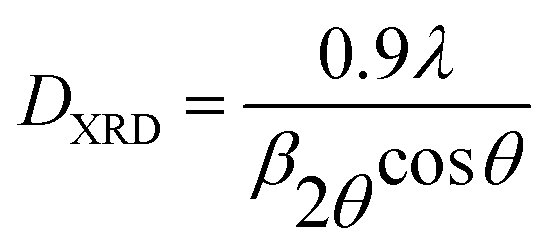
Fig. 4 shows the XRD and magnetization curves fitted with Langevin function at 300 K for the as synthesized AM as well as TMA. The diffraction patterns for all the powder samples are almost identical with the presence of characteristic finger-print reflections of magnetite. The peaks are identified as Fe3O4 with cubic fluorite structure (JCPDS 19-0629) having space group (227) shown as vertical drop lines. However, the peaks were significantly broader due to small crystallite size and associated lattice strain. The DXRD are calculated to be 8.67 and 10.69 nm for AM and TMA respectively.
 | ||
| Fig. 4 (A) XRD patterns of synthetic magnetite crystals. The vertical drop lines are the theoretical Bragg positions for inverse spinel magnetite phase following JCPDS card no. 19-0629. (B) Magnetization versus applied magnetic field profiles with typical Langevin fits represented by solid red lines with the magnetization data as a function of 1/H for (a) AM and (b) TMA are shown in the inset. | ||
The magnetization curves indicate a superparamagnetic behavior for both the studied specimens, as evidenced by both zero coercivity and zero remanence on the magnetization loop. Due to the asymptotic increase of magnetization for high fields (see Fig. 4), the Ms value can be measured from the fitting of the M vs. 1/H curves, extrapolating the magnetization value to 1/H = 0. The saturation magnetization for AM and TMA are 64.68 and 57.92 emu g−1 of sample respectively. The Ms value of TMA is ∼90% of that of AM and is due to (i) lowering the proportion of magnetite by the presence of nonmagnetic organic molecules along with (ii) the smaller particle sizes (Fig. 2). The calculated values of Ms, individual magnetic moment, magnetic size of the NPs and fit results from Langevin function are presented in Table 1.
| Sample | Exp Ms (emu g−1) | Cal Ms (emu g−1) | μ (emu per NP) | R2 | Standard error | DMag (nm) |
|---|---|---|---|---|---|---|
| AM | 64.68 | 65.14 | 1.28 × 10⁻¹⁶ | 0.9989 | ±1.87 | 8.98 |
| TMA | 57.92 | 58.30 | 1.16 × 10⁻¹⁶ | 0.9988 | ±1.74 | 9.02 |
The mean diameter of the superparamagnetic TMA particles can therefore be estimated to be ∼9.02 nm, which is smaller than the average physical size of ∼10.3 nm (magnetic core + non-magnetic outer shell) as determined from TEM measurements. XPS spectrum of TMA powder shown in Fig. 5 was used to study the valence states of Fe in the as-prepared NPs after functionalization. The XPS wide spectrum shows the peaks attributed to the core levels of O (A), Fe (A), Fe 2p and 3p, O 1s, C 1s where the Fe 2p electron core level is characterized by 2p1/2 and 2p3/2 series peaks. Fe 2p3/2 and Fe 2p1/2 double peaks correspond to the binding energies of 709 and 722 eV respectively (Fig. 5B). The double peaks in high resolution Fe 2p scan are broadened due to the appearance of Fe2+ (2p3/2) and Fe2+ (2p1/2) in magnetite which is in agreement with the reported literature.34
 | ||
| Fig. 5 (A) Survey spectrum and high resolution XPS, (B) Fe 2p, and (C) O 1s spectra of TMA sample. The solid red line over a raw data is a typical fit by XPS software. | ||
The relative peak areas of Fe2+ and Fe3+ in the high resolution 2p scan were calculated as 0.33:0.61, close to that of the stoichiometric Fe3O4, which could also be shown as FeO·Fe2O3. The predominant peak at 528.3 eV is attributed to O 1s,35 which can be deconvoluted to the binding energies of 528.3 and 529.6 eV belonging to the lattice oxygen in Fe3O4. The lower intensity of the C 1s peaks compared to that of Fe and O proves that the organic layer around MNPs is substantially thinner. The asymmetric C 1s peak is deconvoluted into three peaks at 284.6, 285.5 and 290 eV (see inset Fig. 5A). The main peak at 284.6 eV is assigned to C–H bonds in methyl group,36 and the second peak centred at 285.5 eV can be attributed to C–N bonds.37 The first two peaks of C 1s belonging to TMA attached electrostatically to the surface hydroxyls and the third very weak peak at 289 eV is ascribed to trace amount of CO2 adsorbed onto the fine NPs surface. The amount of residual TMAH in the dried TMA NPs was too little, so that the N 1s signal obtained by the XPS detector was very weak and almost absent.
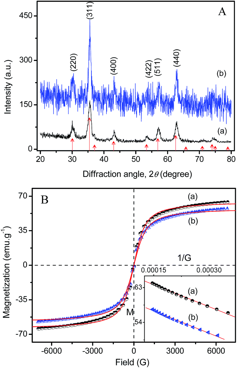
The variations in the zeta potential of uncoated and TMA peptized magnetite as a function of the pH and their phase plots are shown in Fig. 6 and S3 in ESI.† The zeta potential is the electrical potential measured at the shear plane, and represents the portion of the charge that can exert electrostatic attraction and repulsion forces on neighboring particles in suspension. The magnitude of the ζ-potential is proportional to the amount of charge on the NPs surface. The ζ-potential values of AM and TMA suspensions at their natural pH and the suspension stability are shown in Table 2. Our experiments were performed at the natural pH of the AM suspension where no electrostatic repulsion is expected. Bare magnetite nanoparticle surfaces are slightly positively charged (pH 6.7) and the small electrostatic repulsive forces among the particles is not able to prevent particle–particle contact and consequent clustering as was indicated by the zeta potential of 4.9 mV (Table 2). TMA suspension peptized with strong organic base TMAH shows very high zeta potential of −44 mV in spite of dilution (∼1:5) and exhibited ultra stability over 1 year without any apparent precipitation. The +N(CH3)4 cations of TMAH interact electrostatically with the OH groups attached to the bare magnetic NPs surface.
 | ||
| Fig. 6 Variations in the zeta potentials of (a) TMA and (b) AM magnetite suspended in Millipore water as a function of the fluid pH. | ||
| Sample | Zeta (mV) | pH | Stability |
|---|---|---|---|
| AM | +4.9 | 6.7 | 1 h |
| TMA | −44 | 11.75 | >1 year |
This surface structure creates electrostatic interparticle repulsion that can overcome the coagulation forces of magnetic interaction and van der Waals attraction forces among NPs in water. Zeta potentials of the NPs are pH dependent and was observed to flip on both sides of the isoelectric point from positive surface charge (+ve zeta potential) in acidic pH to negatively charged surfaces as reflected in the negative zeta potential at basic pH. As pointed out in Fig. 6, the point of zero charge (PZC) slightly moved from 5.88 for unmodified NPs to higher pH values 5.98. Pristine magnetite NP dispersion exhibited ζ-values in the range of +40.3 to −39.8 mV in the 2 to 13.1 pH range. Adsorption of [(CH3)4N]+ ions tend to increase the negative charge of the magnetite further to −44.2 mV at alkaline pH and increases positive charge to 48.7 mV in the acidic range. The TMA coated magnetite was extremely stable in both acidic and alkaline pH except for the pH close to their PZC between 5 and 7. The higher clustering tendency of NPs in AM can be explained by the bigger particle sizes and as a result higher crystallinity (Fig. 4A). In larger particles of well-crystallized magnetite, the bulk properties supersede surface properties, and they are expected to magnetically attract more strongly each other.38
Moreover, the role of the ionic strength on the particle size also is largely dependent on the nature of the electrolyte. The smallest cations being the best screening ions, their influence on the surface charge is highest. Therefore, the TMA ferrofluid was stable above the pH > 7 and below pH < 5. The phase plot obtained with TMA sample is excellent in quality in all the pH ranges and flipped from negative to positive zeta while varying the pH from 13 to 2 shown in Fig. 6.
The hydrodynamic diameter (Z-average size, Zav) and the corresponding correlation function against time are depicted in Fig. 7. Few snap shots of PCS plots from the machine for TMA slurry at various temperatures 25, 35, 45, 65 °C are shown in Fig. S4.† The overall yield of Fe3O4 into aqueous dispersed MNPs was >95% for TMA. The Zav size was estimated by running the experiment in dynamic auto-mode using Zetasizer Nano ZS in which the machine optimizes all machine parameters to derive the best and reproducible size data. Aqueous dispersion of AM showed NPs in 33–825 nm size range with Zav size of 129 nm with PDI 0.358 indicating polydisperse nature of the distribution. The calculated number distribution, Nav is 43 nm. This is further consolidated by the poor fit to the correlation function (panel A in Fig. 7). The signal correlation due to the random thermal motion of NPs reduced to ∼0.74 after 100 μs in TMA where as the correlation of the signal takes a long time to decay and remained almost unchanged at ∼1.0 at the same point of time (100 μs) in case of AM containing slow moving larger particles. The Zav size for TMA is 28 nm with the presence of particles in the 8.7–91 nm size range which is ∼2.5 times larger than DTEM size of 10.3 nm (Fig. 2). The DPCS of ∼28 nm could be due to tetrahedrally arranged close-packed clusters of a total of maximum 4 particles with average hydrodynamic diameter of ∼2.5 × DTEM = 26 nm which is very close to 28 nm. The algorithm in the Malvern size measurement software converts the intensity of photon signal to size by cumulants analysis. Photon scattering techniques rely on the fact that the intensity of scattered light increases by about a million times if the size of particles responsible for photon scattering increases by one order of magnitude.39 In other words, TMA suspension contains more than 99% 13 nm particles with <0.01% of 28 nm clusters. The PDI (0.228) indicates the complete absence of any larger aggregates as supported by good fit to the correlation data also.
 | ||
| Fig. 7 Hydrodynamic size (Zav) of (A) pure as well as (B) peptized synthetic magnetite. The correlation function of the same is plotted against time. (C) Raw correlation data of the (a) pure and (b) coated magnetite. | ||
To understand the clustering kinetics, hydrodynamic size, PDI, mean photon count in TMA slurry was recorded as a function of NP concentration, temperature and time and is provided in Fig. 8 and S5.† The measurement has been carried out under static mode when, ideally, the number of photons scattered is proportional to the Zav of the scattering NPs. When the concentration of NPs was 25 mM (neat) as well as its 1:1 dilution, the dispersion exhibited the smallest reproducible Zav size of ca. 28 nm which confirmed the excellent colloidal stability of TMA where as its Nav (13 nm), calculated from the intensity distribution, is close to the primary particle size as observed from TEM (10.3 nm). The Zav size increased from 28 to 46 nm when NP concentration was decreased from an initial 25 mM to 4.2 mM with a concomitant decrease in the mean photon count rate from 450 to 260 kcps. However, the TMA suspension was always stable irrespective of MNPCs concentrations in water.
 | ||
| Fig. 8 (A) Zav size and the colloidal stability as a function of temperature for MNPs suspension examined at various concentrations, the mean particle size (dotted line) plotted against mean count rate (solid line) with different concentrations neat (square box), 1:1 (triangle), 1:5 (star) dilutions. (B) Zav and PDI of magnetite dispersion plotted as function of time. Insets are the corresponding TMA suspensions used in the experiments. | ||
When temperature for 25 mM slurry was increased from 25 to 45 °C, the hydrodynamic size 28.3 nm grew to 32.3 nm and decreased down to 27.3 nm when the temperature was further increased from 45° to 65 °C. The increase in size and mean photon counts till 45 °C may be due to the transformation of cluster structure from initial tetrahedral (2.5 × DTEM) to one-shell hexagonal cose-packed (3 × DTEM) and back to the tetrahedral at ∼65 °C. In the process, the count rate changed by ∼22% from initial of 450 kcps in 25 mM suspension. No obvious change in the Zav size (∼28 nm) was observed with ageing time extended even upto 12 months (panel B).
We observed that the ferrofluids are stable on ageing at room temperature over a year even after the temperature treatment, with no evidence of flocculation or settling and there was no change in the hydrodynamic size. These results show that monodispersed nanoscale magnetite crystals have been synthesised in aqueous medium which retained its magnetic properties, with very good water dispersibility. This makes them suitable as a candidate for biomedical applications.
The FTIR and the thermal analysis patterns of magnetite specimens are presented in Fig. 9. The two most intense IR absorption bands in the 630–550 cm−1 range is attributed to the lattice vibrations of Fe–O bonds in tetrahedral and octahedral sites and must have been resulted from the split of the ν1 band at ∼570 cm−1. The band at ∼447 cm−1 is due to the octahedral Fe only and corresponds to the ν2 band of Fe–O of bulk magnetite. FTIR spectra also confirm the presence of fcc magnetite in the materials as was previously confirmed from the TEM and XRD data (Fig. 2 and 4) also. The intensity reduction in TMA with respect to that of AM is probably due to the presence of tetramethylammonium ions on the Fe3O4 NP surface. The presence of O–H stretching vibration at ∼3415 cm−1 and O–H deformed vibration (bending modes) at 1622 cm−1 are attributed to the presence of coordinated OH groups or water molecules with the unsaturated surface Fe atoms.40
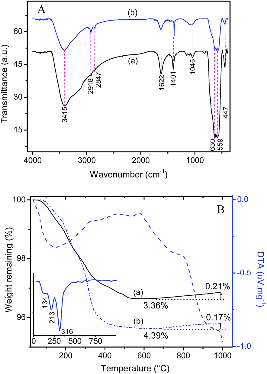 | ||
| Fig. 9 (A) FTIR spectra, and (B) thermogravimetric and DTA profiles of (a) pure and (b) TMA magnetite. Inset in B is the first derivative of TG spectrum of TMA. | ||
C–O stretching vibrations of CO![[double bond, length as m-dash]](https://www.rsc.org/images/entities/char_e001.gif) 3 anion at 1401 cm−1 (ν3) in both the samples are due to atmospheric CO2.41 Small absorption bands at 2918 and 2847 cm−1 are due to the νas(C–H) and νs(–CH3) vibrations of tetramethyl group in TMA sample. The very small hump at ∼2918 cm−1 in AM might have come from some remaining oil impurities from the rust-protecting oil applied to the IR mould. The presence of which was confirmed by XPS also.
3 anion at 1401 cm−1 (ν3) in both the samples are due to atmospheric CO2.41 Small absorption bands at 2918 and 2847 cm−1 are due to the νas(C–H) and νs(–CH3) vibrations of tetramethyl group in TMA sample. The very small hump at ∼2918 cm−1 in AM might have come from some remaining oil impurities from the rust-protecting oil applied to the IR mould. The presence of which was confirmed by XPS also.
The TG profiles of AM and TMA show a total weight loss of 3.15 and 4.22% respectively with similar patterns of three-step decomposition in the temperature range 30–995 °C. The peak centred at ∼130 °C is responsible for the removal of molecular water confined in the pores and or chemisorbed on the crystal lattice. The next exothermic peak at ∼213 °C for the removal of crystalline water and unwashed chlorides from the material followed by the last peak for a huge loss nearly 4% at ∼310 °C is due to burning of carbon products from the decomposition of tetramethyl ammonium group attached to the nanocrystals surfaces in the case of TMA sample. The weight loss increased till ∼565 °C and it could be attributed to the removal of multi-layers of water of hydration from the surface of the NPs as well as dehydration of iron oxyhydroxide (FeOOH) formed in the ambient moisture. A small weight gain of ∼0.2% was observed after 640 °C for AM and 0.21% after 668 °C onwards for TMA sample which is due to oxidation of magnetite to γ-Fe2O3, though the experiment was conducted under continuous nitrogen purge. The TMAH content in TMA magnetite can easily be calculated as 1.1%. When this is applied to the Ms value of AM, one can expect a value of 63.97 emu g−1 for TMA instead of actual 57.92 emu g−1. We now conclude that nearly 9% reduction in the Ms value of TMA is due to its reduced crystal dimension of 10.3 nm compared to that (11.6 nm) in AM in addition to ∼1% reduction due to the nonmagnetic chemical surrounding in TMA.
The pore structure and the BET surface area of the pure and peptized samples were investigated by nitrogen isothermal adsorption and is shown in Fig. 10. The isothermal gas adsorption–desorption exhibits a type IV profile according to the IUPAC nomenclature of mesoporous materials where the lower curve represents the adsorption of N2 gas on the surfaces of the NPs, while the upper curve represents the progressive withdrawal; desorption of the adsorbed N2.42,43 Surface area was estimated from the desorption branch of the isothermal gas-adsorption isotherm which indicated that there is no apparent changes in the pore structure on peptization.
 | ||
| Fig. 10 Nitrogen isotherms and their pore size distribution profiles for magnetite nanocrystals (a) before and (b) after peptization with TMA. | ||
The BET surface area of 106 m2 g−1 in TMA with a pore volume of 0.3051 cm3 g−1, is comparable to that of AM (102 m2 g−1, pore volume 0.3426 cm3 g−1). From the isothermal gas desorption, based on the BJH method, a mononodal pore size distribution of around ∼12 nm was estimated in both the magnetites. The BET SA in TMA (106 m2 g−1) can be equated to DBET of 10.9 nm which is slightly larger than the DTEM (10.3 nm) probably because of presence of surface coatings.2 102 m2 g−1 in AM gives rise to a DBET size of 11.4 nm which very close to the DTEM of 11.6 nm. Probably the creation of SA due to the presence of interparticulate pores just counterbalanced the loss in SA due to clustering.
The magnetite nanocrystal dimensions, determined by TEM, XRD, Langevin fitting magnetization curve, PCS and BET techniques are summarized in Table 3. A very close observation is that the dry nanoparticles characterized by BET, TEM, XRD, magnetization curve, proven the mesoporous single crystalline uniform size distributed, superparamagnetic behavior of magnetite characteristic belongs to FCC inverse spinel crystal system. Size measurement based on the light scattering method is one of the most important techniques by which one can have in the in situ information on the state, clustering and extent of hydration of NPS in suspensions where there are many counter acting forces intercating. The situation becomes more complicated when NPs are dispersed in water. The temperature, concentration and time dependent kinetics of growth of hydrodynamic size confirmed their stability of TMA suspension in this study.
| Sample | DTEM (nm) | DXRD (nm) | DMag (nm) | DPCS (nm) | DBET (nm) |
|---|---|---|---|---|---|
| AM | 11.6 | 8.7 | 8.98 | 43 | 11.4 |
| TMA | 10.3 | 10.7 | 9.02 | 13 | 10.9 |
Conclusions
The nanocrystalline magnetite, used in highly stable TMA nanofluid, have been synthesised by ammonia coprecipitation in physiological saline. HRTEM, SAED and XRD confirmed the magnetite phase belonging to FCC inverse spinel and had a BET SA of 106 m2 g−1. The reduced Ms value of 58 emu g−1 in TMA specimen is due to its reduced crystal dimension along with small contribution of the presence of non-magnetic TMAH. TG-DTA, EDAX, as well as XPS and FTIR spectra confirmed the presence of surface coordinated tetramethyl group in TMA. TG-DTA as well as EDAX also confirmed the successful coating of tetramethyl ammonium hydroxide on nanoparticles in TMA. The reduced Ms value in TMA specimen is more due to its reduced crystal dimension along with small contribution of the presence of non-magnetic TMAH. The size of the magnetite one-shell nanoclusters was controlled within 28.3–32.3 nm range in a wide temperature range. The novelty of this work is that this is an successful attempt to correlate the stability of ultra-stable colloidal suspension of the superparamagnetic NPs against time and temperature by simple photon correlation spectroscopy in conjuction with colloid surface properties. The monodispersity of these MNPs containing aqueous ferrofluid have no effect on heating which has great potential in hyperthermia and may find suitable in various biomedical and industrial applications.Acknowledgements
The authors are grateful to the Director, Central Glass & Ceramic Research Institute, Kolkata for permission and extending facilities to carry out the above work. SJI and MJ acknowledge CSIR and UGC for their fellowships. SJI thanks Mrs Asha Krishnan for deconvoluting the XPS data. Staff members of XPS, XRD and MCID are also sincerely acknowledged. We thank 12 FYP CSIR Network project ESC-0103 for funding the PCS facility.Notes and references
- X. Li, H. Si, J. Z. Niu, H. Shen, C. Zhou, H. Yuan, H. Wang, L. Ma and L. S. Li, Dalton Trans., 2010, 39, 10984 RSC.
- S. J. Iyengar, M. Joy, C. K. Ghosh, S. Dey, R. K. Kotnala and S. Ghosh, RSC Adv., 2014, 4, 64919 RSC.
- S. Garcia-Jimeno and J. Estelrich, Colloids Surf., A, 2013, 420, 74 CrossRef CAS.
- K. L. Lai, W. Jiang, J. Z. Tang, Y. Wu, B. He, G. Wang and Z. W. Gu, RSC Adv., 2012, 2, 13007 RSC.
- Y. Piao, J. Kim, H. Bin Na, D. Kim, J. S. Baek, M. K. Ko, J. H. Lee, M. Shokouhimehr and T. Hyeon, Nat. Mater., 2008, 7, 242 CrossRef CAS PubMed.
- M. Mahmoudi, A. Simchi, M. Imani, M. A. Shokrgozard, A. S. Milani, U. O. Haefeli and P. Stroeve, Colloids Surf., B, 2010, 75, 300 CrossRef CAS PubMed.
- Z. X. Sun, F. W. Su, W. Forsling and P. O. Samskog, J. Colloid Interface Sci., 1998, 197, 151 CrossRef CAS PubMed.
- R.-C. Wang, X.-B. Fu, X. Liu, H.-J. Liu, Y. Chen and J. Cui, RSC Adv., 2013, 3, 17016 RSC.
- C. Galindo-Gonzalez, M. T. Lopez-Lopez and J. D. G. Duran, J. Appl. Phys., 2012, 112, 043917 CrossRef.
- Y.-G. Han, M. Aoyagi, M. Kogiso, M. Asakawa and T. Shimizu, Colloids Surf., A, 2012, 395, 63 CrossRef CAS.
- J. Philip, T. Jaykumar, P. Kalyanasundaram and B. Rai, Meas. Sci. Technol., 2003, 14, 1289 CrossRef CAS.
- Y. Zhang, G. Zhu, J. Lu, Z. Guo and J. Cao, RSC Adv., 2015, 5, 87841 RSC.
- U. Jeong, X. Teng, Y. Wang, H. Yang and Y. Xia, Adv. Mater., 2007, 19, 33 CrossRef CAS.
- J. P. Huang and K. W. Yu, Appl. Phys. Lett., 2005, 86, 041905 CrossRef.
- S. L. Pu, X. F. Chen, L. J. Chen, W. J. Liao, Y. P. Chen and Y. X. Xia, Appl. Phys. Lett., 2005, 87, 021901 CrossRef.
- V. Mahendran and J. Philip, Appl. Phys. Lett., 2012, 100, 073104 CrossRef.
- L. F. Shen, P. E. Laibinis and T. A. Hatton, Langmuir, 1999, 15, 447 CrossRef CAS.
- T. Ninjbadgar and D. F. Brougham, Adv. Funct. Mater., 2011, 21, 4769 CrossRef CAS.
- F. Vereda, J. de Vicente and R. Hidalgo-Alvarez, Colloids Surf., A, 2008, 319, 122 CrossRef CAS.
- D. Ramimoghadam, S. Bagheri and S. B. A. Hamid, J. Magn. Magn. Mater., 2015, 379, 74 CrossRef CAS.
- S. Ghosh, Ph. D. Thesis, Dublin City University, Ireland, 2006.
- P. Majewski and B. Thierry, Crit. Rev. Solid State Mater. Sci., 2007, 32, 203 CrossRef CAS.
- E. Allard-Vannier, S. Cohen-Jonathan, J. Gautier, K. Herve-Aubert, E. Munnier, M. Souce, P. Legras, C. Passirani and I. Chourpa, Eur. J. Pharm. Biopharm., 2012, 81, 498 CrossRef CAS PubMed.
- L. Xiao, J. Li, D. F. Brougham, E. K. Fox, N. Feliu, A. Bushmelev, A. Schmidt, N. Mertens, F. Kiessling, M. Valldor, B. Fadeel and S. Mathur, ACS Nano, 2011, 5, 6315 CrossRef CAS PubMed.
- L. Lartigue, P. Hugounenq, D. Alloyeau, S. P. Clarke, M. Levy, J.-C. Bacri, R. Bazzi, D. F. Brougham, C. Wilhelm and F. Gazeau, ACS Nano, 2012, 6, 10935 CrossRef CAS PubMed.
- Z. Tu, B. Zhang, G. Yang, M. Wang, F. Zhao, D. Sheng and J. Wang, Colloids Surf., A, 2013, 436, 854 CrossRef CAS.
- J. K. Stolarczyk, S. Ghosh and D. F. Brougham, Angew. Chem., Int. Ed., 2009, 48, 175 CrossRef CAS PubMed.
- D. Brougham and S. Ghosh, US Pat., US 8435496 B2, 2013.
- D. Sakellari, S. Mathioudaki, Z. Kalpaxidou, K. Simeonidis and M. Angelakeris, J. Magn. Magn. Mater., 2015, 380, 360 CrossRef CAS.
- C. P. Bean, J. Appl. Phys., 1955, 26, 1381 CrossRef.
- C. P. Bean and I. S. Jacobs, J. Appl. Phys., 1956, 27, 1448 CrossRef CAS.
- C. P. Bean and J. D. Livingston, J. Appl. Phys., 1959, 30, S120 CrossRef.
- R. M. Cornell and U. Schwertmann, The iron oxides: structure, properties, reactions, occurrences and uses, in The iron oxides, VCH, New York, 1996, p. 117 Search PubMed.
- Y. Lai, W. Yin, J. Liu, R. Xi and J. Zhan, Nanoscale Res. Lett., 2010, 5, 302 CrossRef CAS PubMed.
- B. Li, H. Cao, J. Shao, M. Qu and J. H. Warner, J. Mater. Chem., 2011, 21, 5069 RSC.
- D. K. Jha, M. Shameem, A. B. Patel, A. Kostka, P. Schneider, A. Erbe and P. Deb, Mater. Lett., 2013, 95, 186 CrossRef CAS.
- X. Wang, X. Liu, L. Lai, S. Li, J. Weng, Z. Zhou, Q. Cui, X. Chen, M. Cao and Q. Zhang, Adv. Funct. Mater., 2008, 18, 1809 CrossRef CAS.
- T. Hosono, H. Takahashi, A. Fujita, R. J. Joseyphus, K. Tohji and B. Jeyadevan, J. Magn. Magn. Mater., 2009, 321, 3019 CrossRef CAS.
- C. J. Meledandri, J. K. Stolarczyk, S. Ghosh and D. F. Brougham, Langmuir, 2008, 24, 14159 CrossRef CAS PubMed.
- A. Ebrahiminezhad, Y. Ghasemi, S. Rasoul-Amini, J. Barar and S. Davaran, Bull. Korean Chem. Soc., 2012, 33, 3957 CrossRef CAS.
- C. P. Chen, P. Gunawan and R. Xu, J. Mater. Chem., 2011, 21, 1218 RSC.
- F. Liebau, Microporous Mesoporous Mater., 2003, 58, 15 CrossRef CAS.
- L. B. McCusker, F. Liebau and G. Engelhardt, Pure Appl. Chem., 2001, 73, 381 CrossRef CAS.
Footnote |
| † Electronic supplementary information (ESI) available. See DOI: 10.1039/c5ra26488j |
| This journal is © The Royal Society of Chemistry 2016 |