Ying Zhang,
Xuejing Wang,
Shenghua Ma,
Kunpeng Jiang and
Xiaojun Han*
State Key Laboratory of Urban Water Resource and Environment, School of Chemical Engineering and Technology, Harbin Institute of Technology, 92 West Da-Zhi Street, Harbin, 150001, China. E-mail: hanxiaojun@hit.edu.cn
First published on 22nd January 2016
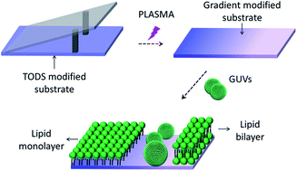
The relationship between surface wetting properties and lipid membrane status formed via giant unilamellar vesicle rupture was investigated using chemical gradient surfaces. Fluorescence microscopy and AFM analysis confirmed that GUVs could form uniform monolayers, monolayer patches and bilayer patches on surface regions with contact angles ranging from 108° to ∼61°, ∼60° to ∼55° and less than 5°, respectively. The intact GUVs stand in the area with contact angle between ∼54° and ∼28°.
The gradient surfaces, including chemical gradients and topography gradients, have been demonstrated to be a powerful system in many studies in chemical, physical, and biological science.5–7 A number of methods have been used to prepare surface gradients. Luk et al. fabricated a gold film with gradient nanotopography using varying angle vapor deposition to investigate the adhesion of mammalian cells.8 Xu et al. prepared mussel-inspired polydopamine (PDA) gradients on different surfaces by simply immersing substrates into a dopamine solution at a tilt angle.9 Moreover, some gradient surfaces could generate a contact angle gradient simultaneously. Among them, the space limited plasma oxidization method could create the substrate with both chemical gradient and contact angle gradient.10
Giant unilamellar vesicles (GUVs) were rarely used to study the process of vesicle fusion method.11–13 Most relevant reports were based on the small unilamellar vesicles (SUVs) rather than GUVs (larger than 1 μm in diameter).14 However, because the size of GUVs is closer to that of mammalian cells, they play a more crucial role in studying the dynamics and structural features of cells, including budding and endocytosis,15–17 etc.
Herein we investigated the influence of surface with different contact angle on supported lipid membrane formation using GUVs, and found out 4 regions with various types of lipid membranes, which may provide clues for the surface wetting property influence on cell immobilization.
In order to form the contact angle gradient substrates, the exposure time in air plasma should be decided firstly. To this aim, the power of the plasma was fixed at 80 W and the exposure time of TODS SAMs modified substrates in air plasma was varied from 10 s to 160 s. From Fig. S1† it is noted that the contact angle decreases from 108° to ∼5° as the exposure time increases to 120 s, and levels off afterwards. Therefore 2 min was chosen as the exposure time.
A freshly prepared SAM modified substrate covered by a silicon wafer with an extremely thin spacer at one end to form a “wedge” shape space, as shown in Fig. 1a, was loaded in the plasma generator for 2 min to create the chemical gradient surface. The contact angle of the resultant surface was measured from position 1 to 3 along the middle line (dash line shown in Fig. 1a) as shown in Fig. 1c. It is clear to see that a gradient surface has been created by this way. The initially hydrophobic surface (108°) became gradually hydrophilic from position of 0 mm to 5 mm (position 1 to 2 in Fig. 1a). The depth of the gradient is about 80°. In the area between 5 mm to 7 mm (position 2 to 3 in Fig. 1a), the contact angles are constant. The images of water droplets in different position, shown in Fig. 1c, also confirmed the formation of gradient surface. The relationship between the contact angle range (region c1–c4 in Fig. 1c) and lipid membrane formation will be illustrated in the following section.
The gradient substrates were incubated in GUVs solution for 2 h, followed by rinsing with abundant water to remove the residual GUVs. Fig. 2a shows the fluorescence microscopy images of the finally substrates along the dash line in Fig. 1a. The perpendicular dark lines in this image are unreal, which are caused by the dim marginal area of the fluorescence microscope. Getting rid of this problem, when we linked the image with the contact angle of the substrate shown in Fig. 1c, it could be found that the substrate can be divided into four parts, including homogeneous area (on the strongly hydrophobic surface with contact angle from 108–61°, Fig. 2a1), patches (on the weakly hydrophobic surface, ∼60° to ∼55°, Fig. 2a2), bright spots (on the surface with contact angle between 54° and 28°, Fig. 2a3) and bright patches (on the surface contact angle less than 5° Fig. 2a4). In order to get more detailed information, higher resolution microscopy images of different areas were taken (Fig. 2b). From these close-up pictures, it can be observed clearly that the bright spots in Fig. 2a3 are GUVs attached on the surface (Fig. 2b3). The features of GUVs observed here are in consistent with those of free GUVs (Fig. S2†), which leads us to propose the membrane states in region c3 are intact GUVs. There is homogenous membrane and bright patches on area of a1 and a4 respectively, because the area a1 is very hydrophobic surface and area a4 is hydrophilic surface. The lipid membranes on them should be lipid monolayer and lipid bilayer respectively. Moreover, according to the fluorescence intensity analysis, under same experiment condition the value of fluorescence intensity in Fig. 2b1 and b2 is 50% of that in Fig. 2b4, because there are only as half number of lipids in the monolayer as that in the bilayer on the same size of area. Meanwhile, the average size of patches in Fig. 2b2 is 36.1 ± 1.8 μm2, which is properly as twice big as that in the Fig. 2b4 (19.7 ± 2.4 μm2). This result also confirms that they are monolayer and bilayer respectively, because the size of monolayer is equal to the size of inner leaflet plus that of the outer leaflet of GUVs. The formations of lipid membranes in different regions were schematically depicted at the bottom row of Fig. 2b.
To further characterize the lipid membranes, AFM images and section profiles were obtained (Fig. 2c). Height of defects in Fig. 2c1 and lipid membrane patch in Fig. 2c2 is 2.45 nm and 2.62 nm respectively, which confirms the lipid membrane formed on the contact angle range of 108° to ∼55° is lipid monolayer. Whereas the height of lipid membrane prepared on the surface with contact angel less than 5 degree is 5.15 nm, which proves the membrane in this area is lipid bilayer. The height analysis is in accordance with the results of fluorescence microscopy images in this study.
Therefore, it can be concluded that lipid monolayer was formed on the surface with strongly hydrophobic property (∼61° to ∼108°); the monolayer patches were formed on the weakly hydrophobic surface (∼55° to ∼60°); GUVs were intactly attached on the surface with contact angle from ∼28° to ∼54°; lipid bilayer patches were formed on the hydrophilic area where the contact angle was less than 5°. It is noteworthy that, the minimum lipid concentration for preparation of uniform monolayer on the surface with contact angle range from ∼61° to ∼108° is 1 μg mL−1.
The physical mechanism behind the formation uniform monolayers with GUVs fusion on hydrophobic surface locally relates the surface energy between the hydrophobic SAM surface and the vesicle solution. The high energy aroused by the exposure of a hydrophobic surface to an aqueous environment is thermodynamically unfavourable, which leads to vesicle rupture and fusion spontaneously to form lipid monolayer on top of SAM surface.12 As for the lipid bilayer formation process, the high curvature caused by the deformation of GUV strongly adsorbed on the hydrophilic surface plays a critical role.11 Obviously, the rupture of GUVs could dissipate the energetically unstable situation provided by the highly curved region. On the other hand, in the medium range of contact angle region (∼28° to ∼54°), vesicles “stood” on the substrate because the surface energy is not sufficient enough to drive vesicles to rupture. As for the region with partially plasma oxidation (∼55° to ∼60°), polar functional groups, including carboxyl, aldehyde and hydroxyl group, could promote H-bond formation between water molecules and substrate interface, consequently to reduce the surface free energy and prevent homogeneous lipid monolayer formation.18
Fluorescence recovery after photobleaching (FRAP) (Fig. 3) was used to measure the diffusion coefficient D of TR-DHPE within the lipid monolayers area (Fig. 2b1). The images in Fig. 3a and b were taken 10 s and 10 min after photobleaching, respectively. A dark spot can be seen in the centre of the square 10 s after photobleaching, but after 10 min the fluorescence intensity is homogeneously distributed. The D value was found to be 1.33 ± 0.12 μm2 s−1 (over three samples) with a mobile fraction of 0.95, and these values are similar to those of the lipid monolayer formed on hydrophobic surfaces (1.0 μm2 s−1).19 The FRAP data confirm sufficient mobility of lipids in supported monolayer.
The membrane formed with LUVs of 200 nm was also studied. On the region with contact angle below 5°, uniform bilayer is formed. The uniform bilayer is supposed to be formed by merging small bilayer patches.11 The LUVs form smaller patch on the surface than GUVs. Smaller patches are thermodynamically unstable than bigger ones, which leads to LUVs rupture at the edge of those patches to form continuous bilayer membrane.11,20 For other areas, the membrane status is in consistent with those prepared with GUVs (Fig. S3†).
In summary, we discovered that the homogenous supported lipid monolayer could be prepared using GUVs fusion method on the substrate with contact angle between ∼61° and 108°. To the best of our knowledge, this is the first report of utilization of GUVs fusion in the creation of uniform supported monolayer system. The relationship between the wetting property of a surface and lipid membrane status was also revealed using a chemical gradient surface. Furthermore, the results also show that the GUVs could be intactly immobilized on the surface with contact angle from ∼28° to ∼54°, which provides an ideal platform for transmembrane protein reconstitution.
Footnote |
† Electronic supplementary information (ESI) available. See DOI: 10.1039/c5ra26205d |
This journal is © The Royal Society of Chemistry 2016 |