DOI:
10.1039/C5RA25514G
(Paper)
RSC Adv., 2016,
6, 12143-12157
An agile, simplified and sonication mediated one-pot aqueous extraction and antibacterial assessment of predominant Korean mushrooms†
Received
1st December 2015
, Accepted 8th January 2016
First published on 14th January 2016
Abstract
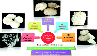
Solvent-based extractions have always held the upper hand when it comes to mushrooms. Assimilating the fact that mushrooms are a part of culinary components cooked in water and not in solvents; solvent-based extraction becomes a priority. Effective water based extraction stretches 24 h, leaving space for prospective improvising through analytical interference. We have demonstrated the effective downsizing of the extraction time from 24 h to 2 min via sonication based extraction strategies. A water bath-based method could achieve effective extraction at 30 min, whereas further enhancement was seen through the use of a probe sonication approach to 2 min. The extraction efficiency was tested based on the antibacterial activity of mushroom extracts against two pathogens, Streptococcus mutans and Pseudomonas aeruginosa. The systematic optimization of the sonication approach and a comparison of their effectiveness versus conventional approaches are demonstrated. The bioactive components in the extracts obtained via the different extractions have been characterized using biochemical characterization as well as GC-MS analysis. The enhanced extraction and potent role of butanoic acid, hexadecanoic acid, octadecanoic acid and 1,2-benzenedicarboxylic acid were confirmed to be behind the success behind the sonication mediated extraction.
Introduction
For time immemorial, mushrooms have been extensively known for their nutritive and medicinal roles. Mushrooms form an integral part of Asian cuisine, dominating countries such as China, Japan and Korea. With increased knowledge on the positive aspect of mushrooms, more recently there has opened a huge market for mushrooms. In terms of not only nutrition but also their specific aroma or texture, mushrooms have risen from a delicacy to a routine everyday regular food. The demands are growing on the basis of the fact that mushroom encompass a huge diversity of biomolecules established for not only nutritional but also their exceptional medicinal properties. Previous studies have reported that mushrooms are rich in proteins, polysaccharides, fibers, and minerals, yet low in lipid and caloric contents.1–3 Mushrooms are also a good source of natural antibiotics, through their low molecular weight (such as terpenes, steroids, and sesquiterpenes) and high molecular weight compounds.4 These bioactive compounds have been proved in the past for their anti-inflammatory, antiviral, antidiabetic, immunomodulatory antioxidant, antitumor and antimicrobial properties.4–8
Researchers have reported that mushroom extracts from either the fruiting body or mycelium show a wide range of antimicrobial and antioxidant activities.9–11 Turkoglu et al. 2007,12 have reported the ethanolic extract of Laetiporus sulphureus (Bull.) Murrill showed antioxidant activity, which positively correlates with their phenolic and flavonoid contents. Their corresponding antimicrobial activity, whereby they strongly inhibit the growth of Gram-positive bacteria and decrease the inhibition of Gram-negative bacteria has also been reported. Numerous organic solvent-based mushroom extracts have been trialed and reported for their antimicrobial activity. These include ethyl acetate and hexane extracts of Agaricus bisporus and reports on their antimicrobial activity against Micrococcus luteus, Bacillus subtilis and Bacillus cereus.13 Furthermore, an aqueous extract of Cordyceps sinensis has been demonstrated to inhibit the growth of Bacillus subtilis and Streptococcus epidermidis.10 Based on the current status of the literature survey on this research area, it appears that the shiitake mushroom (Lentinus edodes) emerges as the most studied mushroom species. Aida et al. 2009,14 reported the correlation that shiitake extracts exhibit higher antibacterial activity than the broad spectrum antibiotic, ciprofloxacin. Hirasawa et al. 1999,15 has further demonstrated that shiitake extracts have pronounced inhibitory action on various groups of bacteria including Streptococcus spp., Actinomyces spp., Lactobacillus spp. and Pseudomonas aeruginosa.
According to Kitzberger et al. 2007,9 extraction techniques play a vital role in the pre-selection of substances or components of interest. Thus far, extraction methods have employed sequential extraction13,15 followed by parallel analysis; some approaches made use of a single solvent-based extraction, while the extraction time was observed to be inconsistent ranging from 5 to 72 h.9,11,16–18 Some approaches use special equipment to assist the extraction process, such as bath sonication19 and supercritical fluid extraction, which are for solute extraction.9 The advantages of these techniques are that they are more effective and use less solvent. It is recognized that high intensity ultrasound can promote the mass transfer of different products and processes.20 Despite the high temperature induction that can lead to the formation of free radicals on the cell membranes, ultrasound sonication probes are proposed for plant extraction processes due to its high efficiency, reproducibility and lower solvent consumption.20–22
Prevalently, classical organic solvents, such as ethanol, methanol, chloroform, ethyl acetate and dichloromethane, have been used for the extraction of active compounds from mushrooms. Organic solvents are reported to be better solvents when it comes to extraction and several researchers have proved that ethanol was the best extractive solvent and shows maximum antimicrobial activity.18,23 However, Bala et al. 2011,19 have reported that the ethanolic and water extracts of two Nigerian edible mushrooms revealed similar antimicrobial activity. In addition, the drawback of organic solvents is their toxicity towards bacterial cells, which will affect the observations on the antimicrobial activity. Moreover, looking at the culinary point of view, people do not cook mushrooms in organic solvents, but water, thus a water-based extraction is inevitable for practical purposes. When compared to organic solvents, the universal solvent water is safer and easier to implement. However, a literature sweep reveals that the water-based extraction of mushrooms for testing their antibacterial properties has rarely been reported nor published.
The present study aims at looking into the antimicrobial activity of five predominantly used Korean mushrooms, which are consumed by a majority of the population as part of their daily diet. These mushrooms are usually consumed as soups or steamed in water; therefore, the extraction process for the extraction of the antimicrobial properties if any, based on water extraction has been worked out in detail. A comparison of the water-based extraction against conventional ethanolic extraction has also been made. The use of an accelerated sonication based extraction methodology based on water bath type sonication and probe type ultrasonication has been established. The antioxidant properties, bioactives, such as phenols, polysaccharides, flavonoids, and proteins, have been estimated and correlated to the success of the extraction process. The antibacterial properties of these extractions have been tested against Streptococcus mutans and Pseudomonas aeruginosa. For the first time, we optimize a sonication based procedure for the aqueous extraction of antimicrobial compounds from mushrooms in less than 5 min compared to the conventional process, which requires more than 24 h.
Materials and methods
Samples of fresh fruiting bodies of 5 predominant commercial mushrooms species used in Korean cuisine, namely, Pleurotus ostreatus, Agaricus bisporus, Lentinula edodes, Pleurotus eryngii, and Flammulina velutipes, were purchased from the local supermarket in Seoul, Korea. These will be referred to by the codes M1, M2, M3, M4 and M5, in the order represented above, in the following sections. After collection, each mushroom was freeze-dried and then ground into a powder. The samples were stored in sealed plastic bags at room temperature for further use.
Reagents and apparatus
Folin–Ciocalteu reagent, gallic acid and rutin were obtained from Sigma-Aldrich. 1,1-Diphenyl-2-picrylhydrazyl (DPPH) was obtained from Wako. Acridine orange was purchased from Alfa Aesar. All the other solvents and reagents were of analytical grade. Water was collected from a Milli-Q water purification system. Water bath sonication and probe ultrasonication were performed using a Bath Sonicator (JAC-2010, 300 W/60 Hz) and Probe Sonicator (Bandelin GM 2200, 200 W/20 kHz), respectively. The absorbance value was measured using a Shimadzu UV-1700 spectrophotometer. Fluorescence imaging was conducted using a Olympus FluoView™ FV1000 confocal laser scanning microscope (CLSM), OLYMPUS AMERICA INC. Corporate Center Drive, Melville, NY, USA. The bacterial cells were observed using field emission scanning electron microscopy (JSM-5410LV). For GC analysis, a Shimadzu GCMS-QP2010 SE with Rts-5MS column (Restek, 30 m × 0.25 mm, id × 0.25 μm film) was used.
Extraction procedures
Ethanol and water extraction
Ethanol is reported to be a promising solvent for conventional extraction of total phenolic compounds from mushrooms.24 Water extraction is also known to contain high amounts of polysaccharides and soluble protein compounds.25 To compare the difference between organic solvent extraction and water extraction, in the present study, both extractions were performed. Samples were prepared for water and ethanol extraction following the procedure of Barros et al., 2006 (ref. 16) with brief modifications. Two grams of lyophilized mushroom powder were immersed in 100 mL of distilled water (water extraction) or 70% ethanol (ethanol extraction) for 24 h at room temperature with magnetic stirring. The mixture was centrifuged at 10
000 rpm for 15 min at 4 °C. The residue was then re-extracted. The supernatants were combined and evaporated on a rotary evaporator at 40 °C, concentrated to approximately 20 mL and made up to 25 mL. The extracts were stored at −20 °C prior to analysis.
Sonication based water extraction
2 g of sample in 100 mL of water was sonicated for different sonication times (30 min, 60 min, and 180 min). The resulting suspension was centrifuged and evaporated as described above.
2 g of the respective mushroom powder was suspended in 100 mL of water and sonicated for 1 min, 2 min and 5 min with varying sonication frequencies of 10%, 20%, 50% and 100%. The resulting suspension was evaporated following the same protocol mentioned above.
Biochemical characterization
Determination of total phenolic compounds
The phenolic compounds in the mushroom water extracts were analyzed based on the spectrophotometric method described by Loots et al., 2007.26 Briefly, 50 μL of mushroom extract was mixed with 1150 μL of distilled water and then 200 μL of dilute Folin–Ciocalteu reagent was added. The mixture was mixed thoroughly and allowed to stand for 7 min at room temperature. Then, 600 μL of 20% sodium carbonate aqueous solution was added to this mixture. The reaction was incubated for 60 minutes and the absorbance measured at 765 nm against the blank using a spectrophotometer. Different concentrations of gallic acid (0.0325–0.5 mg mL−1) were used to construct the calibration curve. The results were expressed as mg of gallic acid equivalents per gram dry weight (mg GAE per g DW).
Determination of total flavonoid compounds
The total flavonoid content in the different mushroom extractions was estimated using a colorimetric assay according to the method of Chang et al., 2002 (ref. 27) with slight modifications. Briefly, 0.5 mL of mushroom extract was mixed with 0.1 mL of 10% aluminum chloride and 4.3 mL of distilled water. The mixture was incubated for 30 min at room temperature. Absorbance was then measured at 415 nm. Rutin was used to obtain the standard curve (15.15–500 μg mL−1). The final results were expressed as μg rutin equivalent per gram dry weight (μg RE per g DW).
Determination of total polysaccharides
The total polysaccharides were also measured using a colorimetric assay based on the procedure of Masuko et al., 2005.28 The stock mushroom extract was diluted 100 times using distilled water. 0.5 mL of the diluted sample was mixed with 1.5 mL of concentrated sulfuric acid and the mixture shaken for 30 min at room temperature. Subsequently, 0.3 mL of 5% phenol solution was added and the mixture heated for 5 min at 90 °C in a water bath, followed by reading the absorption value at 490 nm. The calibration curve was construed using standard D-glucose solutions (7.8125–250 μg mL−1).
Determination of 1,1-diphenyl-2-picrylhydrazyl radical scavenging activity
The free radical scavenging activity of the mushroom extracts was determined following the protocol reported by Chung et al., 2000.29 0.25 mL of each mushroom extract was mixed with 2.5 mL of 0.1 mM DPPH and made up to 3 mL with distilled water. The mixture was vortexed and incubated for 80 min at room temperature in the dark. The absorbance was obtained at a wavelength of 517 nm. The mushroom extract was replaced by distilled water, which served as the control sample. The free radical scavenging activity on DPPH was calculated using the equation as follows:
Free radical scavenging effect% = (1 − Abssample/Abscontrol) × 100 |
Evaluation of the antibacterial activity
The antibacterial activity of the mushroom extracts was determined via turbidometric assay using a spectrophotometer and a total viable count method using the plate count technique. Two bacteria strains, Streptococcus mutans 11823 (ATCC 25175) were purchased from the Korean culture center of microorganisms, Seoul, South Korea and Pseudomonas aeruginosa (KACC 11085), were used as the test organisms. Before, testing the antibacterial activity, all the mushroom extracts were filtered using a 0.20 μm sterilized bacterial filter (Minisart). S. mutans was cultured in Bacto™ Brain Heart Infusion broth and BBL™ Brain Heart Infusion Agar and P. aeruginosa was cultured in Difco™ Nutrient Broth and Difco™ Nutrient Agar.
5 mL quantities of BHI broth/nutrient broth were inoculated with S. mutans or P. aeruginosa, respectively and the mushroom extracts (concentrations varying from 500, 1000, and 2000 μL) were added and incubated in a shaker cum incubator at 35 °C overnight. The absorbance was measured at 600 nm using a spectrophotometer. The total viable count (TVC), indicating the number of bacteria that survived after interaction with the mushroom extract, was enumerated using the plate count method. The TVC was represented as cfu per mL (colony forming unit per mL).30 Fig. 1 gives a schematic of the work-flow used in this study.
 |
| Fig. 1 Schematic of the work flow followed in our study. | |
Post exposure analysis of bacteria using confocal laser scanning microscopy (CLSM)
To 500 μL of the interacted sample, 100 μL of acridine orange (0.1% solution in distilled water) was added and incubated in the dark for 10 min. After 10 min, the unbound stain was removed by centrifugation at 5000 rpm for 10 min. This washing was repeated thrice and the acridine orange stained cells were finally suspended in 500 μL of sterile distilled water. Then, 10 μL of the respective cell suspensions were laid on glass slides, covered with a cover slip and viewed using a fluorescence microscope. Acridine orange (Alfa Aesar, CAS: 10127-02-3), a fluorescent dye, differentially stains single stranded RNA and double stranded DNA, fluorescing orange when intercalated with the former and green when complexing with the latter. Thus, the number of orange fluorescing cells depicts the actively metabolizing cells and the green fluorescing cells depict the dead cells.31
Field emission scanning electron microscopy (FE-SEM)
FESEM (JEOL, JSM-5410LV) at an accelerating voltage of 2 kV was used to image the damage incurred on the bacterial cells following interaction with the M3 mushroom extract. 500 μL of the S. mutans and P. aeruginosa cells were pelleted via low speed centrifugation, the supernatant discarded, the cell pellet washed with sterile water thrice and resuspended in 500 μL of sterile water. 10 μL of each cell pellet suspension was laid on aluminium tape (dried overnight at 50 °C) and mounted on the stubs using carbon tape. The samples were sputter coated with platinum and viewed using FESEM.
Gas chromatography-mass spectrometry (GC-MS) analysis
The chemical constituents in the water extract (WE), bath sonication extract (WEBS) and probe ultrasonication extract (WEPUS) of M3 (which showed the highest antimicrobial activity) were identified using the GC-MS technique.32,33 Our samples were analyzed after TMS derivatization. 1 mL of the water extract samples were mixed with chloroform (1/10, v/v). The organic layer was collected and evaporated to dryness for GC-MS analysis. Another set of samples were prepared in ethanol, which were evaporated to dryness. Then, both the two residues were incubated with 100 μL of bis(trimethylsilyl)trifluoroacetamide (BSTFA) and 900 μL of pyridine at 70 °C for 1 h and analyzed using GC-MS. The injector and detector were set at 275 °C and 300 °C, respectively. Helium was used as the carrier gas. The column temperature was held at 70 °C for 10 min, then increased to 300 °C at a rate of 5 °C min−1 and maintained at this temperature for 20 min. Mass spectra were obtained using the electron impact mode. The identification of compounds was carried out based on the comparison of their fragmentation patterns and retention times consulting the NIST library.
Results and discussion
Antimicrobial activity of conventional extractions
Conventional extraction procedures involving ethanol based extraction and water extraction were used for extracting the bioactive components in five different mushrooms. Following the 24 h extraction procedure, the mushroom extracts were tested for their antibacterial activity against the oral pathogen, S. mutans. Four different concentrations, 30 μL mL−1, 150 μL mL−1, 300 μL mL−1 and 500 μL mL−1 were tested. Fig. 2 gives the results of this study, as observed from the results obtained from the spectrophotometric method, compared to the control, the 300 μL mL−1 concentrations of the M1, M2, M3, M4 and M5 extracts showed significant antimicrobial activity. It can be observed that in some cases the bacterial counts were even more than the control sample. This is because the extracts show no inhibitory properties, rather they serve as an additional nutritional source for the bacteria and therefore they grow better in here than in the control sample. With respect to EE, M1, M3 and M5 showed significantly higher antibacterial activity. In the case of WE, M2, M3 and M5 yielded significant results. However, when compared to EE, WE showed lower antibacterial activity. This has also been reported earlier in the literature. Jonathan and Fasidi, 2003 (ref. 18) have reported the effective extraction of antimicrobial compounds using organic solvents, indicating ethanol to be the most effective amidst the other solvents tested. However, the organic solvents by themselves are toxic to bacteria and are known to disrupt bacterial cell membranes34 and therefore solvent-based extractions for antibacterial studies would have their own limitations. Moreover, with respect to their practical application, the human consumption of mushrooms does not involve the use of solvents, but water. Therefore, to increase the practical implications of the study, it is necessary to look for water-based extraction methods or increase the efficacy of water-based extractions. This would facilitate the harnessing of the full potential of the antimicrobial bioactive compounds contained in these edible mushrooms. Hirasawa et al., 1999 (ref. 15) have shown that the aqueous extract obtained from L. edodes possesses antibacterial activity against S. mutans. Our results also indicate that WE of M3 (which is L. edodes) showed the highest total phenolic compounds and DPPH scavenging activity corresponding to the highest antibacterial activity compared to the other extracts studied.
 |
| Fig. 2 A graph showing a comparison of the antibacterial activity of S. mutans using conventional ethanol extraction (EE) and water extraction (WE) from M1 (Pleurotus ostreatus); M2: Agaricus bisporus; M3: Lentinula edodes; M4: Pleurotus eryngii; M5: Flammulina velutipes. | |
The antibacterial activity of the WE's of the five mushroom samples were further investigated in more detail against another Gram negative pathogen, P. aeruginosa. As observed from Fig. 3A(a and b), M1, M2, M3, M4 and M5 showed significant inhibitory activity on P. aeruginosa compared to S. mutans (Fig. 3B(a and b)). Moreover, it was interesting to note that the variation in the trend observed from the spectrophotometric method compared to the TVC method was distinctly high. The possible colored mushroom extracts and the likelihood that the mushroom extracts did not inhibit growth but rather led to cell death via cell interaction based mechanisms are probable explanations. Moreover, because the spectrophotometric method is a turbidity-based method, it is not a standalone technique for evaluating the live/dead condition of cells (because the dead cells will also add to the turbidity of the assayed suspension). This is the reason why the TVC method was also attempted to validate the results. Using the TVC method, we get more accurate information of the number of living cells following any antibacterial treatment.
 |
| Fig. 3 Antibactericidal activity of the extracts against (A) Pseudomonas aeruginosa and (B) Streptococcus mutans evaluated using (a) spectrophotometric methods and (b) the plate count method. | |
A distinct difference in the antibacterial activity of the mushroom extracts on P. aeruginosa and S. mutans was observed. P. aeruginosa as observed from Fig. 3A(b) was susceptible to all five mushroom extracts, whereas S. mutans (Fig. 3B(b)) appeared to be less susceptible to all the extracts, showing marginal inhibition even at high concentrations such as 500 μL mL−1 in the case of M1, M3 and M5. P. aeruginosa is a Gram-negative common clinical pathogen, whereas S. mutans is a Gram-positive pathogen responsible for dental caries. Fig. S1† shows the total inhibition of P. aeruginosa by the M1, M3 and M5 extracts at a concentration of 300 μL mL−1 (Fig. S1A(a)†), whereas S. mutans (Fig. S1B(b)†) showed only just one order difference even at 500 μL mL−1. Thus, all these results indicate that there is a well-defined specificity in the bactericidal activity exhibited by the mushroom extracts and that most of the mushroom extracts studied did not display broad-spectrum antibacterial properties. Of all the extracts, M3 apparently showed trends close to broad-spectrum antibacterial activity. Previous studies have demonstrated that aqueous extracts of L. edodes (M3 in our study) showed broad-spectrum antimicrobial activity.35 Of the five mushrooms studied, M3 is the most reputed and most characterized mushroom with respect to its antibacterial properties. The water extracts of M3 are well reported for their antibacterial activity. Water-soluble polysaccharides and lenthionine, a cyclic organosulfur compound is believed to be the compounds driving the antibacterial activity of M3.15 Few other researchers have emphasized specifically that bacteria related to oral infectious disease (e.g. S. mutans, Prevotella intermedia) were more sensitive to shiitake extract.15,36 The differences could be ascribed to the differences in the cell wall components of Gram positive and Gram negative bacteria as well as their relative susceptibility to the bioactive compounds in the various extracts,37–44 which will be detailed later.
Water extraction via bath sonication (WEBS)
Other extraction techniques were sought to enhance the extraction efficiency of the WE method, because the conventional WE approach took more than 24 h and therefore was highly time consuming. Two sonication-based techniques were employed for furtherance of the WE technique in terms of time as well as efficiency. Earlier studies have demonstrated that the extraction efficiency varied significantly depending on the sonication time and ultrasound amplitude.45 Therefore, in the current study, WEBS was carried out at different time intervals of 30 min, 1 h and 3 h. The results of the WEBS sonication time optimization are shown in Fig. 4. As can be observed in the case of the M3 extracts against P. aeruginosa, significant extraction of the antimicrobial bioactive compounds was achieved in less than 30 min via the WEBS approach. The extraction efficiency was confirmed by the bacterial inhibition results obtained. Thus, WEBS sonication could bring about a 23.5 h reduction in the extraction time compared to the WE method.
 |
| Fig. 4 Optimization of WEBS using M3 extracts against P. aeruginosa. | |
Water extraction via probe ultrasonication (WEPUS)
Probe-based sonication was also attempted to reduce the extraction time and further increase its efficiency. The sonication time and the frequency were varied from 1 min, 2 min and 5 min with 10%, 20%, 50% and 100% frequency, respectively. Fig. 5 gives the results of the optimization studies for the WEPUS method using P. aeruginosa. As observed from Fig. 5A, the results showed that the extraction efficiency did not increase proportionally with increasing sonication frequency or time. This is shown by the decreasing antibacterial activity with increasing time and sonication frequency. The plate count method (Fig. 5B) also confirmed this trend. With respect to S. mutans, a similar trend was confirmed (Fig. S2†), wherein little improvement in the antimicrobial property was observed upon increasing the frequency or time. Therefore, the optimum conditions for ideal extraction were optimized at 20% for 2 min. The rest of the experiments reported use this optimum WEPUS condition.
 |
| Fig. 5 Optimization of WEPUS approach using M3 against P. aeruginosa evaluated using (A) spectrophotometric methods and (B) the plate count method. | |
Fig. 6 represents the demonstration of the optimized WEBS and WEPUS techniques against the conventional WE method using the M3 extract. In the case of P. aeruginosa, both the WEBS and WEPUS techniques showed results equivalent to that obtained using the 24 h WE method. As observed from Fig. 6A, highly significant antibacterial activity was observed compared to the control sample 1010 cfu mL−1 to 0 cfu mL−1 in the test samples (absolute inhibition). With respect to time, the activity that was observed after a 24 h extraction process using the WE method and after 30 min in the WEBS method was achieved at 2 min using the WEPUS method. Fig. 6B shows the results obtained against S. mutans. Although absolute inhibition was not observed in the case of S. mutans, the WEBS and WEPUS techniques apparently showed an order of magnitude enhanced antibacterial activity compared to the WE method. Thus, the use of sonication-based extraction strategies has unequivocally resulted in reducing the 24 h process to 30 min and 2 min. The most effective extraction method could be described by the following order: WEPUS > WEBS > WE.
 |
| Fig. 6 A graph showing the comparative antimicrobial activity exhibited by WE, WEBS and WEPUS techniques against (A) Pseudomonas aeruginosa and (B) Streptococcus mutans. | |
Antibacterial examination via imaging techniques
The live dead condition of the bacterial cells following treatment with the mushroom extracts using the WE, WEBS and WEPUS methods was studied using the CLSM imaging technique with acridine orange. Acridine orange is a fluorescent dye, which is generally known for its nucleic-acid selective fluorescence characteristics. The fluorophore intercalates into doubled-strand DNA to produce green fluorescence. Simultaneously, it can also stack on the phosphate radical of single-strand DNA or RNA to produce orange to red fluorescence due to electrostatic attractions. The normal cell nucleus will exhibit a red color in the cytoplasm and a green color in DNA, whereas during the process of cell damage leading to cell death, green color fluorescence will be evident.46 The control and WE, WEBS and WEPUS treated P. aeruginosa are shown in Fig. 7A. When compared to the untreated cells (Fig. 7A(a)), the WE (Fig. 7A(b)), WEBS (Fig. 7A(c)) and WEPUS (Fig. 7A(d)) showed decreased cell numbers and also increasing green fluorescence, indicating the increase in dead bacterial cell number upon treatment. Similar trends, however with lesser magnitudes were also observed in S. mutans (Fig. 7B(a–d)). A more advanced imaging method, namely, field emission scanning electron microscopy (FE-SEM), was used to assess the damage at a higher resolution. FE-SEM is known to be an efficient tool providing stereoscopic images, which help us to unravel surface features of the bacteria and cell damage following the treatments more intricately. Previous researchers have illustrated that the mode of action is through (I) interference with the synthesis of the cell wall or proteins, even chromosome replication or through (II) modifying the permeability of plasmatic membranes.46–48 In the present study, FE-SEM was used to explore the damage incurred by the mushroom extract on the bacteria cells. It can be observed from Fig. 8A(a) that compared to the P. aeruginosa control, evident disruption of the cells and cell morphology is evident following WE interaction (Fig. 8A(b)). FESEM, further shows more evidence to the enhanced antibacterial properties of the WEBS and WEPUS approaches, as shown in Fig. 8A(b and c), wherein the total cell shape and integrity have been totally compromised and only cell debris could be observed. In the case of S. mutans, the FESEM results clearly show that the cell damage is comparably lesser than that imaged on P. aeruginosa. This clearly connects the results obtained earlier (indicating the resistance of S. mutans to the mushroom extracts), with those obtained by this imaging method. Fig. 8B(a–d) shows that compared to the control sample and WE, the WEBS and WEPUS approaches showed significant cell damage, as evidenced by the broken and damaged cells, and ruptured cells. Especially with WEPUS interaction (Fig. 8B(d)), extensive damage is evident.
 |
| Fig. 7 Post exposure imaging of (A) Pseudomonas aeruginosa and (B) Streptococcus mutans cells using confocal laser scanning microscopy following incubation with M3 extracts: (a) control; (b) WE; (c) WEBS and (d) WEPUS. The cells were stained with acridine orange; live bacteria are red and dead bacteria are green. | |
 |
| Fig. 8 FE-SEM images of (A) Pseudomonas aeruginosa and (B) Streptococcus mutans treated with M3 extracts: (a) control; (b) WE; (c) WEBS and (d) WEPUS. | |
The CLSM and FESEM imaging studies confirm that the cells have been damaged due to the mushroom extracts. The results show that the M3 extract has significant antibacterial properties and inhibitory properties. The results confirm that the WEBS and WEPUS approaches were not only rapid innovations but also exhibited enhanced antimicrobial activity when compared to the 24 h conventional WE technique.
Characterization of the bioactive compounds
To understand the reason for the enhanced antibacterial activity of the extracts, it is necessary to determine the effective extraction of the bioactive compounds. The total phenols, flavonoids, DPPH and polysaccharides, which are usually behind the antibacterial properties, were assayed. Table 1 clearly reveals that there was a marked increase in the total phenols, flavonoids and DPPH levels in the order: WE < WEBS < WEPUS in the case of M1, M2, M3, M4 and M5. This is the same order of inhibition observed for the bactericidal activity. In L. edodes (M3) polysaccharides, proteins, terpenoids, and phenolics were reported to have an effect on treating different infections as well as on the inhibition of various bacteria. Table 2 shows the consolidated list of the bioactive compounds determined from the WE, WEBS and WEPUS M3 extracts. As observed from Table 2, when compared to WE, the WEBS and WEPUS extracts clearly show the enhanced extraction efficiency (2–3 fold increase compared to WE) of total phenolics, flavonoids, DPPH and polysaccharides. This is actually the reason for the enhanced bioactivity observed in the WEBS and WEPUS methods. Actually, in the present analysis, the total phenolics in the aqueous extract were even higher than in the ethanol extract of M3. Similar results were reported by another group.49 Barros et al., 2007 (ref. 50) reported that the antioxidant and antimicrobial activities of Laetiporus sulphureus could be strongly correlated to the phenol and flavonoid contents. Other studies indicate that the phenolic constituents of medicinal plants play an important role in determining the antimicrobial characteristics, because they can lead to cell membrane lysis and inhibit protein synthesis as well as interact with proteolytic enzymes [Gordanian, 1999].51 It appears that the total phenolic composition determines the antibacterial activity. Other researchers have reported such a positive correlation between total phenolics and antioxidant activity as well as antimicrobial properties.52,53 Polysaccharides are also said to play a vital role in the antimicrobial properties of mushroom extracts. It is reported that the antimicrobial potential of Lentinula edodes was due to the presence of some functional compounds, such as eritadenine and lentinan, a water-soluble polysaccharide. According to Hirasawa et al., 1999,15 the main component in the organic solvent extract from shiitake is lenthionine, a cyclic organosulfur compound, which was identified to inhibit the growth of bacteria.27,54 It is thus confirmed that the WEPUS approach could lead to effective extraction not only in terms of reduced extraction time, but also in the effective extraction of the bioactive components, which accelerates the antimicrobial potential of these mushroom extracts.
Table 1 Characterization of the bioactive compounds isolated from the mushroom extracts
Mushroom extracts |
Total phenolics (mg GAE per g DW) |
Flavonoids (mg RE per g DW) |
DPPH (μmol TE per g DW) |
WE |
M1 |
2.49 ± 0.310 |
0.18 ± 0.006 |
3.48 ± 1.047 |
M2 |
2.55 ± 0.030 |
0.79 ± 0.060 |
7.33 ± 0.230 |
M3 |
3.94 ± 0.040 |
0.18 ± 0.009 |
9.31 ± 0.038 |
M4 |
1.75 ± 0.070 |
0.13 ± 0.009 |
4.18 ± 0.307 |
M5 |
3.08 ± 0.290 |
0.34 ± 0.009 |
16.20 ± 0.269 |
WEBS |
M1 |
3.05 ± 0.600 |
0.60 ± 0.145 |
8.22 ± 0.115 |
M2 |
5.04 ± 0.210 |
1.36 ± 0.047 |
14.30 ± 0.268 |
M3 |
7.49 ± 0.088 |
1.46 ± 0.032 |
8.07 ± 0.663 |
M4 |
1.96 ± 0.100 |
0.26 ± 0.025 |
5.08 ± 0.038 |
M5 |
2.38 ± 0.230 |
0.57 ± 0.063 |
4.73 ± 0.230 |
WEPUS |
M1 |
6.91 ± 0.221 |
1.06 ± 0.101 |
4.33 ± 2.750 |
M2 |
6.69 ± 0.972 |
3.57 ± 0.133 |
3.96 ± 0.988 |
M3 |
7.36 ± 0.114 |
1.39 ± 0.046 |
12.17 ± 0.589 |
M4 |
4.94 ± 0.854 |
0.28 ± 0.026 |
12.50 ± 4.628 |
M5 |
6.78 ± 0.088 |
1.048 ± 0.144 |
1.905 ± 0.468 |
Table 2 A consolidated list of the bioactive compounds found in M3 as a function of the different extraction techniques
Mushroom extracts |
Total phenolics (mg GAE per g DW) |
Flavonoids (mg RE per g DW) |
DPPH (μmol TE per g DW) |
Polysaccharides (mg GE per g DW) |
WE |
3.94 ± 0.040 |
0.18 ± 0.009 |
9.31 ± 0.038 |
0.36 ± 0.005 |
WEBS |
7.49 ± 0.088 |
1.46 ± 0.032 |
8.07 ± 0.663 |
0.61 ± 0.011 |
WEPUS |
7.36 ± 0.114 |
1.39 ± 0.046 |
12.17 ± 0.589 |
0.76 ± 0.026 |
GC-MS analysis of the bioactive compounds obtained from the extracts
To identify the potential antibacterial compounds in M3 (which was found to be the most promising of the five mushroom extracts tested), its extracts were volatilized in two different solvents, namely, chloroform and ethanol. The chemical composition obtained from subjecting the ethanol-based extracts to GC-MS are shown in Fig. 9 for WE (a-1), WEBS (b-1) and WEPUS (c-1). As clearly evident from the GC-MS spectra, significant increases in the peak number and intensity were observed in the case of WEBS and WEPUS when compared to WE. Table S1† details the comprehensive identification of each of the numbered peaks for the WE approach shown in Fig. 9(a-1), Table S2† for WEBS (Fig. 9(b-1)) and Table S3† for WEPUS (Fig. 9(c-1)). Fig. 9(a-2)–(c-2) shows the GC-MS spectra obtained from chloroform for WE, WEBS and WEPUS, respectively and their corresponding peak ids given in Tables S4–S6.†
 |
| Fig. 9 GC-MS of M3 extracted via (a) WE; (b) WEBS (c) WEPUS. (a, b and c-1) The samples were diluted in ethanol. (a, b and c-2) The samples were diluted in chloroform. | |
We selectively put emphasis on the predominant peaks obtained, which have been reported earlier to have relevance on the antimicrobial activity and their effective extraction. Previous researchers have shown that xylitol has beneficial properties, including antibacterial properties with significant inhibitory activity against oral pathogens.55,56 This is the reason why nowadays, it had been used in various medicines and for dental products, gums and mints. In Fig. 9(a-1), the WE extracts peak 13 and peak 14 were identified to belong to xylitol, the WEBS Fig. 9(b-1) and WEPUS Fig. 9(c-1) extracts show similar xylitol peaks at peak positions 18 (WEBS) and 11, 15 (WEPUS), respectively. It is interesting to observe that inspite of the 2 min extraction time, the same peak intensity of xylitol is present in the WEPUS sample as that found using the 24 h WE approach. The other peaks pertain to amino acids, nucleotides and organic acids (malic acid), which give the umami flavor. Sugars such as mannose, glucose, arabinose, and fructose were also present, according to ref. 57, these free sugars contribute to a special flavor of shiitake. Propanonic acid is reported to have more effective antimicrobial activity than lactic acid because of its acidic properties, which can cause intracellular acidification and protein denaturation.58 Propionic acid was identified at the peak 2 positions in WE (Fig. 9(a-1)) and at peak 1 in WEBS (Fig. 9(b-1)).
In the chloroform samples (Fig. 9(a-2)–(c-2)) obtained from WE, WEBS and WEPUS, the major compounds expected are fatty acids, including unsaturated fatty acid. Previous studies have indicated that butanoic acid, 1,2-benzenedicarboxylic acid, hexadecanoic acid and octadecanoic acid are responsible for the potential antimicrobial activity in mushroom extracts.59 Alves et al., 2013,59 have identified 2,4-dihydroxybenzoic acid and protocatechuic acid as phenolic compounds that show relatively higher antibacterial activity against a vast majority of Gram-positive and Gram-negative bacteria. The roles of fatty acids in antimicrobial activity and unsaturated fatty acids showing more inhibition against Gram-positive bacteria have been reported. The researchers explain that the capacity of antibacterial activity depends on the carbon chain length and the concentration of the fatty acid.58
As observed in Fig. 9(a-2) showing the spectra obtained from WE extracts, show low intensity peaks of its non-polar components. Table S4† shows the peak identifications of the WE extracts in chloroform. Peak 1 in Fig. 9(a-2), peak 2 in Fig. 9(b-2) and peak 1 in Fig. 9(c-2) correspond to butanoic acid. Peaks 3, 6 (WE) and peaks 6, 10 (WEBS) 2, and 7 (WEPUS) in Fig. 9(a-2)–(c-2) correspond to hexadecanoic acid. While peak 4, 7 (WE), peaks 7, and 8 (WEBS) and peaks 3, 4, 5, and 8 (WEPUS) in Fig. 9(a-2)–(c-2) belong to octadecanoic acid. Peak 5 in Fig. 9(a-2), peak 9 in Fig. 9(b-2) and peak 6 in Fig. 9(c-2) correspond to 1,2-benzenedicarboxylic acid. All these non-polar moieties hold high reputation as antimicrobial agents. In the WEPUS method, highly effective extraction of the major non-polar groups is confirmed. Thus, from our investigations we could say that the success of the WEPUS method was due to the effective extraction of these bioactive non-polar groups. Moreover, it appears that these non-polar moieties play a distinct role in the antimicrobial activity of mushroom extracts, as evidenced by their domination in the GC-MS analysis.
Conclusions
The WEPUS method has been demonstrated to lead to the highly efficient extraction of antibacterial bioactive compounds from aqueous mushroom extracts. The extraction time has been slashed down to 2 min from the conventional 24 h extraction. A significant enrichment of the bioactive compounds in the extract has been demonstrated to be the reason for the enhanced bioactivity observed using the WEPUS technique. The bioactivity of the M3 extract is ascertained to the non-polar components of the extract based on GC-MS analysis.
Acknowledgements
This study was supported by the KU Research Professor Program of Konkuk University.
References
- L. Barros, T. Cruz, P. Baptista, L. M. Estevinho and I. C. F. R. Ferreira, Food Chem. Toxicol., 2008, 46, 2742–2747 CrossRef CAS PubMed.
- P. Kalac, Food Chem., 2009, 113, 9–16 CrossRef CAS.
- P. Kalac, J. Sci. Food Agric., 2012, 93, 209–218 CrossRef PubMed.
- M. J. Alves, I. C. F. R. Ferreira, J. Dias, A. Martins and M. Pintado, Planta Med., 2012, 78, 1707–1718 CrossRef CAS PubMed.
- E. A. Elsayed, H. Enshasy, M. A. M. Wadaan and R. Aziz, Mediators Inflammation, 2014, 2014, 805–841 Search PubMed.
- I. C. F. R. Ferreira, L. Barros and R. M. V. Abreu, Curr. Med. Chem., 2009, 16, 1543–1560 CrossRef CAS PubMed.
- T. Mizuno, Food Rev. Int., 1995, 11, 7–21 CAS.
- S. Wasser, Appl. Microbiol. Biotechnol., 2002, 60, 258–274 CrossRef CAS PubMed.
- C. S. G. Kitzberger, A. Smania and R. C. Pedrosa, J. Food Eng., 2007, 80, 631–638 CrossRef CAS.
- L. Ren, Y. Hemar, C. O. Perera, G. Lewis, G. W. Krissansen and P. K. Buchanan, Bioact. Carbohydr. Diet. Fibre, 2014, 3, 41–51 CrossRef CAS.
- L. Smolskaite, P. R. Venskutonis and T. Talou, LWT--Food Sci. Technol., 2015, 60, 462–471 CrossRef CAS.
- A. Turkoglu, M. E. Duru, N. Mercan, I. Kivrak and K. Gezer, Food Chem., 2007, 101, 267–273 CrossRef CAS.
- M. Öztürk, M. E. Duru, S. Kivrak, N. Mercan-Doğan, A. Türkoglu and M. A. Özler, Food Chem. Toxicol., 2011, 49, 1353–1360 CrossRef PubMed.
- F. M. N. A. Aida, M. Shuhaimi, M. Yazid and A. G. Maaruf, Trends Food Sci. Technol., 2009, 20, 567–575 CrossRef CAS.
- M. Hirasawa, N. Shouji, T. Neta, K. Fukushima and K. Takada, Int. J. Antimicrob. Agents, 1999, 11, 151–157 CrossRef CAS PubMed.
- L. Barros, R. C. Calhelha, J. A. Vaz, I. C. F. R. Ferreira and P. Baptista, Eur. Food Res. Technol., 2006, 225, 151–156 CrossRef.
- R. Hearst, D. Nelson, G. McCollum, B. C. Millar, Y. Maeda, C. E. Goldsmith, P. J. Rooney, A. Loughrey, J. R. Rao and J. E. Moore, Compl. Ther. Clin. Pract., 2009, 15, 5–7 CrossRef PubMed.
- S. G. Jonathan and I. O. Fasidi, Afr. J. Biochem. Res., 2003, 6, 85–90 Search PubMed.
- N. Bala, E. A. B. Aitken, N. Fechner, A. Cusack and J. K. Steadman, Pharm. Biol., 2011, 49, 492–500 CrossRef PubMed.
- A. R. Jambrak, T. J. Mason, L. Paniwnyk and V. Lelas, J. Food Eng., 2007, 81, 88–97 CrossRef.
- T. J. Klen and B. M. Vodopivec, Food Chem., 2012, 134, 2481–2488 CrossRef PubMed.
- T. J. Mason, L. Paniwnyk and J. P. Lorimer, Ultrason. Sonochem., 1996, 3, 253–260 CrossRef.
- B. Dulger, Fitoterapia, 2004, 75, 395–397 CrossRef PubMed.
- T. Nguyen, R. Nagasaka and T. Ohshima, Int. J. Food Sci. Technol., 2012, 47, 1193–1205 CrossRef CAS.
- F. R. Smiderle, E. R. Carboneroa, G. L. Sassakia, P. A. J. Gorina and M. Iacominia, Food Chem., 2008, 108, 329–333 CrossRef CAS.
- D. T. Loots, F. H. van der Westhuizen and L. Botes, J. Agric. Food Chem., 2007, 55, 6891–6896 CrossRef CAS PubMed.
- R. Chang, Nutr. Rev., 1996, 54, S91–S93 CrossRef CAS PubMed.
- T. Masuko, A. Minami, N. Iwasaki, T. Majama, S. I. Nishimura and Y. C. Lee, Anal. Biochem., 2005, 339, 69–72 CrossRef CAS PubMed.
- I. M. Chung, J. K. Ann, H. Y. Chi and J. O. Lee, Korean Journal of Crop Science, 2000, 45, 328–334 Search PubMed.
- J. Gopal, B. V. R. Tata, R. P. P. George and R. K. Dayal, Surf. Eng., 2008, 24, 447–451 CrossRef CAS.
- J. Gopal, R. P. George, P. Muraleedharan and H. S. Khatak, Biofouling, 2004, 20, 167–175 CrossRef CAS PubMed.
- V. Priya, R. K. Jananie and K. Vijayalakshmi, Int. Res. J. Pharm., 2012, 3, 150–151 CAS.
- C. S. Kim, E. T. Oh, D. H. Lim, D. S. Lim and Y. S. Keum, Biochem. Syst. Ecol., 2015, 59, 190–193 CrossRef CAS.
- S. Isken and J. A. M. de Bont, Extremophiles, 1998, 2, 229–238 CrossRef CAS PubMed.
- T. Ozen, C. Darcan, O. Aktop and I. Turkekul, Comb. Chem. High Throughput Screening, 2011, 14, 72–84 CrossRef CAS PubMed.
- D. H. Tambekar, T. P. Sonar, M. V. Khodke and B. S. Khante, Int. J. Pharm., 2006, 2, 584–587 Search PubMed.
- L. H. Rosa, K. M. G. Machado, C. C. Jacob, M. Capelari, C. A. Rosa and C. L. Zani, Mem. Inst. Oswaldo Cruz, 2003, 98, 967–974 CrossRef PubMed.
- F. Kalyoncu, M. Oskay, H. Saǧlam, T. F. Erdoǧan and A. Ü. Tamer, J. Med. Food, 2010, 13, 415–419 CrossRef CAS PubMed.
- F. Kalyoncu and M. Oskay, Antimicrobial activities of four wild mushroom species collected from Turkey, Proceeding of the 6th International Conference on Mushroom Biology and Mushroom Products, ed. J. I. Lelley, GAMU GmbH, Institut für Pilzforschung, Krefeld, Germany, 2008 Search PubMed.
- L. Barros, B. A. Venturini, P. Baptista, L. M. Estevinho and I. C. F. R. Ferreira, J. Agric. Food Chem., 2008, 56, 3856–3862 CrossRef CAS PubMed.
- M. H. Solak, E. Kalmis, H. Saglam and F. Kalyoncu, Phytother. Res., 2006, 20, 1085–1087 CrossRef PubMed.
- S. Quereshi, A. K. Pandey and S. S. Sandhu, J. Sci. Res., 2010, 3, 9–14 Search PubMed.
- A. I. Cabañero, Y. Madrid and C. Cámara, Anal. Bioanal. Chem., 2005, 381, 373–379 CrossRef PubMed.
- J. G. Bruno, J. Fluoresc., 2015, 25, 211–216 CrossRef CAS PubMed.
- T. E. Cloete, Int. Biodeterior. Biodegrad., 2003, 51, 277–282 CrossRef CAS.
- F. C. Tenover, Am. J. Med., 2006, 119, S3–S10 CrossRef CAS PubMed.
- G. Llauradó, H. J. Morris, L. Ferrera, M. Camacho, L. Castán, Y. Lebeque, Y. Beltrá, P. Cos and R. C. Bermúdez, Innovative Food Sci. Emerging Technol., 2015, 30, 177–183 CrossRef.
- M. M. Cowan, Clin. Microbiol. Rev., 1999, 12, 564–582 CAS.
- Y. S. Velioglu, G. Mazza, L. Gao and B. D. Oomah, J. Agric. Food Chem., 1998, 46, 4113–4117 CrossRef CAS.
- L. Barros, P. Baptista, L. M. Estevinho and I. C. Ferreira, J. Agric. Food Chem., 2007, 55, 8766–8771 CrossRef CAS PubMed.
- B. Gordanian, M. Behbahani, J. Carapetian and M. Fazilati, Res. Pharm. Sci., 2014, 9, 91–96 CAS.
- K. Morita and S. Kobayashi, Chem. Pharm. Bull., 1967, 15, 988–993 CrossRef CAS PubMed.
- H. M. Lien, C. J. Tseng, C. L. Huang, Y. T. Lin, C. C. Chen and Y. Y. Lai, PLoS One, 2014, 9, 1–7 Search PubMed.
- M. E. Peterson, Topics in Companion Animal Medicine, 2013, 28, 18–20 CrossRef PubMed.
- T. Mizuno, Food Rev. Int., 1995, 11, 109–128 CrossRef.
- L. Huang, C. W. Forsberg and L. N. Gibbins, Appl. Environ. Microbiol., 1986, 51, 1230–1234 CAS.
- C. T. Seanego and R. N. Ndip, Molecules, 2012, 17, 6569–6584 CrossRef CAS PubMed.
- G. W. Gould, in Biotechnology and Food Ingredients, ed. I. Goldberg and R. Williams, Van Nostrand Reinhold, New York, 1991, pp. 461–483 Search PubMed.
- M. J. Alves, I. C. F. R. Ferreira, H. J. C. Froufe, R. M. V. Abreu, A. Martins and M. Pintado, J. Appl. Microbiol., 2013, 115, 346–357 CrossRef CAS PubMed.
Footnote |
† Electronic supplementary information (ESI) available. See DOI: 10.1039/c5ra25514g |
|
This journal is © The Royal Society of Chemistry 2016 |
Click here to see how this site uses Cookies. View our privacy policy here.