Han Zhanga,
Menghong Yua,
Aixin Songa,
Yawen Songa,
Xia Xin*ab,
Jinglin Shena and
Shiling Yuan*a
aKey Laboratory of Colloid and Interface Chemistry (Shandong University), Ministry of Education, Shanda nanlu No. 27, Jinan, 250100, P. R. China. E-mail: shilingyuan@sdu.edu.cn; Fax: +86-531-88564750; Tel: +86-531-88365896
bNational Engineering Technology Research Center for Colloidal Materials, Shandong University, Shanda nanlu No. 27, Jinan, 250100, P. R. China. E-mail: xinx@sdu.edu.cn; Fax: +86-531-88361008; Tel: +86-531-88363597
First published on 6th January 2016
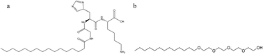
The self-assembly behavior of a nonionic surfactant (n-dodecyl tetraethylene monoether, C12E4) and a peptide amphiphile (PA, C16-GK-3) mixed system was investigated using a combination of microscopic, scattering and spectroscopic techniques, including transmission electron microscopy (TEM), field emission-scanning electron microscopy (FE-SEM), atomic force microscopy (AFM), polarized optical microscopy (POM) observations, small-angle X-ray scattering (SAXS), Fourier transform infrared (FT-IR) spectroscopy, circular dichroism (CD) and rheological measurements. The change of the contents of C16-GK-3 and C12E4 induced the transitions in the nanostructures and simultaneously led to changes in macroscopic properties, i.e., mixtures of C12E4 with C16-GK-3 can be hierarchically self-assembled into various helical nanofibers and then further assembled to dandelion-like and dendrite nanostructures by changing the content of C16-GK-3 and C12E4, which resulted in transitions from solution to two phase, sol and hydrogel states that were noted on increasing the concentration of C16-GK-3 at a fixed concentration of C12E4 or varying C12E4 concentration at a fixed concentration of C16-GK-3. On the basis of a series of characterizations, we proposed a possible mechanism of the self-assembly, for which the hydrogen bonding interaction between the headgroups of C16-GK-3 and between C16-GK-3 and C12E4, as well as hydrophobic interaction between the alkyl chains of C16-GK-3 and C12E4, were the main driving forces.
In general, peptide amphiphiles have biologically relevant lipid chain lengths, in particular palmitoyl (hexadecyl, C16).26 For example, Hest et al. have investigated the influence of mono-alkyl chain length on the self-assembly of PAs containing an octapeptide derived from a protein of the malaria parasite P. falciparum. The results indicated that short chains (C6–C12) can only produce random coil structures, whereas β-sheet ordering structure was only observed for C14 and C16 derivatives and the C16 variant showed the most extended thermal stability range for a β-sheet structure.27 Hotta et al. have discussed the effects of the salt concentrations using sodium dihydrogen orthophosphate (NaH2PO4) on the sol–gel transition behaviors, especially the gelation speed and the gel characteristics, of the designed PA (C16-W3K) hydrogels in an aqueous solution. Their results indicated that the solution exhibited higher gelation speeds and higher mechanical properties at higher salt concentrations, and concurrently, the density, length of wormlike micelles, and the conformational ratio of β-sheets to α-helices in the equilibrium C16-W3K solutions all increased with the increase in the salt concentrations.28 Moreover, the study of the interaction between traditional surfactants and PAs can significantly broaden the research field which can show various phase behaviors and particular self-assembly structures. For example, Hamley et al. have investigated the self-assembly of Pluronic copolymer P123 with the PA C16-KTTKS. The results revealed that the β-sheet structure of C16-KTTKS is retained even with 20 wt% added P123 and the nanotapes comprising peptide bilayers observed for C16-KTTKS itself in an aqueous solution are progressively replaced by cylindrical fibrils upon addition of more P123.29 They have also studied the interaction between the classical anionic surfactant sodium dodecyl sulfate (SDS) and C16-KTTKS. The results indicated that the addition of SDS leads to a transition from tapes to fibrils via intermediate states that include twisted ribbons, and the addition of SDS is also shown to enhance the development of remarkable lateral “stripes” on the nanostructures, which is ascribed to counterion condensation.30
In this article, the binding of a nonionic surfactant (n-dodecyl tetraethylene monoether, C12E4) to a PA, C16-GK-3, containing a cationic peptide headgroup and its effects on the self-assembly behavior of C12E4/C16-GK-3 mixed systems were investigated. Transmission electron microscopy (TEM), field emission-scanning electron microscopy (FE-SEM), atomic force microscopy (AFM), polarized optical microscopy (POM) observations, small-angle X-ray scattering (SAXS), Fourier transform infrared (FT-IR) spectroscopy, circular dichroism (CD) and rheological measurements were used to characterize their physicochemical properties. Our aim is to examine the influence of the uncharged surfactant C12E4 on the aggregation of C16-GK-3 in aqueous solutions to increase and enrich the examples of peptides/surfactants systems.
The FT-IR spectrum was obtained on a VERTEX-70/70v spectrometer (Bruker Optics, Germany). Circular dichroism (CD) spectra were obtained using a JASCO J-810 spectropolarimeter, which was flushed with nitrogen during operation. Wavelength scans were recorded at 0.1 nm intervals from 300 to 180 nm. The hydrogels were determined using a 0.1 mm path length quartz cuvette; the micellar solution used a 1 mm path length quartz cuvette. Small-angle X-ray scattering (SAXS) observations were carried out on a HMBG-SAX X-ray small-angle scattering system (Austria) with a Ni-filtered Cu Kα radiation (0.154 nm) source operating at 50 kV and 40 mA. The distance between the sample and detector was 27.8 cm. Polarized microscopy observations were carried out on an AXIOSKOP 40/40 FL (ZEISS, Germany) microscope and a Nikon Eclipse E400 microscope equipped with a LINKAM THMS 600 heating/cooling stage.
The rheological measurements were carried out on a HAAKE RS75 rheometer with a cone-plate system (Ti, diameter, 35 mm; cone angle, 1°). For the shear-dependent behavior, the viscosity measurements were carried out at shear rates ranging from 0 to 1000 s−1. In oscillatory measurements, an amplitude sweep at a fixed frequency of 1 Hz was performed prior to the following frequency sweep to ensure that the selected stress was in the linear viscoelastic region. The viscoelastic properties of the samples were determined by oscillatory measurements in the frequency range of 0.01–10 Hz. The samples were measured at 20.0 ± 0.1 °C with the help of a cyclic water bath.
Moreover, Fig. 3 shows the phase diagrams of the C16-GK-3/C12E4 system at the fixed concentration of C16-GK-3 (50 g L−1) with increasing the amount of C12E4 (Fig. 3A) and the images for five typical samples (Fig. 3B–F). It can be observed that the color of a 50 g L−1 solution of C16-GK-3 is transparent. Thus, through these experimental results, it can be speculated that the rich phase behavior of the C16-GK-3/C12E4 mixed system can be adjusted by the composition of C12E4 and C16-GK-3, and this is the result of the interaction between C12E4 and C16-GK-3.
![]() | ||
Fig. 4 TEM images of the nanostructures formed with constant cC12E4 (50 g L−1) and varying cC16-GK-3. (A) 0 g L−1 C16-GK-3; (B) 100 g L−1 C16-GK-3; (C) 200 g L−1 C16-GK-3; and (D) 300 g L−1 C16-GK-3. |
The delicate structure of 50 g L−1 C16-GK-3/100 g L−1 C12E4 was further characterized by SEM and AFM (Fig. 5). The dandelion-like morphology consists of left-handed helical nanotapes (Fig. 5C), and some nanotapes further coiled to form a larger-sized superhelix. The appearance of the assemblies shown in AFM images may be more compact than the TEM. It is speculated that the dandelion-like structures with a relative height of 1.35 μm compared to the peripheric surface could come in contact with the tip, leading the nanotapes at the surface somewhat destroyed and even separated from the assemblies; consequently, the remaining structures at the surface could not be supported and collapsed. The AFM images of the dendrite structures formed with cC12E4 = 50 g L−1 and cC16-GK-3 = 200 g L−1 are shown in Fig. 6. The assemblies are exclusively composed of helical nanotapes with left-handed bias (Fig. 5B). Furthermore, some of the nanohelices aggregated into bundles. The measured helical pitch is about 170 nm. From the abovementioned results, a hierarchically self-assembled process can be achieved in which helical nanotapes with left handed direction induced by the chirality of the peptide molecules were first observed, followed by the smaller-sized nanohelices aggregating into larger assemblies. The schematic of the formation of the C16-GK-3/C12E4 nanostructure with the increase in concentration of C16-GK-3 is shown in Scheme 1.
![]() | ||
Scheme 1 The schematic of the formation of C16-GK-3/C12E4 nanostructure with the increase in concentration of C16-GK-3. |
If the concentration of cC16-GK-3 is fixed at 50 g L−1 and the concentration of cC12E4 is changed, the microstructures of the C16-GK-3/C12E4 mixed system were also changed from vesicle to dendrite structures, as shown in Fig. 7. If we further increase the concentration of C12E4 to 300 g L−1 and 400 g L−1, the sample can form liquid crystals (LCs), and it is hard to use TEM to observe its morphology. Thus, POM was used to observe its polarization properties, and the results are shown in Fig. 8. It can be observed that for the pure 200 g L−1 C12E4, it cannot form LCs and there is no polarized texture. However, when 50 g L−1 C16-GK-3 was added, it could form LCs and a polarized texture can be observed. For 300 g L−1 and 400 g L−1 C12E4, the systems are lamellar liquid crystals (LLCs), which showed Maltese cross textures. When 50 g L−1 C16-GK-3 was added, although the lamellar structures of the samples were kept, it changed to an oily texture, indicating the influence of C16-GK-3 on the properties of C12E4 liquid crystals.
![]() | ||
Fig. 7 TEM images of the nanostructures formed with constant cC16-GK-3 (50 g L−1) and varying cC12E4: (A) 50 g L−1 C12E4; (B) 100 g L−1 C12E4; (C) 200 g L−1 C12E4. |
To further obtain the details about the phase behaviors of C16-GK-3/C12E4 LLC composites, SAXS measurements were carried out. Fig. 9 shows the variation of SAXS results of 50 g L−1 C16-GK-3 as a function of the concentration of C12E4 (200–400 g L−1). The SAXS results show two scattering peaks, indicative of the highly ordered supramolecular structures. All the ratios of the two peaks are 1:
2, suggesting a typical lamellar structure of the supra-molecular structures. Moreover, the position of the peaks slightly moved to the left with the increase of C12E4. The d-spacing of the lamellar lattice was calculated by d = 2π/q1 (q1 was the value of the first peak). The calculated inter-planar distances (d) of the typical lamellar structures are 12.1, 9.1, and 6.6 nm for 200 g L−1 C12E4/50 g L−1 C16-GK-3, 300 g L−1 C12E4/50 g L−1 C16-GK-3, and 400 g L−1 C12E4/50 g L−1 C16-GK-3, respectively. Comparing these values with the values of d-spacing for the pure 300 g L−1 (d = 10.55 nm) and 400 g L−1 (d = 8.32 nm) C12E4, it can be observed that the addition of C16-GK-3 decreased the d-spacing of the lamellar lattice, which indicated that the molecular arrangement is more closely packed in C16-GK-3/C12E4 LLC composites.
Rheological measurements can give macro properties of the system, and it is an important method to investigate LLC materials.31–33 The results of the rheological properties of liquid crystal composites as a function of the concentration of C12E4 when cC16-GK-3 = 50 g L−1 are shown in Fig. 10. It can be observed that the increase of cC12E4 greatly increased the range of the linear viscoelastic region of the LLC phase, and the shear modulus increased with the increase of cC12E4. For example, the elastic modulus (G′) of the weak lamellar phase of 200 g L−1 C12E4/50 g L−1 C16-GK-3 was 4.7 Pa, and the value of G′ increased progressively from 120 Pa to 413 Pa when cC12E4 increased from 30 g L−1 to 40 g L−1. For 200 g L−1 C12E4/50 g L−1 C16-GK-3, G′ and the viscous modulus (G′′) are slightly dependent on oscillatory frequency with the same values of about 3.4 Pa at f = 5 Hz, indicating the weak elasticity. When cC12E4 increases to 300 and 400 g L−1, G′ and G′′ are nearly independent of oscillatory frequency but increase to 7.49 and 4.87 Pa for 300 g L−1 C12E4/50 g L−1 C16-GK-3 and 9.11 and 6.03 Pa for 400 g L−1 C12E4/50 g L−1 C16-GK-3, respectively, exhibiting the obvious viscoelasticity and the dominant elastic property. The η* of both systems decreased with the increase of frequency, indicating a shear-thinning behavior (Fig. 10C). Moreover, the value of η* increased with the increase of cC12E4, which indicated that the increase of cC12E4 enhanced the mechanical properties of C16-GK-3/C12E4 LLC composites.
To further characterize the features of the formed nanostructures at various concentrations of C12E4 and C16-GK-3, the FTIR and CD spectra were measured, as shown in Fig. 11. From the IR spectra, it can be observed that for pure C16-GK-3, no β-sheet secondary structure was noticed. The spectra of the C12E4/C16-GK-3 mixtures, however, changed. The spectra displayed a strong peak at 1619 cm−1, which is consistent with the presence of β-sheets, and a second one at 1679 cm−1, which is characteristic of an antiparallel arrangement of the β-sheets.34 An additional contribution at 1650 cm−1 indicates that the peptide region also adopts random coil characteristic.35 The wide peak (∼3400 cm−1) is well-known for symmetric and antisymmetric O–H stretching modes, the stretching vibration of N–H locates at 3300 cm−1 and the peaks at 2920 cm−1 and 2852 cm−1 are the asymmetric and symmetric stretching vibration of the CH2 of C12E4 and C16-GK-3, respectively. For CD analysis, it can be observed that the pure C16-GK-3 solution lacks characteristic structure, whereas no CD signals can be observed in the micelle solution of C12E4. After the interaction between C12E4 and C16-GK-3, the formation of helical dandelion-like or dendrite structures resulting in a remarkable CD signal (Fig. 11d), β-sheets and random coil structures were proved to coexist from the peaks at 215 and 200 nm confirming the helical structure and higher ordered molecular arrangement of C12E4/C16-GK-3.36 It was obvious that the peak for the β-sheet structure was greater than that of the random coil structure, which indicated that more β-sheet structures than random coil structures formed in the obtained nanostructures.
This journal is © The Royal Society of Chemistry 2016 |