DOI:
10.1039/C5RA24938D
(Paper)
RSC Adv., 2016,
6, 7066-7077
Role of manganese-based surfactant towards solubilization and photophysical properties of fluorescein†
Received
24th November 2015
, Accepted 21st December 2015
First published on 28th December 2015
Abstract
Dye–surfactant interactions have been explored from various viewpoints in recent years, in the present work interactions between fluorescein dye and manganese-based, water-soluble surfactant for modulating photophysical properties of photosensitizer in a metal-containing microheterogeneous environment and for evaluating their role in solubilization have been evaluated. For these purposes, manganese (Mn) surfactant complexes were prepared in different ratios of metallic counter ions to numbers of hydrocarbon chains, that is, 1![[thin space (1/6-em)]](https://www.rsc.org/images/entities/char_2009.gif) :1 and 1:2. The formulated complexes were further characterized by diverse techniques such as Fourier-transform infrared spectroscopy (FTIR), elemental analysis, thermogravimetric analysis, and nuclear magnetic resonance (NMR). Thermal stability of manganese surfactant complexes and kinetics of decomposition were investigated. The effect of metallic counter ions and temperature were evaluated on surface activity and aggregation of manganese surfactants, where the presence of metal ions lowered the critical micellization concentration and also affected the packing of adsorbed molecules at the air–solution interface. Our investigation further capitalized on the spectral sensitivity of fluorescein dye in the pre- and post-micellar system of manganese surfactant complexes. The influence of manganese surfactant complexes on the photophysics of fluorescein dye was thoroughly investigated using spectrofluorimetric parameters such as the Stern–Volmer constant, binding constant and anisotropy. NMR was used to locate the binding site of dye molecules in metallomicelles. Fluorescence quantum yield and singlet oxygen quantum yield were estimated using comparative methods, and increased fluorescence and singlet oxygen quantum yields were observed for dye on solubilization in metallomicelles as compared to conventional surfactants.
:1 and 1:2. The formulated complexes were further characterized by diverse techniques such as Fourier-transform infrared spectroscopy (FTIR), elemental analysis, thermogravimetric analysis, and nuclear magnetic resonance (NMR). Thermal stability of manganese surfactant complexes and kinetics of decomposition were investigated. The effect of metallic counter ions and temperature were evaluated on surface activity and aggregation of manganese surfactants, where the presence of metal ions lowered the critical micellization concentration and also affected the packing of adsorbed molecules at the air–solution interface. Our investigation further capitalized on the spectral sensitivity of fluorescein dye in the pre- and post-micellar system of manganese surfactant complexes. The influence of manganese surfactant complexes on the photophysics of fluorescein dye was thoroughly investigated using spectrofluorimetric parameters such as the Stern–Volmer constant, binding constant and anisotropy. NMR was used to locate the binding site of dye molecules in metallomicelles. Fluorescence quantum yield and singlet oxygen quantum yield were estimated using comparative methods, and increased fluorescence and singlet oxygen quantum yields were observed for dye on solubilization in metallomicelles as compared to conventional surfactants.
1. Introduction
Photosensitizers (PSs) are light-absorbing agents that are activated by light of a specific wavelength. They possess a triplet state of higher energy, long triplet-state lifetime, high triplet-state quantum yield and high photoactivity, which make them a promising chemical tool to generate reactive oxygen species (ROS) such as singlet oxygen upon light irradiation. Several metal complexes and organic dyes such as porphyrins,1,2 transition metal complexes,3,4 fluorescent dyes5,6 and phthalocyanine7 are used as sensitizers in various photochemical and phototherapeutic applications. Dyes are among the most effective sensitizers that possess the triplet state of appropriate energy for sensitization of ground-state oxygen. Among xanthene dyes, fluorescein is one of the most common fluorescent probes because of its high molar absorptivity and photostability.8 It can be used (1) as an effective fluorescent probe for detecting HOCl in living cells;9 (2) in interaction with lysosome for detecting of Gram-positive bacteria;10 and (3) in fluorescein-labeled immunoglobulins, which are widely used as proteins in fluorescence microscopy and in immunoassays.11,12 It also acts as a good photosensitizer for singlet oxygen generation in reversed micelles of dodecylammonium propionate13 prepared in cyclohexane and used for anticancer activity. The extent of photodynamic action depends not only on singlet oxygen production but also on the solubilization site, retention and the nature of binding inside the cell in biological systems.
Fluorescent probes are widely studied to examine the physicochemical properties of micelles and environmental effects.14 Surfactants at concentrations higher than the critical micelle concentration (CMC) have been extensively used with dyes/photosensitizers for various applications using spectroscopic (ultra-violet, fluorescence, phosphorescence, atomic spectroscopy), electroanalytical15 and separation methods. There are numerous studies that show the influence of various nonionic, anionic and cationic surfactants on the photophysical properties of dye.16–18 Dye–surfactant interactions are also utilized for the separation of dyes from waste dye-stuffs of diverse textile, paper and pulp industries.19 The results have been interpreted via calculating molar extinction coefficient, empirical fluorescence coefficient and fluorescence quantum yield of dye in various micellar media. However, some dyes show unusual spectral behavior when bound to the synthetic membrane of micelles/vesicles.20 Major challenges are the stability and shelf life of micelles/vesicles bearing these dye molecules. To overcome these problems, it has been suggested that in some cases formation of vesicles containing mixed single-tailed cationic and anionic surfactants result in high encapsulation efficiency of vesicles.21 Recently, dye–surfactant interactions have been explored from various viewpoints.
Transition metals have played a major role in enhancement of fluorescence quantum yield and singlet oxygen production.22 Transition metals, possessing rich and essential geometric, redox, optical and magnetic properties, when incorporated into conventional ligands lead to development of various systems such as metallophotosensitizers,23 metallomesogens,24 metallopolymers25 and metallosurfactants.26 Such systems act as more promising materials for high-end technological applications in photophysics,27 display molecular electronics28 and analyte sensing.29 To cope with increased demand of such materials, testing of new surfactants with diverse functionalities that take advantage of inexpensive and abundant metals, such as iron, cobalt, nickel and copper, aim to assess the strengths, stability and limitations of their use. One such class is metal-functionalized amphiphilic moieties, which are commonly known as “metallosurfactants”. The presence of transition30 and lanthanide metal ions31 in the structure of surfactant (may be at head group, hydrocarbon tail or as counter ions) imparts special properties. Metallosurfactants create supramolecular arrangements such as micelles,32 vesicles,33 reverse vesicles34 and hybrid crystal with layered perovskite structure.35 Both hydrophobic- and hydrophilic-metal embedded surfactants (in head group and as counter ions) have been synthesized and characterized in the past.36,37 It has been observed that the location of metal ions in the metallomicelle is crucial in deciding its further applications; for instance, for an effective catalytic cycle, the metal ion should be at the periphery to facilitate facile transfer rather than present at the micelle core.38 Such abilities prompted us to elicit interactions between fluorescein dye and surfactant-containing metal ions to modulate the photophysical properties of photosensitizers in the microheterogeneous environment.
The present work attempted to synthesize manganese-based metallosurfactants with diverse stoichiometries of metallic counter ions and hydrocarbon chains. Manganese complexes unveil numerous potential applications in electrochemistry39 and exhibit antimicrobial40 and anticancer properties.41 With a Schiff base, the manganese complex also acts as a catalyst in aerobic oxidation of p-xylene to p-toluic acid.42
Therefore, in this work we focus on two aspects: first, the synthesis, characterization and determination of critical micelle concentrations of single and double alkyl-chained manganese surfactant complexes to elucidate the effects of the presence of metallic counter ions (in 1:1 and 1:2 ratios) on micellar properties. Second, we have undertaken the investigation of the influence of cationic surfactant containing manganese metal ions on the photophysical properties of fluorescein (FL), and estimated the solubilization, location/site, binding constant, anisotropy, fluorescence quantum yields and singlet oxygen quantum yields. The results show high fluorescence quantum yields and singlet oxygen quantum yields of fluorescein dye, using manganese surfactant complexes as compared to the parent surfactant, thus highlighting the role of metal counter ions of surfactant complexes in dye–surfactant interactions.
2. Experimental section
2.1. Materials
Manganese chloride anhydrous (purity ≥ 99.0%), hexadecyltrimethylammonium chloride (CTAC) (purity ≥ 98.0%), 1,3-diphenylisobenzofuran (DPBF), D2O (purity ≥ 99.0%) and tris(2,2′-bipyridyl) ruthenium(II) were obtained from Sigma-Aldrich. Fluorescein (FL) was purchased from SD Fine chemical limited (purity ≥ 95%). The materials were used without purification. Deionized distilled water was used for all the studies. NaHPO4, Na2HPO4 and NaOH of analytical grade were used as received. Methanol (purity ≥ 99.0%) and ethanol (purity ≥ 99.9%) was purchased from Fisher Scientific and Changshu Yangyuan Chemical China, respectively.
2.2 Synthesis and characterization of manganese metallosurfactant
The single- and double-chain manganese complexes were synthesized by adding manganese chloride and CTAC in a given amount of ethanol (different molar ratios, i.e., 1:1 for MnC I and 1:2 for MnC II) and refluxed for 2 h at 65 °C. The obtained compounds were filtered, recrystallized in methanol and dried under vacuum.43,44 These manganese complexes were stable in water.44 The prepared complexes were characterized using a PerkinElmer (RX1) spectrometer in the far IR range (200 nm) and IR range (600–3500 nm). For each spectrum, 24 scans were recorded with spectral resolution of 4 cm−1. CHN analyses of the synthesized complexes were performed with an Eager Xperience CHN. All experiments were performed under an inert nitrogen medium. Thermogravimetric analyses were carried out using SDT-Q-600 (TA instruments). A sample mass of approximately 5 mg was transferred to aluminum crucibles and analyzed at room temperature to 1000 °C at a heating rate of 10 °C min−1 under the nitrogen atmosphere.
2.3 Self-aggregation of manganese metallosurfactant
Surface properties and aggregation behavior were estimated using conductivity and surface tension measurements. The conductivity measurements were performed on a Pico Lab India digital conductivity meter using double distilled water (conductivity < 5 μS cm−1). These studies were carried out in the temperature range of 25–40 °C. The measuring cell and sample were dipped into a jacket whose temperature (±0.01 °C) was maintained via thermostat. Surface tension of prepared metal complexes was measured at 25 and 30 °C with a Du Nouy tensiometer (Kruss, type 8451) using double distilled water. The surface tension of the double distilled water used was 71 mN m−1 at 25 °C. Two readings were made on each sample to determine any change over time and to obtain an average value.
2.4 Photophysical properties
UV-visible absorption spectra were recorded on a PerkinElmer spectrophotometer with a matched pair of 1 cm quartz cells fitted in thermostatic cell holder. The concentration of FL used throughout absorption studies was kept at 0.05 mM. In phosphate buffer (0.1 M) at pH 7.2, the wavelength of maximum absorbance of FL was 491 nm. Absorption spectra were carried out between 200 and 800 nm by squirting prepared metallosurfactants in the range of 0.05–2.0 mM into the dye and reference solvent (i.e., buffer). Fluorimetric studies of FL were carried out in the presence of a variable concentration of metallosurfactant on an Hitachi F-7000 Photo luminescence spectrophotometer. Stock solution was prepared by dissolving the dye (0.05 mM) and metallosurfactant (concentration range 0.05–2 mM) in phosphate buffer. The emission and excitation slit widths were kept at 10 nm and scanning speed at 1200 nm min−1. The sample was excited at 460 nm and emissions were recorded from 450–650 nm. The quantum yield of FL (ΦF) in various micellar systems was estimated via a relative method using the quantum yield of FL aqueous solution in the presence of 0.01 M NaOH as a standard solution, which is equal to 0.92. Singlet oxygen quantum yield (ΦΔ1O2) was determined from fluorescence intensity decays of 1,3-diphenylisobenzofuran (DPBF) at 455 nm. Solutions containing 40 μM DPBF and FL in the absence and presence of synthesized metallosurfactant were prepared in methanol and saturated with air for 1 h. DPBF absorbs in the region of dye transparency and rapidly scavenges singlet oxygen generated by photosensitizers. Photosensitizer FL was excited at its λmax and the photooxidation of DPBF was measured for nearly 45 min. The ΦΔ1O2 were calculated by the relative method using tris(2,2′-bipyridyl)ruthenium(II) ([Ru(bpy3)]2+) as the standard (ΦΔ = 0.81 in methanol).
1H NMR spectra were recorded on a Bruker Avance-II spectrometer operating at 400 MHz. Samples of MnC I, MnC II, FL, FL in MnC I and FL in MnC II for 1H NMR were prepared by dissolving in D2O.
3. Results and discussion
3.1 Synthesis and characterization
Micellar moieties have been used in the past to solubilize and stabilize photosensitizers. For these purposes, the manganese-based surfactants in different stoichiometry were prepared by the ligand insertion method. Prepared complexes were characterized using FTIR, elemental analyses and TGA. Structural confirmation of the prepared manganese complexes as provided by elemental analyses are in excellent agreement with the calculated values. For MnC I, the C%, H% and N% observed were 51.18, 9.01, 2.85, respectively, and the calculated figures were 51.55, 9.49, and 3.14, respectively. Similarly, for MnC II, C%, H%, N% values observed and calculated were 58.40, 10.47, 3.33, and 59.55, 10.96, 3.65, respectively. The major vibration peaks appearing in IR region, that is, 2848–2856 and 2915–2925 cm−1 are due to the C–H stretching of methyl and methylene groups of hexadecyl group. An insignificant difference was observed at these positions. The peak at 1260 cm−1 for –C–N stretching in CTAC showed a shift of 10 cm−1 in metal complexes.
On the other hand, for MnCl2 two peaks at 228 and 280 cm−1 corresponded to terminal metal chlorides. In metal complexes along with these, new peaks at 211 and 266 cm−1 were observed that correspond to bridging chloride.45 This confirms that chloride ion of CTAC has coordinated with metal ion. Fig. ES1 and Table ES1 in the ESI† report the major vibrational peaks of the precursor, ligand and prepared metallosurfactants.
1H NMR spectrum of CTAC in D2O solvent is depicted in Fig. ES2(a).† The signal around δ = 0.77 ppm was assigned to the terminal methyl group of the hydrocarbon chain. Next, the signal at δ = 1.65 ppm and at δ = 3.24 ppm were attributed to the α and β methylene protons of CH2CH2N(CH3)3 group, respectively. The methylene groups of hydrocarbon chains appeared at δ = 1.25 ppm; a sharp singlet at δ = 3.03 ppm was assigned to N(CH3)3 protons. As compared to CTAC, there is clear downfield shift in manganese complexes MnC I and MnC II, especially in N(CH3)3 protons, as seen in Fig. ES2(b) and (c).†
Further, the thermal stability and its decomposition kinetics were analyzed by thermogravimetric (TG) analyses for both precursor and metallosurfactants. As seen in Fig. 1, TG curves of complexes demonstrate that the decomposition of both takes place in two steps. Both complexes started to decompose at 247 °C approximately, with initial breakdown of quaternary ammonium moieties that continues up to 450 °C. The second step eventually degrades the metal chloride into metal.46,47 The mass loss percentages obtained experimentally are in good agreement with theoretical values (Table 1). On the basis of TGA studies, a plausible mechanism for the decomposition of manganese complexes is proposed in Scheme 1.
 |
| | Fig. 1 TG/DTG plots of CTAC, MnC I and MnC II. | |
Table 1 TGA data and activation energy calculated for manganese complexesa
| Manganese complex |
CTAC |
MnC I |
MnC II |
| Transition temp. |
Mass loss% |
Transition temp. |
Mass loss% |
Transition temp. |
Mass loss% |
| Cal. |
Obs. |
Cal. |
Obs. |
Cal. |
Obs. |
| R = regression coefficient. |
| Step 1 |
247 |
100 |
98.99 |
344 |
71.84 |
70.84 |
286.3 |
87.57 |
89.07 |
| Step 2 |
— |
— |
— |
575 |
83.61 |
84.47 |
540.5 |
92.87 |
92.77 |
| |
E/kJ mol−1 |
R |
E/kJ mol−1 |
R |
E/kJ mol−1 |
R |
| CR |
23.493 |
0.998 |
32.478 |
0.999 |
51.798 |
0.992 |
| MKN |
23.755 |
0.999 |
33.948 |
0.999 |
52.950 |
0.991 |
| WYHC |
23.833 |
0.999 |
34.006 |
0.998 |
53.186 |
0.993 |
| VK |
27.724 |
0.999 |
36.126 |
0.999 |
57.310 |
0.991 |
| HM |
27.725 |
0.998 |
38.248 |
0.999 |
58.980 |
0.992 |
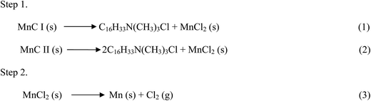 |
| | Scheme 1 Plausible mechanism for thermal decomposition of manganese surfactant (1–3) complexes. | |
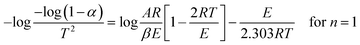
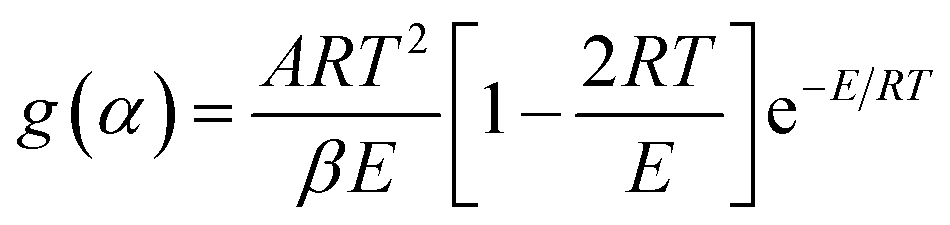
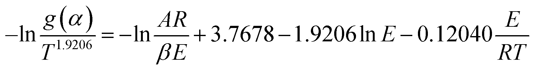
Kinetic and thermodynamic parameters of decomposition for the first step. Five methods based on a single heating rate—Coats–Redfern (CR), Horowitz–Metzger (HM), Madhusudanan Krishnan–Ninan (MKN), van Krevelen (VK) and Wanjun–Yuwen–Hen–Cunxin (WYHC)—were used to calculate the kinetic and thermodynamic parameters for decomposition in step 1. Calculation details are reported in our previous work;48 final equations used are given below. In all the methods, α (degree of reaction) and, in turn, g(α) (integral function of conversion) are estimated under different variation parameters (see below).(1) CR method:
| |
 | (4) |
Taking natural log:
| |
 | (5) |
The fraction mass loss (α) and corresponding (1 − α)n are calculated from TG curves, where n depends on the reaction model.
| |
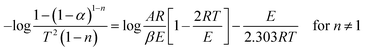 | (6) |
| |
 | (7) |
Plotting the left side of the above equations against 1/T gives the slope (−2.303E/R) and the intercept (A).
(2) MKN method:
| |
 | (8) |
(3) WYHC method:
| |
 | (9) |
(4) VK method:
| |
 | (10) |
(5) HM method:
Parameter T = Tm + θ is used here. If the order of reaction is 1, Tm is defined as the temperature at which (1 − α)m = 1/e = 0.368. Therefore,
| |
 | (11) |
here, symbols
β,
Tm,
E,
A,
R are heating rate, DTG peak temperature, activation energy (kJ mol
−1), pre-exponential factor (min
−1) and gas constant (8.314 J mol
−1 K
−1), respectively. Excellent correlation coefficients, which indicate a good fit of the linear function, were obtained for all methods (Fig. ES3 and ES4 in ESI
†).
E values obtained
via the Coats–Redfern method were used to calculate Δ
H, Δ
S and Δ
G, based on the following equations (data reported in Table S2
†):
| | |
ΔS = 2.303log[Ah/kT]R
| (12) |
where
h is Planck constant,
T is temperature in K,
A is Arrhenius constant or frequency factor and
k represents Boltzmann constant.
Table 1 gives TGA data and the value of activation energy obtained from five methods for the first degradation step of manganese complexes. It was observed that the activation energy of
CTAC was lower than
MnC I and
MnC II, which indicates that synthesized manganese complexes have more ordered systems than the parent system. We concluded that coordination of manganese metal ion with
CTAC is responsible for high thermal stability of metal complexes.
3.2 Surface properties and self-aggregation
The aggregation behavior and surface activity of manganese complexes were evaluated using conductivity and surface tension measurements. In manganese complexes, there is a fixed concentration of metal centers to control the physicochemical properties, that is, hydrophobicity and electrical conductivity. The role played by the incorporation of metal ions to surfactant in lowering the critical micelle concentration (CMC) value is analogue to the role of electrolyte addition to anionic surfactants.49
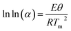
Conductivity measurement. The CMC value of metallosurfactant was determined at four different temperatures. The abrupt change in the slope of plot between concentration versus specific conductance data determines the CMC of the complexes. Fig. 2 shows comparison of variation of CTAC conductivity and prepared metallosurfactants. CTAC is a well-studied surfactant and has CMC in the range of 1.25 to 1.58 mM.50 It was observed that the CMC value of the synthesized metallosurfactant is lower than when compared to CTAC (as seen in Table 2). This shows that coordination of metal with surfactant enhances the micellization property of the surfactant.51 Further, with an increase in temperature, the CMC value for both the manganese complexes increases. This behavior depends on two competing effects. A temperature increase may cause reduced hydration in the hydrophilic group, which favors micellization, or it may cause disruption of water surrounding the hydrophobic group and thus retards micellization. The relative magnitude of these two opposing effects therefore determines the CMC behavior similar to that in conventional surfactants.52
 |
| | Fig. 2 Variation of conductivity (a–c) and surface tension (d–f) of CTAC, MnC I and MnC II with variation in temperature. | |
Table 2 Value of CMC, thermodynamic parameters of micellization and surface properties of synthesized manganese complexes
| Complex |
Temp. (°C) |
CMC (mM) |
β |
ΔGom (kJ mol−1) |
ΔHom (kJ mol−1) |
ΔSom (kJ mol−1) |
Γmax (μmol m−2) |
Amin (nm)2 |
| κ |
γ |
| CTAC |
25 |
1.58 ± 0.2 |
1.55 ± 0.1 |
0.482 |
−39.35 |
−10.19 |
0.090 |
1.53 |
1.08 |
| MnC I |
25 |
0.95 ± 0.1 |
0.95 ± 0.2 |
0.855 |
−30.33 |
−13.86 |
0.055 |
1.123 |
1.47 |
| 30 |
1.04 ± 0.2 |
0.99 ± 0.3 |
0.945 |
−29.03 |
−13.56 |
0.051 |
0.782 |
2.12 |
| 35 |
1.11 ± 0.2 |
|
1.090 |
−25.18 |
−12.08 |
0.043 |
|
|
| 40 |
1.12 ± 0.3 |
|
0.995 |
−28.03 |
−13.67 |
0.046 |
|
|
| MnC II |
25 |
0.53 ± 0.2 |
0.70 ± 0.3 |
0.766 |
−35.33 |
−25.32 |
0.033 |
0.80 |
2.07 |
| 30 |
0.52 ± 0.1 |
0.72 ± 0.1 |
0.768 |
−35.18 |
−26.14 |
0.030 |
0.77 |
2.15 |
| 35 |
0.66 ± 0.2 |
|
0.836 |
−33.61 |
−25.52 |
0.026 |
|
|
| 40 |
0.67 ± 0.1 |
|
0.787 |
−35.51 |
−27.46 |
0.025 |
|
|
The degree of ionization micellization (β) in the present case oscillated with temperature and showed no regular trends. Similar behavior occurred with other surfactants such as cationic octadecyl trimethyl ammonium chloride and anionic sodium dodecyl sulphate.53 Thermodynamic parameters of micelle formation, such as ΔGom (Gibbs free energy), ΔHom (enthalpy) and ΔSom (entropy), were estimated using the following equations:
| | |
ΔGom = (2 − β)RTlnXcmc
| (15) |
| | |
ΔHom = −RT2(2 − β)dlnXcmc/dt
| (16) |
| | |
ΔSom = (ΔHom − ΔGom)/T
| (17) |
where
R,
T,
β and
Xcmc represent gas constant, absolute temperature, degree of ionization and CMC in terms of mole fraction, respectively.
The values obtained for Gibbs free energy (Table 2) were found to be more negative indicating that the overall micellization process is spontaneous and thermodynamically favored. The free energy value (for both single and double chain metal surfactants) falls within the range of conventional ionic and amphoteric surfactants. Hydrophobic and hydrophilic hydration of ionic surfactant monomers play an important role in the micellization process. The negative values of micellization enthalpy infers the exothermic nature of micellization, which is contributed by the hydrocarbon chain transfer of the surfactant monomer from aqueous solution to micelle that ultimately leads to release of solvation water molecules. Moreover, the positive value of entropy of micellization decreases as temperature increases indicating destruction of structured water molecules surrounding the hydrophobic chain due to hydrophobic interactions among surfactant cations.
The free energy, ΔGom, results from entropy–enthalpy contributions (Fig. ES5 in ESI†). We observed that in the micellization process for both manganese complexes, the enthalpic contribution to free energy increases as temperature increases, whereas the entropic contribution decreases. At low temperatures, entropy increases due to the destruction of water molecules around a hydrophobic chain. As temperature increases, the hydrogen bond between water molecules diminishes and less energy was used to destroy the water cluster. Therefore, at high temperature, enthalpy became more significant.
Linear relationships between ΔHom and ΔSom (thermodynamic functions) for various processes are known as compensation phenomena. The slope of the compensation line is referred to as the compensation temperature (Tc), which provides an idea of solute–solvent interactions (solvation region for micellization process). The compensation line is expressed in the following equation:
| | |
ΔSom = ΔHom (1/Tc) + σ
| (18) |
σ (intercept of compensation plot) is considered to be a measure of solute–solute interaction (chemical part of micellization process). Heat capacity was also detected by the slope of Δ
Hom versus temperature plot using
eqn (19). The obtained values of
σ, (
Tc) and
Cp of manganese surfactant complexes are given in Table ES3 (ESI
†).
Surface tension of prepared complexes was measured at 25 and 30 °C and decreased linearly along with increasing concentration of manganese surfactant and then reached a plateau zone. The breakpoint of surface tension was designed as the CMC of the complexes, and the plateau zone was indicative of micelle formation. As shown in Table 2, the CMC values evaluated from surface tension are in good agreement with the values obtained from conductivity studies. Surface excess and area per molecule were calculated from the slope of linear plot of γ versus log(concentration) below the CMC by using Gibbs adsorption equation:
| |
 | (20) |
where
n is the number of ionic species (
ΓCTA,
ΓCl−,
ΓMn+); (d
γ/dlog
C), surface pressure;
R, universal gas constant; and
T, absolute temperature. Further, the minimum surface area occupied by each monomer at the saturated air–solution interface is calculated as follows:
| |
 | (21) |
As compared to CTAC, there are larger counter ions in manganese complexes, which results in a weaker interaction with the hydrophobic head group, and thus lowers surface excess. There is also competitive adsorption between the cationic head group and counter metal ion, which leads to more crowding at the interface and thus increased the Amin value in comparison to CTAC.54 Therefore, it is responsible for lower CMC of manganese complexes in comparison to CTAC. Table 2 reveals that by increasing the temperature, the area per molecule increases, presumably due to increased thermal motion, which causes poorer packing of adsorbed molecules with a consequent decrease in the maximum surface excess (Γmax).
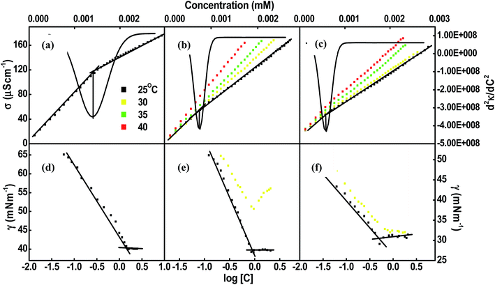
Encapsulation of FL. Interactions between FL and metallosurfactant were estimated by spectroscopic methods via absorption and emission spectroscopy. As discussed earlier, FL is a xanthene dye that is widely used as a fluorophore in biological systems. It exists as dianion molecules in the neutral aqueous solution of pH 7.2. Scheme 2 presents possible existing forms of FL. It is excitated at λ = 490 nm, while the emission is obtained at 523 nm. According to the literature,55 as the concentration of FL in buffer (pH 7.2) increases, the dimerisation process increases through stacking of monomers. Thus, in the present study the maximum concentration of FL (in buffer) is kept at 0.05 mM to prevent aggregate formation.
 |
| | Scheme 2 Possible existing forms of fluorescein. | |
At pre-micellar concentrations of MnC I, there was no change in the absorption and emission spectra of FL, indicating the absence of significant interaction between the FL and manganese surfactant. For MnC II, absorption and fluorescence intensity of FL decreased as manganese surfactant concentration increased (up to 0.05 mM), as shown in Fig. 3(a) and (b). This led to initial charged neutralization (i.e., salt-like pair formation) that induced system protonation. On increasing concentration of manganese surfactant in the post-micellar region (2 × 10⁻³ M), absorption of FL decreases with a red shift (from 491 to 500 nm) for both complexes as shown in Fig. 3(c) and (e). The appearance of red shift and a broad band concomitant with the decrease of absorbance was indicative of molecular organization between the manganese surfactant moieties and FL dye. In the fluorescence spectra of FL, in the postmicellar region a progressive increase in fluorescence intensity with a bathochromic (red) shift from 522 to 533 nm for both the systems (Fig. 3(d) and (f)) was observed. The band present at 522 nm was assigned to the π–π* transition, where π* (excited state) is more polar in nature as compared to π (ground state); hence π* interacts with the polar head group of metallomicelles more than the ground state. Consequently, ΔE (i.e., energy gap) decreases between the ground and excited states, which was attributed to the bathochromic shift in our system.55 From these observations we concluded that the substantial interaction between the dianionic form of dye molecules with the manganese surfactant occurred through a balance of repulsive and attractive actions between hydrophobic and hydrophilic moieties. Hydrophobic dye molecules are expected to solubilize in the hydrophobic core of micelles and lowers the intramolecular motion of dye. More prominent changes were observed in MnC II because it is more hydrophobic. The micelles minimize the action of vibrationally coupled non-radiative deactivation of the excited state, and therefore results in higher fluorescence.54 To understand FL binding and location, quenching experiments were conducted.
 |
| | Fig. 3 Absorbance (a–c) and fluorescence (d–f) spectra of variation of concentration of MnC I and MnC II with FL (0.05 mM). | |
For static quenching, the Stern–Volmer constant was determined by using the following equation:
| |
 | (22) |
where
fo and
f are the fluorescence intensities of FL in the absence and presence of the quencher, respectively.
KSV is the Stern–Volmer quenching constant and [Q] is the quencher concentration. In order to evaluate the
KSV value of manganese surfactant complexes above CMC, a modified Stern–Volmer
eqn (23) was applied:
| |
 | (23) |
where
V is the static quenching constant and
KSV can be obtained from the slope of
 versus
 [Q] plot.
To determine the number of binding sites (n), another modified Stern–Volmer equation was used:
| |
 | (24) |
where
fo and
f are intensity of FL in the absence and presence of the quencher, respectively. [Q] is the quencher concentration,
Ka is the binding constant and
n is the number of binding sites. The binding constant and number of binding sites of manganese surfactant with FL dye is evaluated from the intercept and slope of the linear plot of log[(
fo −
f)/
f]
versus log[Q] (
Fig. 4(a–c)).
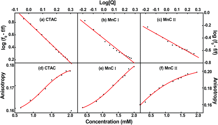 |
| | Fig. 4 Plot of log[(fo − f)/f] versus log[Q] (a–c) and fluorescence anisotropy (d–f) with the variation in concentration of surfactants. | |
Table 3 shows that the binding constant of CTAC with FL is much higher than respective manganese surfactant complexes and the value of n indicates that the surfactant was bound to fluorescein through two binding sites. However, manganese surfactants containing single and double alkyl chains were bound to FL only through a single binding site. This may be because of the bulky nature of counter ions in the case of the manganese surfactant complex, which could bind only to one polar site out of two polar groups of dye. NMR was used to glean information about the FL binding site in manganese complexes. Comparing the 1H-NMR of FL and FL in manganese metallomicelles (Table ES4†), a distinct downfield shift for carbon atoms near the carboxylic group was observed. These observations confirm that FL binding to metallomicelles occurs through a single site, that is, carboxylic group.
Table 3 Stern–Volmer constant, binding constant, number of binding sites and fluorescence quantum yield for different systems
| Complex |
KSV (×103 L mol−1) |
Ka (×103 M−1) |
n |
Concentration (M) |
ΦF |
ΦΔ1O2 |
| CTAC |
2.52 |
6.32 |
2.174 |
0.36 × 10⁻³ |
0.17 ± 0.02 |
— |
| 0.75 × 10⁻³ |
0.31 ± 0.02 |
| 1.89 × 10⁻³ |
0.43 ± 0.01 |
| MnC I |
0.24 |
0.87 |
1.139 |
0.36 × 10⁻³ |
0.30 ± 0.01 |
— |
| 0.75 × 10⁻³ |
0.50 ± 0.02 |
| 2.00 × 10⁻³ |
0.80 ± 0.01 |
| MnC II |
0.13 |
0.38 |
0.938 |
0.36 × 10⁻³ |
0.47 ± 0.03 |
0.68 ± 0.05 |
| 0.75 × 10⁻³ |
0.62 ± 0.01 |
| 0.94 × 10⁻³ |
0.70 ± 0.01 |
3.3 Fluorescence quantum yield (ΦF)
ΦF is an intrinsic property for photoluminescent species and its measurement is a powerful tool for characterizing the photophysics of electronic transitions and scrutinizing the environmental impacts on a chromophore. ΦF is the ratio of photons absorbed to photons emitted through fluorescence. The fluorescence quantum yield can be experimentally approached by two different methods: comparative and absolute. In this work, the first approach was used: a known fluorescent standard (FL aqueous solution in the presence of 0.01 M NaOH) was employed as the reference and the fluorescence quantum yield was obtained by comparison of absorption and emission spectra for samples of the unknown and known standard systems. The ΦF was calculated using the following equation:| |
 | (25) |
where ΦR and ΦF are the fluorescence quantum yield of the sample and reference, respectively; IS and IR are the integrated intensities (areas) of sample and reference spectra, respectively (in units of photons); the refractive indices of the sample and reference solution are ηS and ηR, respectively; and AR and AS are the absorbance of reference and sample, respectively.
The maximum value of the FL fluorescence quantum yield (ΦF) was reported in the basic medium due to the presence of dianion species of FL. The ΦF reported for the FL in CTAC results in a low value.56 However, in the case of manganese surfactant (Table 3), higher values were obtained and it continued increasing as metallosurfactant concentration grew. At high concentrations, the value was approachable to that of NaOH. There may have been interactions between the π-orbital of FL and d-orbital of metal,57 and this interaction restricts the diffusional motion of FL dye (see later in the anisotropy study), resulting in less energy released in non-radiative form that in turn leads to increased fluorescence quantum yield.
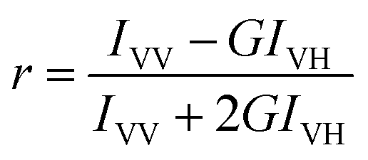
Fluorescence anisotropy, an experimental measure of fluorescence depolarization, is caused by rotational diffusion of the fluorophore using the excited lifetime; this quantity has been extensively used to investigate the binding of macromolecules with proteins, CMC of surfactants58 and stability of mixed micelles at different pH. It was determined by exciting the prepared sample with vertically polarized light and then measuring the vertically and horizontally polarized components of the emission with a polarizer on the emission side. Fluorescence anisotropy (r) is defined by the following expression:
| |
 | (26) |
where
IVV corresponds to the intensity obtained when excitation and emission polarizers are oriented vertically.
IVH is the intensity obtained for the vertically excited polarizer and horizontal emission polarizer.
IHV and
IHH refer to similar parameters as above for the horizontal positions of the excitation polarizer.
G is the correction factor that provides the differences in sensitivity of the detection system in the two polarizing directions
IVV and
IVH.
G will be equal to:
| |
 | (27) |
Fig. 4(d–f) shows the variation of FL fluorescence anisotropy at different concentrations of manganese surfactant and CTAC. Surfactant could significantly alter the anisotropy value of FL in the aqueous phase. Here, when measurements were made for manganese surfactants, the anisotropy value for the micellar state increased as concentration in both systems increased, which reflects a change in environment of FL. In the MnC I system, the anisotropy value was nearly same as that of CTAC, but when compared with MnC II, the r value is higher, which indicates that in the manganese surfactant (1:2) micelle, FL mobility was more restricted and more solubilized at the inner “border” of the Stern region.
Further, the singlet oxygen quantum yield (ΦΔ1O2) of FL was monitored in metallomicellar media by a simple, easy and controllable method based on the dye-sensitized photooxidation of 1,3-diphenylisobenzofuran (DPBF). In this method, the photosensitizer absorbed a light of appropriate wavelength and used that energy to excite the molecular oxygen to its singlet state. For this, 2 mL methanol solution of FL with manganese complexes and DPBF were irradiated. The disappearance of DPBF was followed by monitoring its decrease of fluorescence intensity at the emission maxima (Fig. 5). The singlet oxygen quantum yield of FL in manganese complexes was calculated by using following expressions (28) and (29).
| |
 | (28) |
| |
 | (29) |
 |
| | Fig. 5 (a) Emission spectra of DPBF in presence of FL. (b) FL solubilised in MnC II upon irradiation. Inset: DPBF consumption percentage as a function of irradiation time in air-equilibrated methanol solution. | |
Iin is the incident monochromatic light intensity, Io and I are the fluorescence intensity of DPBF before and after irradiation, respectively. Φab is the light-absorbing efficiency of the photosensitizer; Φr, the reaction quantum yield of 1O2 with DPBF; and t, the irradiation time in minute. Further, plotting (Io − I)/I versus t (inset in Fig. 5) give a straight line and the quantum yield of singlet oxygen generation of the photosensitizer can be deduced from the slope ratio (eqn (29)), in which k is the slope and superscript “S” stands for standard. Using [Ru(bpy3)]2+ as the standard whose ΦΔ = 0.81 in methanol, the ΦΔ of FL with MnC II complex was found to be 0.68, which was higher than the ΦΔ = 0.51 of FL in methanol. No effect was observed in singlet oxygen scavenging of DPBF in the presence of FL with MnC I and CTAC.
4. Conclusions
In this article, we described the formation of single- and double-tail surfactant complexes with manganese chloride as counter ions. After characterization via different techniques, the effect of metal counter ions and different ratios of counter ions to hydrocarbon chains was evaluated on thermal stability, surface properties and aggregation behavior. The observations revealed enhancement of surfactant micellization properties through coordination of metal ions with surfactants and the behavior was more prominent for the double-chained metallosurfactant. Regarding solubilization of FL, close-packed dye surfactant ion pairs were formed between FL and manganese complexes during pre- and post-micellar concentration phases. In micelles, there was a strong synergism between the FL excited state and the polar head group of complexes that resulted in diminishing the vibrationally coupled non-radiative deactivation of excited state. This work provides valuable information about the binding constant and number of binding sites of FL in metallomicelles. Evaluation of NMR spectra shows that FL is coordinated with metallomicelles through the carboxylic group. Furthermore, ΦF of FL in metallomicelles was evaluated using ruthenium dye as the standard. The singlet oxygen quantum yield (ΦΔ1O2) of FL in metallomicellar media was monitored using a method based on the dye-sensitized photooxidation of DPBF. Our observations revealed higher estimated values of ΦF and ΦΔ1O2 of FL in manganese complexes compared to CTAC.
Acknowledgements
P. G. is grateful to University Grant Commission-India for a junior research fellowship. G. K., to Department of Science and Technology for an Inspire Faculty award (IFA-12-CH-41); and Ganga Ram Chaudhary acknowledges Science and Engineering Research Board – Department of Science and Technology India SB/EMEQ-166/2013 for financial support.
Notes and references
- S. Fukuzumi, K. Ohkubo, X. Zheng, Y. Chen, R. K. Pandey, R. Zhan and K. M. Kadish, Metal Bacteriochlorins Which Act as Dual Singlet Oxygen and Superoxide Generators, J. Phys. Chem. B, 2008, 112, 2738–2746 CrossRef CAS PubMed.
- H. Weitman, S. Shatz and B. Ehrenberg, Complexation of Mg–tetrabenzoporphyrin with pyridine enhances singlet oxygen generation and affects its partitioning into apolar microenvironments, Photochem. Photobiol., 2009, 203, 7–12 CrossRef CAS.
- S. M. Pradeepa, H. S. B. Naik, B. V. Kumar, K. I. Priyadarsini, A. Barik, T. R. R. Naik and M. C. Prabhakara, Metal based photosensitizers of tetradentate Schiff base: promising role in anti-tumor activity through singlet oxygen generation mechanism, Spectrochim. Acta, Part A, 2013, 115, 12–21 CrossRef CAS PubMed.
- M. Selke, W. L. Karney, S. I. Khan and C. S. Foote, Reactions of Singlet Oxygen with Organometallic Complexes. 3. Kinetics and Scope of the Oxidative Addition Reaction of Singlet Oxygen with Iridium(I), Rhodium(I), and Platinum(II) Complexes, Inorg. Chem., 1995, 34, 5715–5720 CrossRef CAS.
- T. F. Schmidt, L. Caseli, O. N. Oliveira and R. Itri, Binding of Methylene Blue onto Langmuir Monolayers Representing Cell Membranes May Explain Its Efficiency as Photosensitizer in Photodynamic Therapy, Langmuir, 2015, 31, 4205–4212 CrossRef CAS PubMed.
- R. W. Redmond and J. N. Gamlin, A compilation of singlet oxygen yields from biologically relevant molecules, Photochem. Photobiol., 1999, 70, 391–475 CrossRef CAS PubMed.
- J. R. Darwent, P. Douglas, A. Harriman, G. Porter and M. C. Richoux, Metal phthalocyanines and porphyrins as photosensitizers for reduction of water to hydrogen, Coord. Chem. Rev., 1982, 44, 83–126 CrossRef CAS.
- S. Robert, J. Nygren and M. Kubista, Absorption and fluorescence properties of fluorescein, Spectrochim. Acta, Part A, 1995, 5, 17–21 Search PubMed.
- W. Yin, H. Zhu and R. Wang, A sensitive and selective fluorescence probe based fluorescein for detection of hypochlorous acid and its application for biological imaging, Dyes Pigm., 2014, 107, 127–132 CrossRef CAS.
- M. Arabski, I. Konieczna, E. Tusinska, S. Wasik, I. Relich, K. Zajac, Z. J. Kaminski and W. Kaca, The use of lysozyme modified with fluorescein for the detection of Gram-positive bacteria, Microbiol. Res., 2015, 170, 242–247 CrossRef CAS PubMed.
- J. Slavik, Fluorescent probes in cellular and molecular biology, CRC Press, Boca Raton, FL, 1994 Search PubMed.
- V. J. Allan, Protein localization by fluorescence microscopy, in The practical approach series, ed. B. D. Hames, New York, Oxford University Press, 2000 Search PubMed.
- N. Miyoshi, G. Tomita and Z. Naturforsch, Singlet Oxygen Production Photosensitized by fluorescein in Reversed Micellar Solutions, Chem. Sci., 1980, 35, 731–735 Search PubMed.
- M. Khamis, B. Bulosb, F. Jumeanc, A. Manassraa and M. Dakiky, Azo dyes interactions with surfactants, determination of the critical micelle concentration from acid–base equilibrium, Dyes Pigm., 2005, 66, 179–183 CrossRef CAS.
- M. M. Rahman, M. Y. A. Mollah, M. M. Rahman and A. B. H. Susan, Electrochemical Behavior of Malachite Green in Aqueous Solutions of Ionic Surfactants, ISRN Electrochem., 2013, 2013, 839498 Search PubMed.
- S. Acharya and B. Rebery, Fluorescence spectrometric study of eosin yellow dye–surfactant interactions, Arabian J. Chem., 2009, 2, 7–12 CrossRef CAS.
- N. Zaghbani, A. Hafiane and M. Dhahbi, Separation of methylene blue from aqueous solution by micellar enhanced ultrafiltration, Sep. Purif. Technol., 2007, 55, 117–124 CrossRef CAS.
- A. R. Bagha and K. Holmberg, Solubilization of Hydrophobic Dyes in Surfactant Solutions, Materials, 2013, 6, 580–608 CrossRef.
- M. K. Purkait, S. DasGupta and S. De, Removal of dye from wastewater using micellar-enhanced ultrafiltration and recovery of surfactant, Sep. Purif.
Technol., 2004, 37, 81–92 CrossRef CAS.
- H. Qi, G. Li, W. Xiao, Q. Wang, T. Zhu and G. Li, Fluorescence resonance energy transfer mediated by vesicles containing naphthalene moiety, Dyes Pigm., 2007, 74, 454–457 CrossRef CAS.
- X. Wang, E. J. Danoff, N. A. Sinkov, J. Lee, S. R. Raghavan and D. S. English, Highly Efficient Capture and Long-Term Encapsulation of Dye by Catanionic Surfactant Vesicles, Langmuir, 2006, 18, 6461–6464 CrossRef PubMed.
- H. Ali and J. E. van Lier, Metal Complexes as Photo- and Radiosensitizers, Chem. Rev., 1999, 99, 2379–2450 CrossRef CAS PubMed.
- S. M. Pradeepa, H. S. B. Naik, V. Kumar, K. I. Priyadarsini, A. Barik, T. R. R. Naik and M. C. Prabhakara, Metal based photosensitizers of tetradentate Schiff base: promising role in anti-tumor activity through singlet oxygen generation mechanism, Spectrochim. Acta, Part A, 2013, 115, 12–21 CrossRef CAS PubMed.
- U. Caruso, R. Diana, B. Panunzi, A. Roviello, M. Tingoli and A. Tuzi, Facile synthesis of new Pd(II) and Cu(II) based metallomesogens from ligands containing thiophene rings, Inorg. Chem. Commun., 2009, 12, 1135–1138 CrossRef CAS.
- J. Zhu, J. J. Walsh, A. M. Bond, T. E. Keyes and R. J. Forster, Ruthenium Metallopolymer: Dawson Polyoxomolybdate α-[Mo18O54(SO4)2]4− Adduct Films: Sensitization for Visible Photoelectrocatalysis, Langmuir, 2012, 28, 13536–13541 CrossRef CAS PubMed.
- D. A. Jaeger, M. F. Peacock and D. S. Bohle, A Surfactant Transition Metal Chelate, Langmuir, 2003, 19, 4859–4862 CrossRef CAS.
- A. Guerrero-Martınez, Y. Vida, D. Domínguez-Gutierrez, R. Q. Albuquerque and L. D. Cola, Tuning Emission Properties of Iridium and Ruthenium Metallosurfactants in Micellar Systems, Inorg. Chem., 2008, 47, 9131–9133 CrossRef PubMed.
- L. Dennany, G. G. Wallace and R. J. Forster, Luminescent Metal Complexes within Polyelectrolyte Layers: Tuning Electron and Energy Transfer, Langmuir, 2009, 25, 14053–14060 CrossRef CAS PubMed.
- R. J. Carlton, J. T. Hunter, D. S. Miller, R. Abbasi, P. C. Mushenheim, L. N. Tan and N. L. Abbott, Chemical and biological sensing using liquid crystals, Liq. Cryst. Rev., 2013, 1, 29–51 CrossRef CAS PubMed.
- C. R. Brom, M. Wagner, V. Enkelmann, K. Landfester and C. K. Weiss, Alkylsulfides of Ag(I) and Au(I) as Metallosurfactants, Langmuir, 2010, 26, 15794–15801 CrossRef PubMed.
- F. P. Pereira, M. J. Tapia, A. J. M. Valente, R. C. Evans and H. D. Burrows, On the flocculation and re-dissolution of trivalent lanthanide metal ions by sodium dodecyl sulfate in aqueous solutions, J. Colloid Interface Sci., 2011, 354, 670–676 CrossRef PubMed.
- G. A. Koutsantonis and G. L. Nealon, Wormlike Micelles from a Cage Amine Metallosurfactant, Langmuir, 2007, 23, 11986–11990 CrossRef CAS PubMed.
- P. Mahato, S. Saha, S. Choudhury and A. Das, Solvent-dependent aggregation behavior of a new Ru(II)-polypyridyl based metallosurfactant, Chem. Commun., 2011, 47, 11074–11076 RSC.
- R. Dong and J. Hao, Reverse Vesicles of Ferrum Laurate Metallosurfactant in Non-Aqueous Solution Dried to Produce Solid Shells, ChemPhysChem, 2012, 13, 3794–3797 CrossRef CAS PubMed.
- Y. Y. Zheng, G. Wu, M. Deng, H. Z. Chen, M. Wang and B. Z. Tang, Preparation and characterization of a layered perovskite-type organic–inorganic hybrid compound (C8NH6–CH2CH2NH3)2CuCl4, Thin Solid Films, 2006, 514, 127–131 CrossRef CAS.
- R. Kaur, S. Gupta, S. K. Mehta, Y. Imai, T. Takiue, H. Matsubarab and M. Aratono, Probing the self-aggregation behavior and counter ion distribution of a copper surfactant complex, New J. Chem., 2014, 38, 3925–3932 RSC.
- G. R. Chaudhary, P. Singh, G. Kaur, S. K. Mehta, S. Kumar and N. Dilbaghi, Multifaceted Approach for the Fabrication of Metallomicelles and Metallic Nanoparticles Using Solvophobic Bisdodecylaminepalladium(II) Chloride as Precursor, Inorg. Chem., 2015, 54, 9002–9012 CrossRef CAS PubMed.
- T. Yagyu, M. Tonami, K. Tsuchimoto, C. Takahashi and K. Jitsukawa, Preparation of palladium(II) complexes with long alkyl chain ligand incorporated in micelle, Inorg. Chim. Acta, 2012, 392, 428–432 CrossRef CAS.
- J. Sodtipinta, W. Pon-on, W. Veerasai, S. S. Smith and P. Pakawatpanurut, Chelating agent- and surfactant-assisted synthesis of manganese oxide/carbon nanotube composite for electrochemical capacitors, MRS Bull., 2013, 48, 1204–1212 CrossRef CAS.
- M. Zampakou, M. Akrivou, G. Eleni and P. Catherine, Structure, antimicrobial activity, DNA- and albumin-binding of manganese(II) complexes with the quinolone antimicrobial agents oxolinic acid and enrofloxacin, J. Inorg. Biochem., 2013, 121, 88–99 CrossRef CAS PubMed.
- M. Li, C. Chen, D. Zhang, J. Niu and B. Ji, Mn(II), Co(II) and Zn(II) complexes with heterocyclic substituted thiosemicarbazones: synthesis, characterization, X-ray crystal structures and antitumor comparison, Eur. J. Med. Chem., 2010, 45, 3169–3177 CrossRef CAS PubMed.
- J. Yan, J. Li and K. Li, Synthesis and study of mono-Schiff base Mn(III) complexes with aza-crown or morpholino pendant as catalyst in aerobic oxidation of p-xylene to p-toluic acid, Transition Met. Chem., 2006, 31, 286–292 CrossRef CAS.
- A. M. Badawi, M. A. Mekawi, A. S. Mohamed, M. Z. Mohamed and M. M. Khowdairy, Surface and Biological Activity of Some Novel Cationic Surfactants, J. Surfactants Deterg., 2007, 10, 243–255 CrossRef CAS.
- C. Paraggio, V. Salerno, V. Busico and M. Vacatello, The thermal behaviour of mixed long chain alkylammonium tetrachloromanganates(II), Thermochim. Acta, 1980, 42, 185–191 CrossRef CAS.
- K. W. Wellington, P. T. Kaye and G. M. Watkins, Designer ligands. Part 14. Novel Mn(II), Ni(II) and Zn(II) complexes of benzamide- and biphenyl-derived ligands, ARKIVOC, 2008, 17, 248–264 Search PubMed.
- H. R. C. Ouriques, M. F. S. Trindade, S. Prasad, P. F. A. Filho and A. G. Souza, Kinetics of decomposition of alkylammonium salts, J. Therm. Anal. Calorim., 2004, 75, 569–576 CrossRef CAS.
- R. Keuleers, J. Janssens and H. O. Desseyn, Thermal analysis and vibrational spectroscopy of Mn(II)–urea–halide complexes: comparative study of the metal–ligand bond strength, Thermochim. Acta, 2000, 354, 125–133 CrossRef CAS.
- G. Kaur, G. Karir and S. K. Mehta, Studies on thermogravimetric analysis and solvophobic interactions of micellization of Pd(II) complex in non aqueous solvents, Colloids Surf., A, 2013, 434, 25–34 CrossRef CAS.
- P. Brown, A. Bushmelev, C. P. Butts, J. Cheng, J. Eastoe, I. Grillo, R. K. Heenan and A. M. Schmidt, Magnetic Control over Liquid Surface Properties with Responsive Surfactants, Angew. Chem., Int. Ed., 2012, 51, 1–4 CrossRef.
- J. Yan, D. Wang, F. Bu and F. F. Yang, Investigation of the Thermodynamic Properties of the Cationic Surfactant CTAC in EG + Water Binary Mixtures, J. Solution Chem., 2010, 39, 1501–1508 CrossRef CAS.
- S. K. Mehta, R. Kaur and G. R. Chaudhary, Self aggregation and solution behavior of copper and nickel based surfactants, Colloids Surf., A, 2012, 403, 103–109 CrossRef CAS.
- S. Chandar, N. Caleb, D. Sangeetha and M. N. Arumugham, Synthesis, structure, CMC values, thermodynamics of micellization, steady-state photolysis and biological activities of hexadecylamine cobalt(III) dimethyl glyoximato complexes, Transition Met. Chem., 2011, 36, 211–216 CrossRef CAS.
- K. H. Kang, H. Kim and K. Lim, Effect of temperature on critical micelle concentration and thermodynamic potentials of micellization of anionic ammonium dodecyl sulfate and cationic octadecyl trimethyl ammonium chloride, Colloids Surf., A, 2001, 189, 113–121 CrossRef CAS.
- M. A. Rather, G. M. Rather, S. A. Pandit, S. A. Bhat and M. A. Bhat, Determination of cmc of imidazolium based surface active ionic liquids through probe-less UV-vis spectrophotometry, Talanta, 2015, 131, 55–58 CrossRef CAS PubMed.
- S. K. Ghosh, M. Ali and H. Chatterjee, Studies on the interaction of fluorescein isothiocyanate and its sugar analogues with cetyltrimethylammonium bromide, Chem. Phys. Lett., 2013, 561, 147–152 CrossRef.
- J. Hadjianestis and J. Nikokavouras, Luminal chemiluminescence in micellar media II: energy transfer to fluorescein, J. Photochem. Photobiol., A, 1993, 69, 337–343 CrossRef.
- Z. Jin, X. Zhang, G. Lu and S. Li, Improved quantum yield for photocatalytic hydrogen generation under visible light irradiation over eosin sensitized TiO2—investigation of different noble metal loading, J. Mol. Catal. A: Chem., 2006, 259, 275–280 CrossRef CAS.
- S. Mondal and S. Ghosh, Role of curcumin on the determination of the critical micellar concentration by absorbance, fluorescence and fluorescence anisotropy techniques, J. Photochem. Photobiol., B, 2012, 115, 9–15 CrossRef CAS PubMed.
Footnote |
| † Electronic supplementary information (ESI) available: Spectral and graphical representation of FTIR, TGA and conductivity along with respective data in tabular form. See DOI: 10.1039/c5ra24938d |
|
| This journal is © The Royal Society of Chemistry 2016 |
Click here to see how this site uses Cookies. View our privacy policy here. 


 versus [Q] plot.