An ultrafast hydrogel photocrosslinking method for direct laser bioprinting

Zongjie Wanga, Xian Jina, Ru Daiab, Jonathan F. Holzmana and Keekyoung Kim*a
aSchool of Engineering, University of British Columbia, Kelowna, BC V1V 1V7, Canada. E-mail: keekyoung.kim@ubc.ca
bWest China Hospital, West China Medical School, Sichuan University, Chengdu, 610041, China

Received 24th November 2015 , Accepted 15th February 2016

First published on 15th February 2016


Abstract

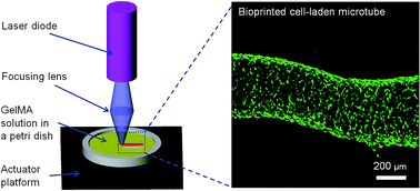
Photocrosslinking is a widely-used method to generate cell-laden hydrogels for tissue engineering. At present, it usually takes more than 30 seconds to crosslink hydrogels using UV illumination, and this delay makes it more likely that damage will occur in the DNA. With this in mind, we introduce an ultrafast photocrosslinking method using a low-cost blue laser diode. Experimental results show that a hydrogel with a diameter of 8 mm can be crosslinked using this process within 10 seconds with over 90% cell viability. Moreover, it is shown that the laser can be focused for the implementation of bioprinting. A microscale cell-laden microtube was successfully fabricated with this laser-based system, demonstrating its feasibility for bioprinting.

Introduction

Photocrosslinkable hydrogels have been used in a number of tissue engineering applications, including cell differentiation, injectable tissue generation, bioprinting, and organ-on-a-chip.1–9 Photocrosslinking has many advantages over conventional physical- or chemical-crosslinking methods. The advantages include spatial and temporal control in the polymerization process, fast crosslinking rates (several seconds to a few minutes) at room temperature, as well as minimal heat production.4 To achieve the fast photocrosslinking of hydrogels, many researchers have applied expensive high-intensity light sources, such as the OmniCure Series 2000 (Excelitas Technologies, Waltham, USA), with wavelengths spanning the 100 to 400 nm ultraviolet (UV) spectrum.9,10 However, it is still difficult to crosslink cell-laden hydrogels within 30 seconds even with these UV light sources (the crosslinking time is often several minutes when using a standard handheld UV lights). At the same time, it has been proposed that exposure to UV light can induce DNA damage11–13 or the cancerization of cells.14,15 Thus, it is highly desirable to find a crosslinking method for the polymerization of cell-laden hydrogels that is both fast and not reliant upon UV radiation.

In this work, we present a simple, ultrafast crosslinking method to create cell-laden hydrogels using a low-cost (off-the-shelf) laser diode at a wavelength of 405 nm. The developed method is shown to complete the crosslinking process within 10 seconds, and the cell viability right after crosslinking is found to be higher than 90%. Cells could also attach and proliferate inside the laser-crosslinked hydrogels, demonstrating that the laser-based crosslinking method has a high potential for tissue engineering. A hydrogel pattern is printed for this study to demonstrate that the proposed direct laser crosslinking method.

To achieve a fast crosslinking speed, the applied light source should have a sufficiently high intensity at the absorption peak of the photoinitiator. Moreover, the light source should ideally have a wavelength that is higher than the UV-B band, i.e., higher than 315 nm, because radiation in the UV-B band has been reported to induce serious DNA damage and apoptosis.11,13 Given these considerations, a laser is an ideal source for crosslinking, as a laser can output the desired high-intensity radiation with a narrow linewidth (at the absorption peak of the photoinitiator). Moreover, the advent of GaN-based laser diodes has introduced a new generation of inexpensive and compact light sources with wavelengths in the proximity of 405 nm. Such laser diodes can match the absorption peaks of many biocompatible photoinitiators.

There are many biocompatible photoinitiators for cell encapsulation, such as 2-hydroxy-4-(2-hydroxyethoxy)-2-methylpropiophenone (Irgacure 2959).16 However, it is difficult to directly apply this photoinitiator to laser crosslinking, since the absorption peak of Irgacure 2959 is at a wavelength of approximately 260 nm.17 Irgacure 2959 under such 265 nm irradiation can crosslink hydrogels very fast, i.e., within a few seconds, but this UV-B irradiation can readily induce DNA damage and apoptosis of cells, as stated above.11,13 It has been reported that Irgacure 2959 also functions under UV-A irradiation, at a wavelength of approximately 365 nm, which is safer in comparison to UV-B irradiation, but it takes a long time to complete the crosslinking process18 due to the relatively low molar extinction coefficient (being equivalent to the absorption coefficient) of the long-wave UV-A light.19 According to our investigation, Irgacure 2959 is not activated at a wavelength of 405 nm, and this result is in agreement with previous reports that the molar extinction coefficient of Irgacure 2959 reaches zero at wavelengths higher than 370 nm.19 Thus, it is concluded that Irgacure 2959 cannot be used for the laser-based photocrosslinking at a wavelength of 405 nm.

The photoinitiator, 2,2′-azobis[2-methyl-n-(2-hydroxyethyl)propionamide] (VA-086), was recently introduced as a promising photoinitiator for tissue engineering.20 Its absorption peak is at approximately 385 nm. This is in the long-wave UV-A range, which allows for activation by laser diodes with short crosslinking times.21 It is also worth noting that VA-086 is more biocompatible than Irgacure 2959, which contributes to high cell viability after crosslinking.21,22 The work presented here shows that the hydrogel with VA-086 can be crosslinked with illumination by a 405 nm laser diode (Fig. 1A shows the crosslinking mechanism). Such a system can be accelerated, by applying illumination for 10 seconds over an 8 mm-diameter beam spot, to achieve the so-called “ultrafast” crosslinking.


image file: c5ra24910d-f1.tif
Fig. 1 Laser-based crosslinking and bioprinting system set-up. (A) Laser-based photocrosslinking mechanism. GelMA hydrogel network was formed in the presence of VA-086 photoinitiator and the blue (λ = 405 nm) laser irradiation. (B) A schematic of the simple laser crosslinking system. (C) A photograph of the crosslinking system setup. (D) A schematic of the direct laser patterning system shows that the focused laser spot prints a hydrogel microtube by the movement of a linear actuator. (E) A photograph of the laser printing system setup.

Materials and methods

Light source

The critical component of the ultrafast crosslinking method is the laser diode. A 405 nm blue laser diode with a peak power of 300 mW (Armlaser, San Francisco, CA, USA) was employed for this. The laser diode was mounted 10 cm above a Petri dish. A DC power supply (CPS-3205, Gopher Technology, Guangzhou, China) was connected to the laser diode, and its voltage was adjusted to control the current supplied to the laser diode—which ultimately controlled the power being emitted by the laser diode. A schematic and photograph of the crosslinking system are given in Fig. 1B and C, respectively. The power of the laser was measured by an optical power meter (Model 1916R, Newport, Irvine, CA, USA). Three laser powers were applied, over a circular beam spot with a diameter of 8 mm, yielding laser intensities of 1 mW mm−2, 2 mW mm−2, and 5 mW mm−2, respectively.

Hydrogel synthesis

The hydrogel prepolymer solution for the experiment is based on a gelatin methacrylate (GelMA) hydrogel. The GelMA was synthesized using a previously reported method.16 Briefly, we dissolved gelatin (from porcine skin) in dimethyl sulfoxide (DMSO) solvent at 50 °C with stirring to have a 10% w/v gelatin in the DMSO solution. Then, 4-dimethylaminopyridine (DMAP) and glycidyl methacrylate were added to the solution to form a mixture with 0.6% w/v DMAP and 7.4% v/v glycidyl methacrylate. The mixture was stirred for two days at 50 °C. Subsequently, the mixture was dialyzed against reverse osmosis (RO) water at room temperature for a week. The RO water was changed twice a day. After dialysis, the GelMA sample was freeze-dried via lyophilization. For the experiments, a photocrosslinkable GelMA prepolymer solution was prepared by dissolving 10% w/v freeze-dried GelMA and 1% w/v VA-086 photoinitiator (Wako Chemical, Osaka, Japan) in phosphate buffered saline (PBS). Unless stated otherwise, all materials were purchased from Sigma-Aldrich, St. Louis, MO, USA.

Bioprinting system setup

To build a bioprinting system, we integrated the laser diode with a spherical lens (f = 50 mm) to achieve a focused laser spot with a diameter of approximately 400 μm. A one-axis linear actuator (L12R, Firgelli Tech., Victoria, BC, Canada) was employed as a stage on which to move the Petri dish. The setup of the modified system is shown in Fig. 1D and E. With the bioprinting system setup, the focused laser spot can be used to selectively crosslink the hydrogel prepolymer solution during motion of the stage. In this way, the laser-based platform can print cell-encapsulated microtube patterns in one direction.

Before bioprinting, the 10% w/v GelMA solution was mixed with NIH 3T3 fibroblast cells (8 × 10⁶ cells per mL). Then, 2 mL of GelMA solution was pipetted and dispensed onto the Petri dish (being 3.5 cm in diameter) to form a thin layer of GelMA solution. The laser diode was then switched on to generate the focused laser spot. At the same time, the linear actuator was used to apply motion at a constant speed of 3 mm s−1. With the movement of the stage, the laser spot crosslinked a line of hydrogel microtubes on the Petri dish. After bioprinting, the uncrosslinked hydrogel was removed. The microtubes were washed twice with PBS. Subsequently, fresh media were added to the Petri dish and the drop was cultured inside the incubator.

Cell culture, encapsulation, and viability assessment

NIH 3T3 fibroblast cells were cultured at 37 °C with 5% CO2. The cell media consists of Dulbecco's Modified Eagle Medium (DMEM) with 10% v/v fetal bovine serum and 1% v/v penicillin streptomycin. All the materials were purchased from ThermoFisher Scientific, Carlsbad, CA, USA. The cell medium was changed every three days.

Experiments were carried out to investigate the biocompatibility of the laser-based crosslinking within various parameters. Before crosslinking, the 10% GelMA prepolymer solution was mixed with NIH 3T3 fibroblast cells (8 × 10⁶ cells per mL). Then, 100 μL drops of the 10% GelMA prepolymer solution were pipetted onto the Petri dish. The drops containing cells were then illuminated and crosslinked by the laser beam. After crosslinking, the drops were washed twice with PBS. Subsequently, fresh media were added to the Petri dish and the cell-laden GelMA drops were cultured inside the incubator. In order to verify the biocompatibility of the laser crosslinking, we checked the cell viability and proliferation at day 0, day 2, and day 4 using a LIVE/DEAD assay (Biotium, Hayward, CA, USA). Samples were washed three times with PBS and treated with the assay for 30 minutes. The images of the assayed samples were taken by a confocal fluorescence microscope (FV1000, Olympus, Tokyo, Japan).

To analyze the cell viability, images taken with the microscope were converted to a 16 bit gray value format, and the cell numbers were counted manually using cell counter plugin provided by FIJI ImageJ (NIH, Bethesda, MD, USA). In the end, the cell viability was calculated with the following formula:

image file: c5ra24910d-t1.tif

In addition, we compared the cell density (being the total number of total cells per mm2) at day 0, day 2, and day 4 to quantify the proliferation of cells. The cell density was calculated as follows:

image file: c5ra24910d-t2.tif

Statistical analysis

A one-way analysis of variance (ANOVA analysis) function in MATLAB 2014b (MathWorks, Natick, MA, USA) was used to statistically analyze all the data. The results are presented as the average ± standard deviation. The sampling size (the time of replicates) was three.

Results and discussion

Laser-based cell-laden hydrogel crosslinking

Occhetta et al. reported that the VA-086-based photocrosslinking process generated nitrogen gas during crosslinking, making the hydrogels cloudy.20 Thus, to find the optimal crosslinking time, we illuminated the GelMA prepolymer solution with the laser until the bubble generation reached a saturation point—which was assumed to be the point at which the GelMA achieves a fully-crosslinked state. As shown in Fig. 2A, the hydrogel was free of bubbles before crosslinking. With the increase of the crosslinking time, bubble generation inside GelMA hydrogel continually increased, until it reached the fully-crosslinked state at which bubble generation stopped. We found that the minimum times needed for crosslinking 10% GelMA hydrogel with the 1% VA-086 photoinitiator at 50 mW, 100 mW, and 250 mW laser powers were 30 seconds, 20 seconds, and 10 seconds, respectively. Based on the minimal crosslinking time, we defined three combinations of laser powers and crosslinking time, 50 mW-30 seconds, 100 mW-20 seconds, and 250 mW-10 seconds, to conduct cell encapsulation experiments to examine the cell viability after crosslinking.
image file: c5ra24910d-f2.tif
Fig. 2 Pictures of the crosslinked hydrogel drops. (A) Pictures of hydrogel with different crosslinking time. The power of the laser was 50 mW. (B) Confocal fluorescence images of the crosslinked cell-laden GelMA at day 0, day 2, and day 4. Scale bar = 100 μm. (C) Phase contrast images of the crosslinked cell-laden GelMA with 250 mW-10 seconds at day 0, day 2, and day 4. Scale bar = 100 μm.

The cell encapsulation results are presented in Fig. 2B. The green dots (calcein AM) represent live cells, and the red dots (ethidium homodimer III) represent dead cells. It can be seen that at day 0 (right after crosslinking), most of the cells were still alive. After two days' culturing, some of the cells attached and started to elongate inside the microenvironment of the hydrogels. At day 4, more cells were elongated compared to day 2, however, the number of dead cells increased slightly. As mentioned above, VA-086-based photocrosslinking has the disadvantage that it generates bubbles inside the crosslinked hydrogel. Fig. 2C shows that there were many bubbles inside the hydrogels at day 0. However, the bubbles disappeared as the cells grew and the hydrogel degraded. At day 4, there were a negligible number of bubbles inside the hydrogel. This observation is similar to the results reported by Occhetta et al.21

Fig. 3A presents the results of the cell viability analysis. It can be concluded that the ultrafast crosslinking process has high biocompatibility. At day 0 and day 2, the viabilities of all samples were higher than 90%, while the viability at day 4 was significantly decreased. The decreased viability at day 4 can be explained by the relatively high cell density inside the hydrogel after four days of culturing. The cell proliferation analysis, seen in Fig. 3B, reveals that the cell density continued to increase up to day 4, at which point it may become difficult for some cells to obtain enough nutrition to live.23 However, even at day 4, the cell viability was greater than 80%. As shown in Fig. 3A, it was found that the p-value between the viabilities of 50 mW and 250 mW at day 4 was 0.059, which is not considered to be statistically significant. Thus, the cell viability was not affected to a great extent by the chosen combinations of crosslinking time and laser power. However, in terms of practicality, it can be seen that the higher power laser diode (250 mW) is seen to offer improved performance by way of its shorter crosslinking time.


image file: c5ra24910d-f3.tif
Fig. 3 Statistial analysis of the cell-laden GelMA. (A) Viability, and (B) cell density of the cells cultured inside the crosslinked hydrogel drops. Error bars indicate the standard deviation (n = 3, *p < 0.05).

Bioprinting application

The high cell viability of the encapsulated cells and the short crosslinking time led us to employ the developed laser-based photocrosslinking method for bioprinting applications. With the bioprinting system setup illustrated in Fig. 1D and E, the focused laser spot can be used to selectively crosslink the hydrogel prepolymer solution during motion of the stage. In this way, the laser-based platform can print cell-encapsulated microtube patterns in one direction.

The phase contrast and fluorescence images of a hydrogel microtube crosslinked by the laser spot are presented in Fig. 4A and B, respectively. The cells attached and grew inside the microtube after three days of culturing, with a cell viability of over 90%. Three-dimensional images of bioprinted microtubes were visualized by BioView3D (version 0.1.18, Center for BioImage Informatics, University of California, Santa Barbara, CA, USA). The constructed three-dimensional image in Fig. 4C shows that the laser-based crosslinking method is a promising solution for bioprinting. It is also worth noting that the use of a focused laser spot reduces the generation of bubbles. According to our observation, with a laser spot having a 400 μm diameter, there are no bubbles generated in the microtube after printing. Even smaller laser spot diameters can be applied by focusing via microscope objectives, and this can enable laser-based bioprinting and competitive cell patterning on a microscale.


image file: c5ra24910d-f4.tif
Fig. 4 Direct laser bioprinted microtube sample. (A) A phase contrast image, (B) fluorescence live/dead assayed image (scale bar = 200 μm), and (C) 3-D reconstructed image of the printed GelMA microtube on day 3 (scale bar = 215.5 μm).

It is important to note that the demonstrated laser-based bioprinting technique differs from existing laser-assisted bioprinting techniques.24–26 Laser-assisted bioprinting typically utilizes an energy-absorbing layer, in response to the stimulus of lasers, with the cells suspended below the absorbing layer. Once the small area of the absorbing layer is illuminated by the laser, a high-pressure bubble is generated, propelling the suspended cells below that area. This forms a droplet for collection by the receiving board.27,28 In this study, by contrast, the laser directly illuminates and crosslinks the desired area to pattern the hydrogel scaffolds. There is no droplet formation process and no supporting layer. Thus, we have named our technique “direct laser bioprinting”. More research will be carried out in the near future to investigate the fabrication of complex three-dimensional cell-laden scaffolds using the direct laser bioprinting.

Conclusion

We have presented an ultrafast photocrosslinking method using a low-cost laser diode and a VA-086 photoinitiator. The developed technique was able to crosslink the hydrogel within 10 seconds, and the cell viability after patterning was over 90%. By implementing this technique with a focusing lens, we established a bioprinting system for spatially-resolved photocrosslinking. We found that microscale ultrafast crosslinking avoided bubble generation during crosslinking. The successful fabrication of a 400 μm hydrogel microtube demonstrated the potential of this direct laser bioprinting system.

Acknowledgements

This work was supported by the Natural Sciences and Engineering Research Council of Canada (NSERC) Discovery Grant (RGPIN-2014-04010).

References

  1. A. Khademhosseini, R. Langer, J. Borenstein and J. P. Vacanti, Proc. Natl. Acad. Sci. U. S. A., 2006, 103, 2480–2487 CrossRef CAS PubMed.
  2. P. Zorlutuna, N. E. Vrana and A. Khademhosseini, IEEE Reviews in Biomedical Engineering, 2013, 6, 47–62 CrossRef PubMed.
  3. Z. Wang, B. Calpe, J. Zerdani, Y. Lee, J. Oh, H. Bae, A. Khademhosseini and K. Kim, Biotechnol. Bioeng., 2015 DOI:10.1002/bit.25905.
  4. K. T. Nguyen and J. L. West, Biomaterials, 2002, 23, 4307–4314 CrossRef CAS PubMed.
  5. Z. Wang, R. Abdulla, B. Parker, R. Samanipour, S. Ghosh and K. Kim, Biofabrication, 2015, 7, 045009 CrossRef PubMed.
  6. K. C. Hribar, P. Soman, J. Warner, P. Chung and S. Chen, Lab Chip, 2014, 14, 268–275 RSC.
  7. Z. Wang, R. Samanipour, K. Koo and K. Kim, Sens. Mater., 2015, 27, 487–506 Search PubMed.
  8. Z. Wang, R. Samanipour and K. Kim, in Biomedical Engineering: Frontier Research and Converging Technologies, Springer International Publishing, 2015, pp. 209–233 Search PubMed.
  9. C. Cha, J. Oh, K. Kim, Y. Qiu, M. Joh, S. R. Shin, X. Wang, G. Camci-unal, K. Wan, R. Liao and A. Khademhosseini, Biomacromolecules, 2014, 15, 283–290 CrossRef CAS PubMed.
  10. J. Jung and J. Oh, Biomicrofluidics, 2014, 8, 036503 CrossRef PubMed.
  11. J. Cadet, E. Sage and T. Douki, Mutat. Res., Fundam. Mol. Mech. Mutagen., 2005, 571, 3–17 CrossRef CAS PubMed.
  12. R. P. Sinha and D. P. Häder, Photochem. Photobiol. Sci., 2002, 1, 225–236 CAS.
  13. D. Kulms, E. Zeise, B. Pöppelmann and T. Schwarz, Oncogene, 2002, 21, 5844–5851 CrossRef CAS PubMed.
  14. B. K. Armstrong and A. Kricker, J. Photochem. Photobiol., B, 2001, 63, 8–18 CrossRef CAS.
  15. F. R. De Gruijl, H. J. Van Kranen and L. H. F. Mullenders, J. Photochem. Photobiol., B, 2001, 63, 19–27 CrossRef CAS.
  16. J. W. Nichol, S. T. Koshy, H. Bae, C. M. Hwang, S. Yamanlar and A. Khademhosseini, Biomaterials, 2010, 31, 5536–5544 CrossRef CAS PubMed.
  17. W. A. Green, Industrial Photoinitiators: A Technical Guide, CRC Press, Boca Raton, 2010 Search PubMed.
  18. J. R. Tse and A. J. Engler, Current Protocols in Cell Biology, 2010, 1–16 Search PubMed.
  19. Y. H. Roh, J. H. Park, J. J. Ye, J. E. Lee and D. Luo, ChemPhysChem, 2012, 13, 2517–2521 CrossRef CAS PubMed.
  20. P. Occhetta, N. Sadr, F. Piraino, a. Redaelli, M. Moretti and M. Rasponi, Biofabrication, 2013, 5, 035002 CrossRef CAS PubMed.
  21. P. Occhetta, R. Visone, L. Russo, L. Cipolla, M. Moretti and M. Rasponi, J. Biomed. Mater. Res., Part A, 2015, 103, 2109–2117 CrossRef CAS PubMed.
  22. A. D. Rouillard, C. M. Berglund, J. Y. Lee, W. J. Polacheck, Y. Tsui, L. J. Bonassar and B. J. Kirby, Tissue Eng., Part C, 2011, 17, 173–179 CrossRef CAS PubMed.
  23. S. Ozturk and W.-S. Hu, Cell Culture Technology for Pharmaceutical and Cell-Based Therapies, CRC Press, Boca Raton, 2005 Search PubMed.
  24. F. Guillemot, a. Souquet, S. Catros, B. Guillotin, J. Lopez, M. Faucon, B. Pippenger, R. Bareille, M. Rémy, S. Bellance, P. Chabassier, J. C. Fricain and J. Amédée, Acta Biomater., 2010, 6, 2494–2500 CrossRef CAS PubMed.
  25. B. Guillotin, A. Souquet, S. Catros, M. Duocastella, B. Pippenger, S. Bellance, R. Bareille, M. Rémy, L. Bordenave, J. Amédée and F. Guillemot, Biomaterials, 2010, 31, 7250–7256 CrossRef CAS PubMed.
  26. V. Keriquel, F. Guillemot, I. Arnault, B. Guillotin, S. Miraux, J. Amédée, J.-C. Fricain and S. Catros, Biofabrication, 2010, 2, 014101 CrossRef PubMed.
  27. S. V. Murphy and A. Atala, Nat. Biotechnol., 2014, 32, 773–785 CrossRef CAS PubMed.
  28. C. Mandrycky, Z. Wang, K. Kim and D.-H. Kim, Biotechnol. Adv., 2015 DOI:10.1016/j.biotechadv. 2015.12.011.

This journal is © The Royal Society of Chemistry 2016
Click here to see how this site uses Cookies. View our privacy policy here.