DOI:
10.1039/C5RA24843D
(Paper)
RSC Adv., 2016,
6, 5990-5998
The cadmium binding characteristics of a lactic acid bacterium in aqueous solutions and its application for removal of cadmium from fruit and vegetable juices
Received
23rd November 2015
, Accepted 4th January 2016
First published on 8th January 2016
Abstract
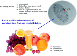
Heavy metal cadmium (Cd) is an environmental pollutant that causes adverse health effects in humans. This toxic metal has been detected in a wide range of fruit and vegetables. A strain of lactic acid bacteria, Lactobacillus plantarum CCFM8610, was screened out for its good ability to bind Cd, and this study was designed to investigate the Cd binding properties of this bacterium, and to evaluate its use for removal of Cd from fruit and vegetable juices. Electron microscopy observations and energy dispersive X-ray analysis confirmed that the majority of the Cd was bound to the surface of the bacterial cell. The Cd biosorption of L. plantarum CCFM8610 was strongly pH dependent, and carboxyl and amino groups of the bacterial surface molecules are important in the binding process. The biosorption was fast and efficient, and could be well explained by the Langmuir–Freundlich dual isotherm model (R2 = 0.99) and the pseudo second-order kinetic model (R2 = 0.99). After a 2 h incubation and a simple centrifugation, L. plantarum CCFM8610 treatment removed 67% to 82% of the Cd from nine types of fruit and vegetable juices. Long-period fermentation by L. plantarum CCFM8610 (36 h) also significantly decreased Cd concentrations in the juices (56% to 81%). Our results show that this food-grade bacterial strain could be used as a potential probiotic for Cd removal from fruit and vegetable juices.
1. Introduction
Heavy metal cadmium (Cd) is a ubiquitous environmental pollutant that can contribute to a broad range of adverse health effects in humans. In recent years, the risk of human exposure to Cd has increased with the increasing contamination of the food chain and inadequate protection from Cd-contaminated food.1 Due to its high rate of soil-to-plant transfer, Cd accumulation has been observed in a variety of fruit and vegetables such as apples, strawberries, pears, tomatoes, and cucumbers.2–4 Even if the Cd levels in fruit and vegetables are normally low, a high consumption of fruit and vegetables and their juices can result in an accumulative contribution to Cd exposure.5 The Joint FAO/WHO Expert Committee on Food Additives has set a tolerable weekly intake (TWI) of Cd at 7 μg kg−1 body weight.6 The Integrated Risk Information System (IRIS) of United States Environmental Protection Agency (EPA) set the threshold of oral Cd dose at 1 μg per kg per day.7 To date, no specific methods for Cd removal from fruit and vegetable juices have been developed and chemical processes, such as chemical deposition and ion exchange, have efficacy and safety issues.8,9
Several studies have revealed that some lactic acid bacteria (LAB), including Lactobacillus rhamnosus, L. plantarum, and L. reuteri, are capable of binding and removing heavy metals such as Cd and lead in vitro.10,11 Oral administration of L. rhamnosus can also prevent the increases in mercury and arsenic blood levels in pregnant women suspected to have high toxic metal exposures.12 In our previous study, a specific lactic acid bacterium, L. plantarum CCFM8610, was identified for its excellent Cd removing ability.13,14 In vivo studies also confirmed that this strain can sequester Cd in the intestines, thus decreasing the Cd absorption of mice subjected to chronic dietary Cd exposure.15 However, the mechanism of the efficient Cd binding process of this strain have not yet to be elucidated.
L. plantarum strains are widely used in the food industry and are generally regarded as safe.16 A considerable number of studies have shown that L. plantarum strains can be used for lactic acid fermentation and to enhance the nutritional, sensory and shelf life properties of fruit and vegetables.17–19 However, to our knowledge, no studies have been carried out so far to evaluate the effects of LAB strains on heavy metal removal from foodstuffs, such as fruit and vegetable juices.
The objective of this study was to investigate the Cd binding properties of L. plantarum CCFM8610 and to assess the potential application of this LAB strain for removal of Cd from fruit and vegetable juices.
2. Materials and methods
2.1. Bacterial strains and culture
The L. plantarum CCFM8610 strain was obtained from the in-house Culture Collections of Food Microbiology (CCFM), Jiangnan University (Wuxi, China). It was cultured in de Man, Rogosa and Sharpe (MRS) broth (Hopebio Company, Qingdao, China) at 37 °C for 18 h. All of the bacteria were subcultured twice before the experiment.
2.2. Cd binding assay
The Cd binding experiment was carried out as previously described.13 Briefly, L. plantarum CCFM8610 cell pellets were suspended in ultrapure water containing 50 mg L−1 of Cd as cadmium chloride, to give a final bacterial density of 1 g L−1 (wet weight). The cell pellets were resuspended in sterile saline at an identical bacterial concentration, as an untreated control. The bacterial suspension samples were then incubated for 1 h at 37 °C, with the pH adjusted to 6.0. The suspension was then centrifuged at 8000 × g for 20 min and a sample was taken from the supernatant for analysis of the residual Cd concentration by flame atomic absorption spectrophotometry (Spectr AA 220; Varian). The Cd binding ability of the strains was expressed as the percentage of Cd removed (bound by the bacteria) as follows,
Removal (%) = 100% × [(C0 − C1)/C0], |
where C0 and C1 are the initial Cd concentration and the residual Cd concentration after removal, respectively.
2.3. Electron microscopy and energy dispersive X-ray analysis
The samples for transmission electron microscopy (TEM) observation were prepared as previously described.20 After the Cd binding assay, bacterial cells were harvested by centrifugation at 8000 × g for 20 min, washed with phosphate buffer solution (PBS, pH 7.2), and resuspended in 1 mL of PBS. One hundred microliters of 25% glutaraldehyde was added to the bacterial suspension and the cells were left to fix for 1.5 h. The suspensions were centrifuged and washed three times in sodium cacodylate buffer (0.05 M). The cell pellets were then mixed at a 1
:
1 ration with molten 2% low melting point agarose, which was solidified by chilling and then chopped into small pieces (approximately 1 mm3). These samples were left overnight in a 2.5% glutaraldehyde/0.05 M sodium cacodylate buffer (pH 7.2) and then transferred to a Leica EM TP tissue processor (Leica Microsystems UK Ltd., Milton Keynes) where they were washed, post-fixed and dehydrated with 1 h between each change. The samples were then infiltrated with a resin (London Resin Company Ltd.) and ethanol mixture, and the tissue blocks of the samples were placed into gelatine capsules containing fresh resin and polymerized overnight at 60 °C. The sections (approximately 90 nm thick) were cut using an ultramicrotome (Ultracut E, Reichert-Jung), and collected on film/carbon-coated copper grids. The sections were examined and imaged using a FEI Tecnai G2 20 Twin TEM at 200 kV.
The samples for scanning electron microscopy (SEM) observation were prepared as previously described, with minor modifications.21 After the Cd binding assay, the bacterial cells were harvested by centrifugation at 8000 × g for 20 min, washed with PBS (pH 7.2), and fixed with glutaraldehyde (2.5% v/v) for 4 h. The cells were then washed with PBS (pH 7.2) three times and dehydrated with graded alcohols. An identical volume of isoamyl acetate was used to displace the graded alcohols. The bacterial samples were lyophilized and a Hitachi S-3400N SEM was used to observe the cellular morphology.
The TEM and SEM were equipped with energy dispersive X-ray (EDX; Edax) microanalysis systems and the elemental composition of the selected areas observed using the electron microscope was analyzed.
2.4. Characterization of the cellular components involved in Cd binding
The Cd binding abilities of the different cellular components of L. plantarum CCFM8610 were determined as previously described, with minor modifications.22,23 After the Cd binding assay with an initial pH of 6.0, the Cd bound to exopolysaccharides, the external surface of the cell wall, the interior surface of the cell wall and the external surface of the cell membrane, and the interior of the protoplast was eluted separately. The samples were transferred to metal-free digestion vessels (Omni; CEM) and digested in concentrated HNO3 using the Microwave Digestion System (MARS; CEM). The Cd concentration of each cellular component was detected by flame atomic absorption spectrophotometry and the Cd binding ability was expressed as follows,
Cd bound by each cellular component (%) = C1/C0 × 100%, |
where C0 and C1 are the Cd concentrations of the intact cell and the cell component, respectively.
2.5. Characterization of the functional groups involved in Cd binding
The bacterial cells were harvested and subjected to chemical treatments to modify the functional groups.24–26 Briefly, the carboxyl groups were neutralized in methanol with HCl (0.1 M), the amino groups were methylated in formaldehyde and formic acid solution (1
:
2 v/v), while the phosphoryl groups were esterified in triethyl phosphite and nitromethane solution (4
:
3 v/v). The treated samples were subjected to the Cd binding assay as described above, with an initial bacterial density of 10 g L−1 (dry weight) and an initial pH of 6.0.
The effect of the initial pH (2.0–7.0) on the Cd binding of L. plantarum CCFM8610 was tested using the same method of Cd binding assay described above, but with an initial Cd concentration of 5 mg L−1.
2.6. Cd binding isotherm study
According to previously reported methods,27 the harvested cells were suspended in ultrapure water containing 0.06 to 90 mg L−1 Cd as cadmium chloride, to give a final bacterial concentration of 1 g L−1 (dry weight). The Cd binding assay was then conducted with an initial pH of 6.0 and the equilibrium content of Cd bound by the bacterium was expressed as follows,
qe (Cd content bound by the biomass) = [(Ci − Ce) × V]/m, |
where Ci and Ce are the initial Cd concentration and the equilibrium Cd concentration, respectively, and V/m = 1 L/g.
The acquired experimental data were fitted to different isotherm models including the Langmuir,28 the Freundlich,28 and the Langmuir–Freundlich dual models.29
2.7. Cd binding kinetic study
In accordance with previously reported methods,27 the harvested cells were suspended in ultrapure water containing 30 mg L−1 of Cd as cadmium chloride, to give a final bacterial concentration of 1 g L−1 (dry weight). The Cd binding assay was then conducted with an initial pH of 6.0 and the concentration of Cd in the supernatant was detected at different time intervals up to 360 min. The acquired experimental data were fitted to different kinetic models (Table 1).
Table 1 Kinetic models used in the present studya
Kinetic models |
|
qt (mg g−1) represents the Cd content bound by Lactobacillus plantarum CCFM8610 biomass at time point “t”. qe (mg g−1) represents the equilibrium content of the Cd bound by the biomass. K1 and K2 are the biosorption constants in the pseudo first-order kinetic model and the pseudo second-order kinetic model, respectively. |
(1) Pseudo first-order kinetic model |
qt = qe − [qe × exp(−K1t)] |
(2) Pseudo second-order kinetic model |
t/qt = 1/K2qe2 + t/qe |
2.8. Cd removal from fruit and vegetable juices
Fresh apples, tomatoes, and cucumbers were collected from three local markets in Jiangsu, Jiangxi, and Hunan Province in China, respectively. These fruit and vegetables were washed, chopped, and added to ultrapure water (1
:
2 w/v). Juice was obtained by pressing the mixture in a juice extractor. The juice was then centrifuged at 10
000 × g for 20 min, sterilized at 105 °C for 10 min, and glass-bottled. The juices were labeled as AJ1, AJ2, AJ3 (apple juices), TJ1, TJ2, TJ3 (tomato juices), CJ1, CJ2, CJ3 (cucumber juices).
The viable L. plantarum CCFM8610 cells were harvested, washed with sterilized saline, and used to inoculate the fruit and vegetable juices at an inoculum level of 4% (v/v), corresponding to ca. 107 cfu mL−1.17,30 The juice without the bacterial inoculum was used as a control. All of the juices were incubated at 37 °C for 2 h or 36 h. After the incubation, the bacterial growth was measured by colony counting. The juices were then centrifuged at 8000 × g for 20 min to remove the Cd-bound bacterial biomass and the supernatant was collected for biochemical analysis.
The antioxidative activities of the juices were measured using a DPPH (1,1-diphenyl-2-picrylhydrazyl) scavenging assay.31 The juices were digested in concentrated HNO3 with the Microwave Digestion System, and the Cd concentration was measured by flame atomic absorption spectrophotometry.
2.9. Statistical analysis
Data are expressed as the mean ± the standard error of the mean (SEM). A minimum of three independent experiments were carried out for each assay. The differences between the means of the test were analyzed using one-way analysis of variance, followed by Tukey's post hoc test. A p value of <0.05 was considered to be statistically significant. The statistical analysis of the data obtained was performed using SPSS 16.0 for Windows software. For the isotherm and kinetic studies, all of the equation parameters were evaluated using MATLAB R2010b software and a correlation coefficient (R2) test was used to measure the fitness of the data to the models.
3. Results
3.1. Electron microscopy observation and EDX analysis
TEM micrographs of L. plantarum CCFM8610 before and after the Cd binding are presented in Fig. 1. Obvious deposits of Cd were observed on the surface of the cells after binding (Fig. 1B), whereas no Cd was visible in the micrographs of untreated cells (Fig. 1A). For the EDX analysis, no Cd signal could be detected in the control sample (Fig. 1C), but a clear peak for Cd was observed in Cd treated cells (Fig. 1D), indicating the presence of Cd due to biosorption.
 |
| Fig. 1 Transmission electron microscopic images of Lactobacillus plantarum CCFM8610 before and after Cd binding. (A) Untreated biomass. (B) Biomass after Cd binding. (C) Energy dispersive X-ray (EDX) spectra of untreated biomass. (D) EDX spectra of biomass after Cd binding. Scale bar = 100 nm. The experiment was carried out in aqueous solution containing Cd as cadmium chloride. | |
The SEM micrographs revealed that Cd exposure caused anomalous aggregation of the L. plantarum CCFM8610 cells (Fig. 2B), whereas no morphological changes in the control biomass were observed (Fig. 2A). The EDX spectra also confirmed an additional Cd peak of Cd-treated cells (Fig. 2D), which was absent in the control sample (Fig. 2C).
 |
| Fig. 2 Scanning electron microscopic images of L. plantarum CCFM8610 before and after Cd binding. (A) Untreated biomass. (B) Biomass after Cd binding. (C) Energy dispersive X-ray (EDX) spectra of untreated biomass. (D) EDX spectra of biomass after Cd binding. Scale bar = 10.0 μm. The experiment was carried out in aqueous solution containing Cd as cadmium chloride. | |
3.2. Cd binding abilities of different cell components
The exopolysaccharides fraction of L. plantarum CCFM8610 did not bind Cd (Fig. 3). The greatest amount of Cd accumulated on the external surface of the cell wall (40.54% ± 0.80%) and in the space between the cell wall and the plasma membrane (54.30% ± 2.93%). Only approximately 7% of the Cd entered the protoplast.
 |
| Fig. 3 Cd binding ability of the cellular component of L. plantarum CCFM8610. Values are mean ± SEM of three determinations. Significant differences (p < 0.05) between the cellular components are indicated with different letters above the bars. The experiment was carried out in aqueous solution containing Cd as cadmium chloride. | |
3.3. Effects of functional groups on Cd binding
When the amino groups of L. plantarum CCFM8610 cell surface molecules were methylated, the bacterial cells' ability to bind Cd was hampered and the removal of Cd decreased from 48.16% to 5.95% (Fig. 4). There was also a decrease in Cd removal when carboxyl groups were neutralized (p < 0.05). In contrast, the chemical modification of phosphoryl groups did not cause a significant difference in Cd removal (p > 0.05).
 |
| Fig. 4 Effects of chemical treatments of amino, carboxyl and phosphoryl groups on the Cd binding ability of L. plantarum CCFM8610. Values are mean ± SEM of three determinations. Significant differences (p < 0.05) between the different chemically treated groups are indicated with different letters above the bars. The experiment was carried out in aqueous solution containing Cd as cadmium chloride. | |
The initial pH was critical to the Cd binding ability of L. plantarum CCFM8610 (Fig. 5). The Cd removal remained negligible when the pH was below 2.0, but increased significantly with the incremental rise in pH. The results for pH values over 7.0 were excluded from the analysis because Cd2+ was found to precipitate under high pH conditions.
 |
| Fig. 5 Effects of initial pH on Cd binding ability of L. plantarum CCFM8610. Values are mean ± SEM of three determinations. The experiment was carried out in aqueous solution containing Cd as cadmium chloride. | |
3.4. Cd binding isotherm
As shown in Fig. 6, the removal of Cd by L. plantarum CCFM8610 was evaluated by plotting the amount of Cd bound by the bacteria (qe) against the equilibrium Cd concentration of the metal (Ce). The Cd binding ability increased with an increase of Cd concentration in the solution, ultimately reaching the equilibrium value. Data were further analyzed using different isotherm models (Table 2). Considering the highest R2 value, the Cd biosorption by the strain showed best fit to the Langmuir–Freundlich dual isotherm model (R2 = 0.9928).
 |
| Fig. 6 Adsorption isotherm of Cd binding by L. plantarum CCFM8610. Values are mean ± SEM of three determinations. EXP, experimental data acquired in the present study; LM, Langmuir model; FM, Freundlich model; LFM, Langmuir–Freundlich model. The experiment was carried out in aqueous solution containing Cd as cadmium chloride. | |
Table 2 Biosorption constants from simulations with different isotherm modelsa
Isotherm models |
Constants |
|
qe (mg g−1) represents the equilibrium content of the Cd bound by the biomass. Ce (mg L−1) represents the equilibrium Cd concentration. The experiment was carried out in aqueous solution containing Cd as cadmium chloride. |
(1) Langmuir  |
Qmax |
24.69 (mg g−1 dry biomass) |
bL |
0.2494 |
R2 (nonlinear) |
0.9679 |
(2) Freundlich qe = KFCe1/nF |
KF |
6.575 |
nF |
3.006 |
R2 (nonlinear) |
0.9834 |
(3) Langmuir–Freundlich dual  |
KLF |
7.941 |
aLF |
0.2038 |
n |
1.926 |
R2 (nonlinear) |
0.9928 |
3.5. Cd binding kinetics
As indicated in Fig. 7, the Cd binding process of L. plantarum CCFM8610 was efficient and fast. Approximately 90% of the binding process was completed in less than 100 min, and the saturation value was reached by about 300 min. Data were further analyzed by the pseudo first and second-order rate models (Table 1), with the latter model showing better fitness (R2 = 0.9954) than the former (R2 = 0.9749).
 |
| Fig. 7 Cd binding of L. plantarum CCFM8610 at different time points. Values are mean ± SEM of three determinations. The experiment was carried out in aqueous solution containing Cd as cadmium chloride. | |
3.6. Removal of Cd from fruit and vegetable juices by L. plantarum
As shown in Table 3, a significant decrease in the pH value was observed in all of the juices after L. plantarum CCFM8610 treatment. The 36 h fermentation caused more significant changes in the pH than the 2 h treatment but the extent of reduction was also dependent on the nature of the juice. There was no statistically significant difference in the viable bacterial cell numbers between the 0 h and 2 h time points (p > 0.05). For the 36 h treatment, the viable count of the strain reached over 1 × 10⁸ cfu mL−1 in each juice (except for CJ3, with a count of 0.95 × 10⁸ cfu mL−1), which is significantly higher than that at 0 h and 2 h time points.
Table 3 Effects of L. plantarum CCFM8610 treatment (2 h and 36 h) on pH and viable cell number of the juicesa
Juices |
pH |
Viable counts (107 cfu mL−1) |
0 h |
2 h |
36 h |
0 h |
2 h |
36 h |
Values are mean ± SEM of three determinations. The superscript letters indicate statistically significant differences at a p value of <0.05 in comparisons between different time points for each juice group. AJ, TJ and CJ indicate apple juices, tomato juices and cucumber juices, respectively. |
AJ1 |
3.96 ± 0.003a |
3.87 ± 0.003b |
3.46 ± 0.007c |
1.30 ± 0.04a |
1.42 ± 0.02a |
14.87 ± 0.19b |
AJ2 |
3.94 ± 0.003a |
3.90 ± 0.003b |
3.39 ± 0.003c |
1.38 ± 0.06a |
1.57 ± 0.03a |
15.63 ± 0.19b |
AJ3 |
3.98 ± 0.009a |
3.90 ± 0.012b |
3.40 ± 0.006c |
1.30 ± 0.03a |
1.63 ± 0.02a |
13.97 ± 0.22b |
TJ1 |
4.36 ± 0.021a |
4.20 ± 0.008b |
3.17 ± 0.009c |
1.39 ± 0.06a |
1.80 ± 0.11a |
27.37 ± 0.70b |
TJ2 |
4.33 ± 0.009a |
4.23 ± 0.000b |
3.20 ± 0.007c |
1.30 ± 0.02a |
1.64 ± 0.02a |
20.70 ± 0.57b |
TJ3 |
4.39 ± 0.006a |
4.24 ± 0.009b |
3.18 ± 0.017c |
1.43 ± 0.02a |
1.62 ± 0.03a |
21.70 ± 0.72b |
CJ1 |
5.13 ± 0.000a |
4.90 ± 0.006b |
3.68 ± 0.015c |
1.39 ± 0.02a |
1.44 ± 0.02a |
10.87 ± 0.19b |
CJ2 |
5.09 ± 0.003a |
4.93 ± 0.029b |
3.61 ± 0.012c |
1.25 ± 0.03a |
1.35 ± 0.03a |
14.27 ± 0.56b |
CJ3 |
5.13 ± 0.009a |
4.95 ± 0.003b |
3.64 ± 0.019c |
1.37 ± 0.03a |
1.46 ± 0.02a |
9.50 ± 0.35b |
The effects of L. plantarum CCFM8610 treatment on Cd removal from juices are shown in Tables 4 and 5 at 2 h and 36 h time points respectively. The presence of Cd was observed in all juices collected from different regions of China. Compared with the control groups, the treatment of L. plantarum CCFM8610 significantly decreased the Cd levels in each juice (p < 0.05). After 2 h, the minimum Cd removal (67.12%) was found in the tomato juice and the maximum removal (82.87%) was observed in the cucumber juice (Table 4). For the 36 h treatment, Cd removal rate varied between 56.18% and 81.79% depending on the type of juice (Table 5).
Table 4 Cd removal from juices by L. plantarum CCFM8610 treatment (2 h)a
Juices |
Cd concentration (μg mL−1) |
Cd removal (%) |
Control |
L. plantarum CCFM8610 treated |
Values are mean ± SEM of three determinations. The superscript letters indicate statistically significant differences at a p value of <0.05 in comparisons between control and L. plantarum CCFM8610 treated samples for each juice group. AJ, TJ and CJ indicate apple juices, tomato juices and cucumber juices, respectively. |
AJ1 |
0.51 ± 0.006a |
0.095 ± 0.014b |
81.42 ± 2.74 |
AJ2 |
1.21 ± 0.033a |
0.27 ± 0.005b |
78.08 ± 0.16 |
AJ3 |
0.72 ± 0.013a |
0.13 ± 0.002b |
82.28 ± 0.33 |
TJ1 |
0.50 ± 0.005a |
0.13 ± 0.002b |
74.59 ± 0.21 |
TJ2 |
0.58 ± 0.006a |
0.16 ± 0.009b |
71.71 ± 1.30 |
TJ3 |
0.59 ± 0.012a |
0.19 ± 0.003b |
67.12 ± 0.64 |
CJ1 |
0.70 ± 0.008a |
0.15 ± 0.005b |
78.10 ± 0.62 |
CJ2 |
1.71 ± 0.013a |
0.29 ± 0.006b |
82.87 ± 0.25 |
CJ3 |
2.90 ± 0.008a |
0.52 ± 0.005b |
82.07 ± 0.22 |
Table 5 Cd removal from juices by L. plantarum CCFM8610 treatment (36 h)a
Juices |
Cd concentration (μg mL−1) |
Cd removal (%) |
Control |
L. plantarum CCFM8610 treated |
Values are mean ± SEM of three determinations. The superscript letters indicate statistically significant differences at a p value of <0.05 in comparisons between control and L. plantarum CCFM8610 treated samples for each juice group. AJ, TJ and CJ indicate apple juices, tomato juices and cucumber juices, respectively. |
AJ1 |
0.51 ± 0.005a |
0.097 ± 0.007b |
81.16 ± 1.26 |
AJ2 |
1.25 ± 0.036a |
0.26 ± 0.013b |
78.89 ± 1.30 |
AJ3 |
0.70 ± 0.017a |
0.17 ± 0.004b |
75.54 ± 0.82 |
TJ1 |
0.51 ± 0.002a |
0.19 ± 0.016b |
62.98 ± 0.21 |
TJ2 |
0.58 ± 0.004a |
0.26 ± 0.014b |
56.18 ± 2.62 |
TJ3 |
0.61 ± 0.005a |
0.22 ± 0.012b |
64.35 ± 1.97 |
CJ1 |
0.72 ± 0.011a |
0.18 ± 0.004b |
75.44 ± 0.25 |
CJ2 |
1.62 ± 0.055a |
0.32 ± 0.010b |
80.21 ± 0.97 |
CJ3 |
2.95 ± 0.030a |
0.54 ± 0.002b |
81.79 ± 0.25 |
It was also noticed that the 36 h treatment significantly increased the free radical scavenging ability of most of the juices (Table 6), whereas the 2 h treatment only increased such ability in only two of the tomato juices.
Table 6 Effects of L. plantarum CCFM8610 treatment (2 h and 36 h) on DPPH scavenging ability of the juicesa
Juices |
Scavenging rate of DPPH (%) |
0 h |
2 h |
36 h |
Values are mean ± SEM of three determinations. The superscript letters indicate statistically significant differences at a p value of <0.05 in comparisons between different time points for each juice group. AJ, TJ and CJ indicate apple juices, tomato juices and cucumber juices, respectively. |
AJ1 |
63.09 ± 1.51 ab |
59.72 ± 0.86a |
67.90 ± 1.29b |
AJ2 |
58.48 ± 0.83a |
58.87 ± 1.34a |
61.48 ± 1.44a |
AJ3 |
64.79 ± 1.75 ab |
59.38 ± 2.41a |
70.28 ± 0.52b |
TJ1 |
72.98 ± 0.81a |
71.10 ± 0.61a |
78.51 ± 0.76b |
TJ2 |
62.39 ± 0.85a |
69.52 ± 0.30b |
72.69 ± 0.33c |
TJ3 |
58.83 ± 1.60a |
67.20 ± 0.70b |
69.12 ± 0.59b |
CJ1 |
52.39 ± 2.32a |
51.01 ± 0.11a |
54.33 ± 1.55a |
CJ2 |
46.80 ± 0.81a |
48.04 ± 0.68a |
52.69 ± 0.73b |
CJ3 |
54.78 ± 1.94a |
51.22 ± 0.50a |
62.75 ± 0.75b |
4. Discussion
As a consequence of environmental Cd contamination, this hazardous heavy metal has become a threat to food safety. In China, Cd has been detected in a wide range of fruit and vegetables.32–34 Our data on the levels of Cd in nine fruit and vegetables collected from different regions in China (Tables 4 and 5) further support this finding. As Cd is a non-essential but extremely toxic metal for humans, the concentrations of Cd in fruit and vegetables should be controlled to a minimum. The present study confirmed that a lactic acid bacteria strain, L. plantarum CCFM8610, can be used effectively to decrease the Cd levels in fruit and vegetable juices. This represents the first study identifying the use of a food-grade microorganism to reduce Cd levels in foodstuffs. After exposure of juices to L. plantarum CCFM8610, it was possible to remove over 56% of the Cd in the juices, which can help to keep the oral intake of Cd of consumers under the limits set by FAO/WHO or EPA.6,7
The Cd binding characteristics of L. plantarum CCFM8610 were investigated first. Compared with the control samples, additional Cd signals were observed in the EDX spectra after Cd exposure, confirming the presence of Cd in the cell biomass due to biosorption (Fig. 1 and 2). The morphological alterations of the strain after Cd binding, observed in the SEM micrographs, may be a result of the change in surface charge and the degeneration of the surface proteins caused by Cd exposure, leading to the anomalous aggregation and enhanced Cd binding ability of the cells. A previous study reported similar phenomenon in an Acidiphilium symbioticum strain after Cd binding and indicated such a mechanism as a form of self-protection by the bacterium.27 The TEM micrographs showed that the majority of the Cd passes through the surface polysaccharides of the strain and deposits on the surface of the cell, which is consistent with the differential Cd binding capacities of specific cell components (Fig. 3). It was noted that the deposition of Cd is discontiguous, indicating the involvement of specific biosorption sites on the cell surface. Similar deposition of Cd on the perimeter of the cell was also observed in a sulfate-reducing bacterium.35
To evaluate the possible functional groups involved in Cd binding, chemical modification was used to block the carboxyl, amino and phosphoryl groups on the surface structures of L. plantarum CCFM8610. The chemical modifications of the former two groups reduced the Cd binding ability of the strain by 18% and 42%, respectively, indicating that these two functional groups are important in the Cd binding process (Fig. 4). These results are consistent with previous studies on the effects of functional groups of A. symbioticum and Bacillus subtilis on heavy metal binding and the possible ion exchange mechanisms involved could be as follows,27,36
R–NH2 + CdX → R–NH2·Cd2+–X, |
R–COOH + Cd2+ ↔ (R–COO)2–Cd + 2H+, |
where R represents other molecular components on the cell surface and X represents compounds that can complex with Cd
2+.
As the electronegative carboxyl groups are abundantly available on the cell surface, they actively participate in the binding process of Cd2+ with simultaneous release of protons.27 The nitrogen atom of the amino groups can bind to Cd following electron pair sharing. Nitrogen donates a lone pair of electrons in the process of Cd binding, which facilitates the formation of stable metal complex.27,37 Previous research has shown that Cd removal by L. fermentum decreased significantly after blocking of the phosphoryl groups.25 However, the chemical modification of the phosphoryl groups in this study did not cause significant changes in Cd removal by L. plantarum CCFM8610. The Cd biosorption of L. plantarum CCFM8610 was strongly pH-dependent and the optimal pH for Cd binding was between 6.0 and 7.0 (Fig. 5). Similar results have been reported for other lactic acid bacteria strains, indicating that ion exchange is at least partly responsible for the binding process.38,39
The isotherm and kinetic models were established to further understand the binding characteristics of L. plantarum CCFM8610 (Tables 1 and 2). The isotherm experimental data of Cd binding fit the Langmuir model well (R2 = 0.97), and the theoretical monolayer Cd biosorption capacity (Qmax) of the strain was calculated as 24.69 mg g−1 (dry biomass), which is higher than previously reported commercial LAB strains such as L. casei Shirota (12.1 mg g−1) and L. rhamnosus GG (13.2 mg g−1).38 Similar differences in Cd binding ability between LAB strains, including L. plantarum CCFM8610 and L. rhamnosus GG, were also identified in our previous study.13 Considering the highest R2 value, the biosorption showed the best fit to the Langmuir–Freundlich dual model (R2 = 0.99). This is in accordance with the heavy metal binding characterization of other bacteria, indicating the contribution of both physical and chemical binding mechanisms.27,40 The kinetic analysis suggests a rapid binding process that best fits the pseudo second-order rate model, which is consistent with previous reports.27,38 The proper correlation with pseudo second-order rate model also indicates the involvement of chemical absorption during the binding process.41,42
The potency of this bacterium as a food-grade Cd absorbent was examined using 9 different Cd containing fruit and vegetable juices obtained from different regions of China. After exposure of these juices to L. plantarum CCFM8610 for only 2 h followed by a simple centrifugation to remove the Cd-bound bacterial biomass, it was possible to remove between 67% to 82% of the Cd present in the juices. The strain was also tested as a starter culture and the juices were fermented for 36 h. This treatment also significantly decreased Cd concentration of the juices (56% to 81%). The dramatic decrease in the pH of the juices after the 36 h incubation (Table 3) could have caused negative effects on the binding ability of the bacterium. However, as the viable cell number significantly increased after the 36 h incubation, more biomass was involved in Cd binding than in the 2 h treatment, enhancing the Cd removal and offsetting the reduced efficiency due to lower pH. The 36 h incubation also improved the antioxidant ability of the juices compared with the 2 h incubation (Table 6), which could be due to the increase of total flavanones in the juices after a long fermentation by this bacterium.31,43 As oxidative stress has been reported to be an important toxic mechanism of Cd exposure,44 fermentation could enhance the antioxidant ability of juices against Cd-induced toxicity. Therefore, although such fermentation processes will add the cost of juice production, the improved safety by Cd reduction and enhanced antioxidant properties of the vegetable and fruit juices may make such applications still worthwhile. A further study to investigate the organoleptic properties of these probiotic-treated juices is now in progress, to fully evaluate the consumer's acceptance of such products.
5. Conclusions
The Cd binding characteristics of a food-grade microorganism, L. plantarum CCFM8610, and the application of this strain to Cd removal from fruit and vegetable juices were evaluated in this study. The Cd biosorption by L. plantarum CCFM8610 was a fast, efficient, and pH-dependent process, which followed the Langmuir–Freundlich dual isotherm model and showed the best fit to the pseudo second-order rate kinetic model. The L. plantarum CCFM8610 treatment (both 2 h and 36 h) significantly decreased the Cd concentration in nine types of juices. Our results show that this strain could be used as a potential probiotic for Cd removal from fruit and vegetable juices.
Acknowledgements
This work was supported by the National Natural Science Foundation of China Key Program (No. 31530056), the National Natural Science Foundation of China (No. 31470161, 31371721), the BBSRC Newton Fund Joint Centre Award, the 111 Project B07019, and the Program for Changjiang Scholars and Innovative Research Team in University (IRT1249).
References
- G. F. Nordberg, K. Nogawa, M. Nordberg and L. Friberg, in Handbook on the Toxicology of Metals, ed. G. F. Nordberg, B. A. Fowler, M. Nordberg and L. Friberg, Academic Press, Burlington, MA, 3rd edn, 2011, ch. 23, pp. 446–451 Search PubMed
. - F. L. Coco, P. Monotti, F. Cozzi and G. Adami, Food Control, 2006, 17, 966–970 CrossRef
. - O. Muñoz, J. M. Bastias, M. Araya, A. Morales, C. Orellana, R. Rebolledo and D. Velez, Food Chem. Toxicol., 2005, 43, 1647–1655 CrossRef PubMed
. - Z. Krejpcio, S. Sionkowski and J. Bartela, Pol. J. Environ. Stud., 2005, 14, 877–881 CAS
. - R. Tahvonen, Food Addit. Contam., 1998, 15, 446–450 CrossRef CAS PubMed
. - FAO/WHO, Evaluation of certain food contaminants: 64th report of the Joint FAO/WHO Expert Committee on Food Additives, WHO Technical Report Series 930, Geneva, 2006, http://apps.who.int/iris/bitstream/10665/43258/1/WHO_TRS_930_eng.pdf, accessed December, 2015 Search PubMed
. - Integrated Risk Information System of United States Environmental Protection Agency, http://cfpub.epa.gov/ncea/iris/iris_documents/documents/subst/0141_summary.pdf, accessed November, 2014.
- E. Maliou, M. Malamis and P. Sakellarides, Water Sci. Technol., 1992, 25, 133–138 CAS
. - W. W. Ngah and M. Hanafiah, Bioresour. Technol., 2008, 99, 3935–3948 CrossRef PubMed
. - T. Halttunen, M. Collado, H. El-Nezami, J. Meriluoto and S. Salminen, Lett. Appl. Microbiol., 2008, 46, 160–165 CrossRef CAS PubMed
. - J. Bhakta, Y. Munekage, K. Ohnishi and B. Jana, Int. J. Environ. Sci. Technol., 2012, 9, 433–440 CrossRef CAS
. - J. E. Bisanz, M. K. Enos, J. R. Mwanga, J. Changalucha, J. P. Burton, G. B. Gloor and G. Reid, mBio, 2014, 5, e01580 CrossRef CAS PubMed
. - Q. Zhai, R. Yin, L. Yu, G. Wang, F. Tian, R. Yu, J. Zhao, X. Liu, Y. Q. Chen and H. Zhang, Food Control, 2015, 54, 23–30 CrossRef CAS
. - Q. Zhai, G. Wang, J. Zhao, X. Liu, F. Tian, H. Zhang and W. Chen, Appl. Environ. Microbiol., 2013, 79, 1508–1515 CrossRef CAS PubMed
. - Q. Zhai, G. Wang, J. Zhao, X. Liu, A. Narbad, Y. Q. Chen, H. Zhang, F. Tian and W. Chen, Appl. Environ. Microbiol., 2014, 80, 4063–4071 CrossRef PubMed
. - B. Foligné, C. Daniel and B. Pot, Curr. Opin. Microbiol., 2013, 16, 284–292 CrossRef PubMed
. - P. Filannino, L. Azzi, I. Cavoski, O. Vincentini, C. G. Rizzello, M. Gobbetti and R. di Cagno, Int. J. Food Microbiol., 2013, 163, 184–192 CrossRef CAS PubMed
. - R. di Cagno, R. Coda, M. de Angelis and M. Gobbetti, Food Microbiol., 2013, 33, 1–10 CrossRef CAS PubMed
. - P. Filannino, Y. Bai, R. di Cagno, M. Gobbetti and M. G. Gänzle, Food Microbiol., 2015, 46, 272–279 CrossRef CAS PubMed
. - E. Dertli, I. J. Colquhoun, A. P. Gunning, R. J. Bongaerts, G. Le Gall, B. B. Bonev, M. J. Mayer and A. Narbad, J. Biol.
Chem., 2013, 288, 31938–31951 CrossRef CAS PubMed
. - R. Chakravarty and P. C. Banerjee, Extremophiles, 2008, 12, 279–284 CrossRef CAS PubMed
. - A. M. Massadeh, F. A. Al-Momani and H. I. Haddad, Biol. Trace Elem. Res., 2005, 108, 259–269 CrossRef CAS PubMed
. - S. Deb, S. Ahmed and M. Basu, Bull. Environ. Contam. Toxicol., 2013, 90, 323–328 CrossRef CAS PubMed
. - G. Panda, S. Das, S. Chatterjee, P. Maity, T. Bandopadhyay and A. Guha, Colloids Surf., B, 2006, 50, 49–54 CrossRef CAS PubMed
. - H. Teemu, S. Seppo, M. Jussi, T. Raija and L. Kalle, Int. J. Food Microbiol., 2008, 125, 170–175 CrossRef CAS PubMed
. - S. S. Majumdar, S. K. Das, T. Saha, G. C. Panda, T. Bandyopadhyoy and A. K. Guha, Colloids Surf., B, 2008, 63, 138–145 CrossRef CAS PubMed
. - R. Chakravarty and P. C. Banerjee, Bioresour. Technol., 2012, 108, 176–183 CrossRef CAS PubMed
. - K. Vijayaraghavan, T. Padmesh, K. Palanivelu and M. Velan, J. Hazard. Mater., 2006, 133, 304–308 CrossRef CAS PubMed
. - S. Chatterjee, S. K. Das, R. Chakravarty, A. Chakrabarti, S. Ghosh and A. K. Guha, J. Hazard. Mater., 2010, 174, 47–53 CrossRef CAS PubMed
. - A. L. F. Pereira, T. C. Maciel and S. Rodrigues, Food Res. Int., 2011, 44, 1276–1283 CrossRef CAS
. - B. Escudero-López, I. Cerrillo, G. Herrero-Martín, D. Hornero-Méndez, A. Gil-Izquierdo, S. Medina, F. Ferreres, G. Berná, F. Martín and M.-S. Fernández-Pachón, J. Agric. Food Chem., 2013, 61, 8773–8782 CrossRef PubMed
. - G. Wang, M.-Y. Su, Y.-H. Chen, F.-F. Lin, D. Luo and S.-F. Gao, Environ. Pollut., 2006, 144, 127–135 CrossRef CAS PubMed
. - H. Zhang, J. Chen, L. Zhu, G. Yang and D. Li, PLoS One, 2014, e108572 Search PubMed
. - X. Bi, L. Ren, M. Gong, Y. He, L. Wang and Z. Ma, Geoderma, 2010, 155, 115–120 CrossRef CAS
. - N. Naz, H. K. Young, N. Ahmed and G. M. Gadd, Appl. Environ. Microbiol., 2005, 71, 4610–4618 CrossRef CAS PubMed
. - T. Beveridge and R. Murray, J. Bacteriol., 1980, 141, 876–887 CAS
. - S. K. Das, A. R. Das and A. K. Guha, Environ. Sci. Technol., 2007, 41, 8281–8287 CrossRef CAS PubMed
. - T. Halttunen, S. Salminen and R. Tahvonen, Int. J. Food Microbiol., 2007, 114, 30–35 CrossRef CAS PubMed
. - F. Ibrahim, T. Halttunen, R. Tahvonen and S. Salminen, Can. J. Microbiol., 2006, 52, 877–885 CrossRef CAS PubMed
. - W. Huang and Z.-m. Liu, Colloids Surf., B, 2013, 105, 113–119 CrossRef CAS PubMed
. - V. C. Srivastava, M. M. Swamy, I. D. Mall, B. Prasad and I. M. Mishra, Colloids Surf., A, 2006, 272, 89–104 CrossRef CAS
. - B. Bueno, M. Torem, F. Molina and L. De Mesquita, Miner. Eng., 2008, 21, 65–75 CrossRef CAS
. - E.-J. Yang, S.-I. Kim, S.-Y. Park, H.-Y. Bang, J. H. Jeong, J.-H. So, I.-K. Rhee and K.-S. Song, Food Chem. Toxicol., 2012, 50, 2042–2048 CrossRef CAS PubMed
. - J. Liu, W. Qu and M. B. Kadiiska, Toxicol. Appl. Pharmacol., 2009, 238, 209–214 CrossRef CAS PubMed
.
|
This journal is © The Royal Society of Chemistry 2016 |
Click here to see how this site uses Cookies. View our privacy policy here.