DOI:
10.1039/C5RA24521D
(Paper)
RSC Adv., 2016,
6, 14171-14177
Synthesis of luminescent squaramide monoesters: cytotoxicity and cell imaging studies in HeLa cells†
Received
19th November 2015
, Accepted 22nd January 2016
First published on 26th January 2016
Abstract
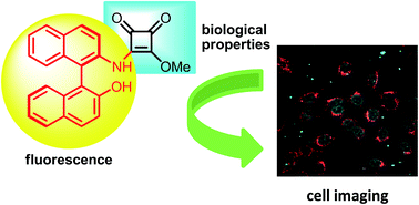
Novel luminescent squaramide monoesters functionalised with different fluorophore groups have been synthesised and explored in cell imaging for the first time. Cytotoxicity studies performed in HeLa cervical cancer cells revealed high activity for some of these novel structures, highlighting the importance of the fluorescent fragment in the efficiency of these promising anticancer agents. In addition, fluorescence cell microscopy disclosed the different biodistribution behaviour depending on the fluorescent moiety, and the possibility of nuclear localisation of chiral non planar squaramide monoesters.
Introduction
In the last decade, squaramides have received special attention since they exhibit unique and promising properties that make them appealing for many different areas of research, such as medicine,1 catalysis2 and synthesis.3
One of the most important features that these scaffolds display is their ability to selectively bind through cooperative hydrogen bonds. This interesting property has been extensively investigated in the area of asymmetric organocatalysis,2 since the pioneering work reported by Rawal and co-workers in 2008.4 Thus, the use of squaramides as excellent hydrogen bond donor catalysts has been experiencing an impressive growth. Beyond their application in catalysis, the squaramides capacity as receptors for molecular recognition5 is currently being explored in medicinal chemistry.6
The facility of different active compounds to penetrate into the cells could be decisive for the effectiveness of drug delivery. However, for that purpose, many times these compounds must be transported by using additional carriers, which often exhibit diverse toxicity levels with the corresponding cell damage. In contrast, other promising drug candidates can effectively cross the cell membrane by the formation of hydrogen bond and electrostatic interactions between the compound and a cell membrane receptor or the cell membrane surface.
The squaramides have an interesting dual behaviour, as they are able to act as good hydrogen bond donors and acceptors, which made them suitable for both anion and cation recognition, respectively. There are some examples of receptors based on the squaramide motif, i.e. Costa's group has provided evidence that squaramide-based molecules are able of binding both cations7 and anions,8 by means of different computational and experimental methods. More recently, Taylor,9 Fabbrizzi,10 and co-workers among other research groups11 have employed squaramide-based receptors for the recognition of various anions.
Moreover, squaramide motifs have been the focus of an increased interest in medicine being considered as promising candidates for drug design.1,12 However, to the best of our knowledge the study of squaramide monoesters as cytotoxic agents or for cell imaging has been overlooked in the literature so far, and no examples as potential anticancer agents have been previously reported. In this respect, we envisioned that the ability of squaramide monoesters to generate hydrogen bonds and electrostatic interactions could be an interesting option to design specific recognition systems, for their consequent internalisation into the cell. All these, together with the interest of searching for new antitumor agents, encouraged us to explore the antitumor activity of a variety of squaramide monoesters in HeLa cells. Herein, we report on the synthesis of new luminescent squaramide monoesters and a squaramide derivative with anticancer activity together with their exploration in fluorescence cell microscopy.
Results and discussion
Taking into the account all the unique properties of squaramides and the lack of background of squaramide monoesters, we hypothesised that these species could form plausible interactions or covalent bonds with internal amine groups belonging to biological molecules in the cells, generating the resulting squaramide, or could be anchored by acidic organelles to help in the internalisation into the cells (Scheme 1).
 |
| Scheme 1 Synthesis of squaramide monoesters and possible reaction/interaction of squaramide monoesters into the cells (green helix: biological material containing an amine group). | |
Consequently, we started preparing a battery of squaramide monoesters following the optimised reaction conditions recently reported by us.13 In this manner, squaramide monoesters 1–7 and squaramide 8 containing several fluorophore groups were synthesised, with the aim to make them useful for cell imaging studies (Fig. 1).
 |
| Fig. 1 Model squaramide monoester and squaramide structures 1–8 tested. | |
With these model structures, the photophysical and biological properties of all these new compounds were examined. UV-visible absorption, emission and excitation spectra of compounds 1–8 were recorded in a dimethylsulfoxide solution at 298 K and the most significant data are collected in Table 1.
Table 1 Absorption bands, excitation and emission values (1 × 10⁻⁵ M, DMSO, 298 K) and quantum yields (2 × 10⁻⁵ M, DMSO, 298 K) for compounds 1–8a
|
λmax/nm (ε/dm3 mol−1 cm−1) |
λexc/nm |
λem/nm |
Φ |
sh = shoulder. |
1 |
292 (22 500) sh, 353 (54 000) |
450 |
515 |
0.2 |
2 |
262 (40 200), 274 (37 100), 319 (16 900), 383 (3400) |
414 |
474 |
27.9 |
3 |
259 (25 100), 337 (14 800), 483 (14 000) |
514 |
591 |
45.7 |
4 |
283 (27 800), 318 (21 500), 337 (18 500) sh, 378 (7020) sh |
375 |
490 |
0.7 |
5 |
266 (76 200), 348 (18 000), 416 (3300) |
490 |
616 |
0.6 |
6 |
334 (32 300), 394 (5400) sh |
440 |
508 |
0.5 |
7 |
280 (14 900) sh, 332 (30 000), 434 (11 000) |
457 |
525 |
2.3 |
8 |
280 (32 600), 374 (88 900), 430 (7600) |
434 |
525 |
7.3 |
Photophysical properties of the specific fluorophores, benzidine, 9-aminophenanthrene, 3-aminofluoranthene, NOBIN, 6-aminochrysene and 2-aminofluorene were also analyzed for comparison purposes (Fig. S1 and S2†).
Squaramide monoesters 1–7 and squaramide 8 had similar absorption spectra pattern to that of their correspondent fluorophores. Specifically compounds 2, 4 and 5 presented a π → π* transition at high energy, <300 nm, followed by a less intense n → π* transition and going in line with the UV-vis spectra of their fluorophores. In the case of compounds 1, 3, 6, 7 and 8, there is a clear red shift in the absorption bands in comparison with that of the free fluorophore. Probably, the linearity between the squarate skeleton with that of the fluorophores enables an increase of the electronic delocalisation between both fragments, and thus, a stabilisation of the LUMO orbitals. Furthermore, compound 5, 7 and 8 showed an additional absorption band at lower energies, 416, 434 and 430 nm, respectively, that could suggest the deprotonation of the NH group by the DMSO used as solvent. A similar effect has already been observed by Taylor and co-workers for N,N-diarylsquaramides, when changing the solvent of the UV-visible analysis from CH3CN to DMSO a new absorption band appeared over 400 nm.9a
Luminescence spectra of all the synthesised species showed a broad emission band between 475 and 615 nm which was tentatively attributed to IL transition processes among the fluorophores and the squaramide (Fig. 2).
 |
| Fig. 2 Normalised emission spectra of compounds 1–8 recorded in DMSO solution at 298 K. | |
In concordance with the UV-vis results, compounds 1, 3, 6, 7 and 8 presented a high red shift of the emission maxima in comparison with that observed for their fluorophores. Conversely, compound 5 also presented a red shifted emission of ca. 200 nm that contrasted with the similarity of the absorption spectra of 5 and its fluorophore, indicating the high influence of the substituents on the emissive properties.14 Even though all of them displayed a highly intense luminescence, compound 8 stands out as the most emissive species, possibly because of the presence of two fluorophores within its structure. Stokes shifts vary from 25 to 150 nm, which denotes a fluorescence nature for the emission.
The quantum yields for compounds 1–8 have been measured in DMSO solution. Significant values were obtained for compounds 2 and 3 with quantum yields of 27.9 and 45.7%, respectively, and a smaller value of 7.3 for 8 and 2.3% for 7. Considerably lower values, <1%, were obtained for the rest of derivatives.
In view of the good emission properties of compounds 1–8, a series of experiments were undertaken to test their cytotoxic activity and viability as specific cell imaging agents in human HeLa cervical cancer cells. Cytotoxic studies were performed by a MTT assay and revealed that compounds 1, 3 and 6–8 possess the highest cytotoxic activity (Table 2). These compounds correspond to the benzidine, aminofluoranthene, and aminofluorene derivatives, and all of them are known to be useful platforms for designing antitumoral compounds.15 The aminochrysene species 5 has a moderate cytotoxic activity of 25.3 μM. These results suggest that the fluorophores which contain a cyclopentadiene moiety are the most active species, followed by the benzidine fluorophore which has two connected phenyl units, whereas those with fused phenyl rings are less cytotoxic.
Table 2 Values of IC50 (μM) for species 1–8 in HeLa cells
Comp. |
1 |
2 |
3 |
4 |
IC50 |
2.25 ± 1.1 |
115.6 ± 13.9 |
0.88 ± 0.22 |
46.7 ± 0.3 |
Comp. |
5 |
6 |
7 |
8 |
IC50 |
25.3 ± 11.4 |
1.02 ± 0.07 |
1.17 ± 0.07 |
1.18 ± 0.23 |
On the basis of the luminescence and cytotoxic activity results of compounds 1–8, fluorescence confocal microscopy was used with selected species to assess their suitability as cell imaging agents. Among them, species 1, 3, and 6–8 were discarded for this analysis because they displayed high cytotoxicity, and some of them low solubility in the buffer media, features to avoid in the design of bioprobes for cell imaging applications. Therefore, selected squaramide monoesters 2, 4 and 5 were incubated with HeLa cells using loading concentrations below their IC50 to avoid cellular death and the subsequent possible changes of localisation.16 A known lysosomal co-staining fluorescent dye, LisoTracker Red DND-99, was used as internal standard to ascertain the localisation. Confocal fluorescent images were taken after excitation at either 405 or 473 and 578 nm. When the cells were irradiated at 405 or 473 nm, only the emission from the synthesised compounds was observed, whereas exciting at 578 nm, the displayed emission would be that of the commercial fluorescent dye, which is localised in the lysosomes. Specifically, the laser of 405 nm was used to excite 2 and 4, and species 5 was visualised upon excitation at 473 nm, going in line with the different photophysical behaviour (Table 1). Superimposition of the images obtained upon excitation at either 405 or 473 nm with that obtained upon excitation at 578 nm for the internal standard gave crucial information to elucidate the possible localisation pattern (Fig. 3 and 4).
 |
| Fig. 3 Fluorescence microscopy images of HeLa cells incubated with compounds 2 and 5 at a concentration of 50 μM and 12.5 μM respectively for 12 h at 37 °C; (A) images upon excitation at either 405 or 473 nm; (B) images upon excitation at 578 nm for visualizing the internal standard (IS); (C) superimposed image of (A) and (B). | |
 |
| Fig. 4 Fluorescence microscopy images of HeLa cells incubated with compound 4 at a concentration of 35 μM for 12 h at 37 °C; (A) image upon excitation at 405 nm; (B) image upon excitation at 578 nm; (C) superimposed image of (A) and (B). | |
Hence, both 2 and 5 showed an emission coming from the cytoplasm area and a more specific granulated accumulation in the perinuclear region. Remarkably, the superimposition image (Fig. 3C), showed that both species, compounds 2 and 5 seemed to have some accumulation in the same organelles than the internal standard i.e., the LisoTracker Red DND-99 that is retained in the lysosomes. Consequently, it could be reasonable to think that 2 and 5 could be localised in acidic vesicles such as endosomes as well as in lysosomes. Specifically, late endosomes bear an acidic environment (pH = 4.7) and they move towards the perinuclear region after their formation. They arise from the maturation of early endosomes, which in turn are formed by an endocytosis process and, therefore, pointing out to an energy-dependent pathway as the internalisation process.17
Costa, Rotger and co-workers report in the examination of fluorescent cyclosquaramides6b that internalisation of the probe into the cell takes place through endocytotic receptor-mediated mechanisms and remains sequestered in late endosomes. The authors attributed this to the strong affinity of cyclosquaramides for anionic phosphate compounds present on the cell membrane. In the present case, compounds 2 and 5 have also the ability to form covalent bonds with biological molecules bearing amine groups in the cell membrane, which could be the pathway to enter the cell.
To support the assumption that the internalisation mechanism occurs via an energy-dependent pathway, the evaluation of the uptake of compound 5 in HeLa cells, at a concentration of 100 μM for 2 h at 37 °C and at 4 °C, was evaluated via flow cytometry (see Fig. S3 in ESI†). Under these conditions, the uptake was significantly reduced at 4 °C compared to that observed at 37 °C. Although the solubility of the probe may changes with the temperature, the fact that no uptake is observed at 4 °C, together with the lysosomal localisation obtained for this compound in HeLa cells, suggest that internalisation is an energy dependent process, indicating that the mechanism might be a receptor-mediated endocytotic process.
In addition, negligible emission from the nucleus itself indicates that both compounds 2 and 5 remained only in the outer part. In contrast, when the same experiment was performed with 4, a completely different bio-distribution pattern was observed (Fig. 4).
Interestingly, compound 4 entered the cell and accumulated in the nuclear region. A closer look revealed that the nuclear distribution was not homogenous, thus, presenting a granulated emission (Fig. 4C). The fact that 4 was able to permeate the nucleus indicates that either its acidic character is different from that of 2 and 5 and, consequently, they can escape from the internalisation in the endosomes and lysosomes, or that the cellular internalisation mechanism did not occur via an endocytotic process. An additional factor to take into account for the different biodistribution is the chirality and non-planarity of the fluorophore fragment. Previous work published by Seitz and co-workers highlights that even though the typical base surrogates are planar to help the π-stacking as well as hydrophobic interactions with the DNA bases, it is possible to intercalate non-planar aromatic structures such as binaphthyl derivatives.18 They assume that the torsional flexibility of the binaphthyl hinge is assisting the stacking interactions to take place at the interior and exterior of the DNA duplex. Considering the chemical similarity between binaphthyl and NOBIN, the fluorophore appended in compound 4, it might be reasonable to think that a similar process is occurring in this case. In addition, the chirality of the probe may lead also to specific DNA binding and chiral recognition, a key feature for designing selective bioprobes.19
Conclusions
In summary, novel luminescent squaramide monoesters have been explored as bioactive agents and in cell imaging for the first time. Cytotoxicity studies performed in HeLa cells revealed a high activity for some of these species, where the selection of the fluorescent fragment seems to be decisive for the efficiency of these promising anticancer agents in HeLa cells. Fluorophores containing the cyclopentadiene unit or without fused phenyl rings are the most active. In addition, fluorescence cell microscopy pointed out the different biodistribution behaviour depending on the fluorescent moiety. Both lysosomal and nuclear localisation has been observed, which highlights the possibility for chiral and non-planar bioprobes, as in the case of complex 4, which accumulates in the nuclear area to interact with DNA. Since the squaramide functionality provides a way to increase the transport ability of a receptor without significantly increasing the lipophilicity, it offers an ideal platform for designing future anion transporters. More complex squaramide-containing transporters are currently being investigated in our laboratory and will be published in due course.
Experimental section
General experimental methods and instrumentation
Purification of reaction products was carried out either by filtration or by flash chromatography using silica-gel (0.063–0.200 mm). Analytical thin layer chromatography was performed on 0.25 mm silica gel 60-F plates. ESI ionisation method and mass analyser type MicroTof-Q were used for the ESI measurements. 1H and 13C{1H}-APT NMR were recorded at room temperature on a BRUKER AVANCE 400 spectrometer (1H, 400 MHz; 13C, 100.6 MHz) or on a BRUKER AVANCE II 300 spectrometer (1H, 300 MHz; 13C, 75.5 MHz) in DMSO-d6 as solvent. Chemical shifts were reported in the δ scale relative to residual DMSO (2.50 ppm) for 1H NMR and to the central line of DMSO (39.43 ppm) for 13C NMR. Room temperature steady-state emission and excitation spectra were recorded with a Jobin-Yvon-Horiba fluorolog FL3-11 spectrometer fitted with a JY TBX picosecond detection module. UV/vis spectra were recorded with a 1 cm quartz cells on an Evolution 600 spectrophotometer. The quantum yields were measured in a Hamamatsu Photonics KK in DMSO solution.
Representative procedure for synthesis of squaramide monoesters 1–7 and squaramide 8
To a mixture of 3,4-dimethoxy-3-cyclobutene-1,2-dione (0.2 mmol) in MeOH (0.5 mL), the corresponding amine (0.2 mmol) was added in one portion at room temperature. After a reaction time, the mixture was cooled in the freezer (30 min) and the product was purified by filtration and washed with cold MeOH (2 mL) or by column chromatography when necessary. Yields are reported below and pure compounds were obtained as stable solids.
4,4′-([1,1′-Biphenyl]-4,4′-diylbis(azanediyl))bis(3-methoxycyclobut-3-ene-1,2-dione) (1). Following the general procedure, compound 1 was obtained after 64 h of reaction at room temperature as a yellow solid in 75% yield (61 mg). M.p. > 260 °C decomp. 1H NMR (300 MHz, DMSO-d6, 60 °C) δ 10.84 (s, 2H), 7.67 (d, J = 8.6 Hz, 4H), 7.44 (d, J = 7.6 Hz, 4H), 4.41 (s, 6H). 13C-APT NMR (75 MHz, DMSO-d6, 60 °C) δ 136.9 (2C), 134.7 (2C), 126.5 (4C), 119.7 (4C), 60.2 (2C). IR (KBr film) (cm−1) ν 3250, 3192, 3095, 2923, 2853, 1797, 1706, 1616, 1569, 1514, 1456, 1392, 1377, 1080, 926, 846, 733, 457. MS (ESI+) 405.2 [M + H]. HRMS (ESI+) calcd for C22H16N2NaO6 427.0901; found 427.0896 [M + Na].
3-Methoxy-4-(phenanthren-9-ylamino)cyclobut-3-ene-1,2-dione (2). Following the general procedure, compound 2 was obtained after 64 h of reaction at room temperature as a white solid in 58% yield (35.5 mg). M.p. 188–190 °C. 1H NMR (400 MHz, DMSO-d6) δ 11.03 (s, 1H), 8.93–8.88 (m, 1H), 8.86–8.81 (m, 1H), 8.18 (dd, J = 8.0, 1.4 Hz, 1H), 7.95 (dd, J = 7.6, 1.7 Hz, 1H), 7.82–7.63 (m, 5H), 4.30 (s, 3H). 13C-APT NMR (100 MHz, DMSO-d6) δ 188.6 (1C), 184.7 (1C), 178.8 (1C), 171.2 (1C), 131.1 (1C), 130.9 (1C), 130.3 (1C), 128.5 (1C), 128.4 (1C), 127.4 (1C), 127.4 (1C), 127.1 (1C), 127.0 (1C), 126.8 (1C), 123.6 (1C), 123.3 (1C), 122.9 (1C), 121.1 (1C), 60.4 (1C). IR (KBr film) (cm−1) ν 3214, 2953, 2924, 2853, 1812, 1694, 1621, 1454, 1362, 1231, 936, 766, 749, 727, 420. MS (ESI+) 326.1 [M + Na]. HRMS (ESI+) calcd for C19H13NNaO3 326.0788; found 326.0778 [M + Na].
3-(Fluoranthen-3-ylamino)-4-methoxycyclobut-3-ene-1,2-dione (3). Following the general procedure, compound 3 was obtained after 93 h of reaction at room temperature and purified by column chromatography, as a red solid in 49% yield (32.2 mg). M.p. > 260 °C decomp. 1H NMR (400 MHz, DMSO-d6) δ 11.24 (s, 1H), 8.16 (d, J = 8.1 Hz, 2H), 8.11 (d, J = 7.5 Hz, 1H), 8.06–8.01 (m, 2H), 7.74 (dd, J = 8.2 Hz, J = 7.1 Hz, 1H), 7.45 (d, J = 7.5 Hz, 1H), 7.44–7.39 (m, 2H), 4.35 (s, 3H). 13C-APT NMR (100 MHz, DMSO-d6) δ 188.2 (1C), 184.8 (1C), 179.0 (1C), 170.3 (1C), 138.5 (1C), 138.1 (1C), 136.0 (1C), 133.5 (1C), 133.3 (1C), 131.8 (1C), 128.1 (1C), 127.9 (1C), 127.3 (1C), 123.8 (1C), 123.0 (1C), 121.9 (1C), 121.6 (1C), 121.5 (1C), 121.1 (1C), 120.9 (1C), 60.4 (1C). IR (KBr film) (cm−1) ν 3322, 3285, 2923, 2853, 1801, 1711, 1583, 1453, 1362, 1282, 1257, 940, 849, 778, 756, 750, 456. MS (ESI+) 350.1 [M + Na]. HRMS (ESI+) calcd for C21H13NNaO3 350.0788; found 350.0780 [M + Na].
3-([(R)-2′-Hydroxy-{1,1′-binaphthalen}-2-yl]amino)-4-methoxycyclobut-3-ene-1,2-dione (4). Following the general procedure, compound 4 was obtained after 120 h of reaction at room temperature and purified by column chromatography (Hex
:
EtOAc 7
:
3 to 3
:
7), as a yellow solid in 76% yield (60 mg). M.p. 142–144 °C. [α]24D = +152.7 (c 0.27, CHCl3). 1H NMR (400 MHz, DMSO-d6) δ 9.97 (br s, 1H), 9.66 (br s, 1H), 8.04 (d, J = 9.2 Hz, 1H), 8.01 (d, J = 9.7 Hz, 1H), 7.93 (d, J = 8.9 Hz, 1H), 7.88 (d, J = 8.1 Hz, 1H), 7.59 (d, J = 8.7 Hz, 1H), 7.50–7.44 (m, 1H), 7.35 (d, J = 8.9 Hz, 1H), 7.33–7.23 (m, 2H), 7.19–7.14 (m, 1H), 7.06 (d, J = 8.5 Hz, 1H), 6.84 (d, J = 8.3 Hz, 1H), 4.23 (s, 3H). 13C-APT NMR (100 MHz, DMSO-d6) δ 188.3 (1C), 183.9 (1C), 178.3 (1C), 170.5 (1C), 153.2 (1C), 134.1 (1C), 133.6 (1C), 132.3 (1C), 131.5 (1C), 129.7 (1C), 128.0 (1C), 127.9 (1C), 127.9 (1C), 126.6 (1C), 126.4 (1C), 126.1 (1C), 125.7 (1C), 125.3 (1C), 124.0 (1C), 123.6 (1C), 122.5 (1C), 118.4 (1C), 113.7 (1C), 60.1 (1C). IR (KBr film) (cm−1) ν 2924, 2854, 1800, 1711, 1604, 1574, 1463, 1377, 814, 434. MS (ESI+) 396.3 [M + H]. HRMS (ESI+) calcd for C25H17NNaO4 418.1050; found 418.1046 [M + Na].
3-(Chrysen-6-ylamino)-4-methoxycyclobut-3-ene-1,2-dione (5). Following the general procedure using 1 mL of MeOH and Et3N (0.2 mmol), compound 5 was obtained after 60 h of reaction at room temperature as a yellow paled solid in 76% yield (53.7 mg). M.p. > 260 °C decomp. 1H NMR (400 MHz, DMSO-d6) δ 11.20 (br s, 1H), 9.00 (d, J = 8.0 Hz, 1H), 8.92 (d, J = 8.1 Hz, 1H), 8.86 (d, J = 9.1 Hz, 1H), 8.69 (br s, 1H), 8.32 (d, J = 7.6 Hz, 1H), 8.10 (d, J = 8.5 Hz, 2H), 7.88–7.59 (m, 4H), 4.34 (s, 3H). 13C-APT NMR (100 MHz, DMSO-d6) δ 188.7 (1C), 184.9 (1C), 178.9 (1C), 170.8 (1C), 131.8 (1C), 131.7 (1C), 130.4 (1C), 129.8 (1C), 128.3 (1C), 127.4 (1C), 127.3 (1C), 127.1 (1C), 126.9 (1C), 126.8 (1C), 126.7 (1C), 126.5 (1C), 126.0 (1C), 123.7 (1C), 123.6 (1C), 123.4 (1C), 121.1 (1C), 116.0 (1C), 60.3 (1C). IR (KBr film) (cm−1) ν 3225, 2923, 2853, 1797, 1720, 1592, 1509, 1486, 1403, 809, 748, 447. MS (ESI+) 354.2 [M + H]. HRMS (ESI+) calcd for C23H15NNaO3 376.0944; found 376.0934 [M + Na].
3-([9H-Fluoren-2-yl]amino)-4-methoxycyclobut-3-ene-1,2-dione (6). Following the general procedure using 2 mL of MeOH, compound 6 was obtained after 90 min of reaction at room temperature as a yellow solid in 77% yield (45 mg). M.p. > 250 °C decomp. 1H NMR (400 MHz, DMSO-d6) δ 10.86 (br s, 1H), 7.89–7.83 (m, 2H), 7.61–7.54 (m, 2H), 7.42–7.34 (m, 2H), 7.32–7.26 (m, 1H), 4.40 (s, 3H), 3.92 (s, 2H). 13C-APT NMR (75 MHz, DMSO-d6) δ 183.8 (1C), 144.0 (1C), 142.7 (1C), 140.5 (1C), 137.2 (1C), 136.8 (1C), 126.9 (1C), 126.3 (1C), 124.9 (1C), 120.3 (1C), 119.6 (1C), 118.4 (1C), 116.3 (1C), 60.5 (1C), 36.4 (1C). IR (KBr film) (cm−1) ν 3245, 3198, 2923, 2853, 1795, 1709, 1606, 1584, 1522, 1519, 1467, 1389, 1077, 1016, 764, 730. MS (ESI+) 314.0 [M + Na]. HRMS (ESI+) calcd for C18H13NNaO3 314.0788; found 314.0781 [M + Na].
3-([9H-Fluoren-2-yl]amino)-4-propoxycyclobut-3-ene-1,2-dione (7). Firstly, 3,4-dipropoxycyclobut-3-ene-1,2-dione was prepared with 3,4-dimethoxy-3-cyclobutene-1,2-dione at reflux in 1-propanol during 24 h. Following the general procedure and using the resulting squarate 3,4-dipropoxycyclobut-3-ene-1,2-dione, compound 7 was obtained after 72 h of reaction at room temperature as a yellow solid in 68% yield (43.7 mg). M.p. > 250 °C decomp. 1H NMR (400 MHz, DMSO-d6) δ 10.85 (br s, 1H), 7.89–7.82 (m, 2H), 7.62–7.54 (m, 2H), 7.42–7.25 (m, 3H), 4.75–4.68 (m, 2H), 3.92 (br s, 2H), 1.89–1.74 (m, 2H), 1.08–0.94 (m, 3H). 13C-APT NMR (75 MHz, DMSO-d6) δ 183.7 (1C), 169.2 (1C), 143.9 (1C), 142.7 (1C), 137.2 (1C), 136.8 (1C), 126.7 (1C), 126.3 (1C), 125.0 (1C), 120.3 (1C), 119.6 (1C), 118.5 (1C), 116.4 (1C), 74.6 (1C), 36.4 (1C), 22.8 (1C), 9.7 (1C). IR (KBr film) (cm−1) ν 3233, 3122, 3067, 2923, 2853, 1806, 1710, 1587, 1495, 1458, 1414, 1364, 828, 764, 727. MS (ESI+) 342.1 [M + Na]. HRMS (ESI+) calcd for C20H17NNaO3 342.1101; found 342.1090 [M + Na].
3,4-Bis([9H-fluoren-2-yl]amino)cyclobut-3-ene-1,2-dione (8). Following the general procedure using 2 mL of MeOH and Et3N (0.2 mmol), compound 8 was obtained after 72 h of reaction at room temperature as a yellow solid in 23% yield (20 mg). M.p. > 250 °C decomp. 1H NMR (400 MHz, DMSO-d6) δ 10.01 (s, 2H), 7.90 (d, J = 8.2 Hz, 2H), 7.85 (d, J = 7.4 Hz, 2H), 7.78 (s, 2H), 7.57 (d, J = 7.4 Hz, 2H), 7.49 (dd, J = 8.2 Hz, J = 2.1 Hz, 2H), 7.41–7.35 (m, 2H), 7.32–7.26 (m, 2H), 2.08 (s, 4H). 13C-DEPT NMR (100 MHz, DMSO-d6) δ 126.5 (2C), 126.1 (2C), 124.7 (2C), 120.1 (2C), 120.0 (2C), 119.4 (2C), 36.2 (2C). IR (KBr film) (cm−1) ν 3142, 2923, 2853, 1786, 1670, 1600, 1576, 1561, 1556, 1461, 1453, 952, 871, 819, 765, 730, 442. MS (ESI+) 441.2 [M + H]. HRMS (ESI−) calcd for C30H19N2O2 439.1452; found 439.1456 [M − H].
Cell culture
HeLa (cervical cancer) cells were maintained in glucose DMEM (Dulbecco's Modified Eagle's Medium) supplemented with 5% fetal bovine serum (FBS), 200 U mL−1 penicillin, 100 μg mL−1 streptomycin and 2 mM L-glutamine. Cultures were maintained in a humidified atmosphere of 95% air/5% CO2 at 37 °C.
Cytotoxicity assay by MTT
The MTT assay was used to determine cell viability as an indicator for cells sensitivity to the complexes. Exponentially growing cells were seeded at a density of approximately 104 cells per well in 96-well flat-bottomed microplates and allowed to attach for 24 h prior to addition of compounds. The complexes were dissolved in DMSO and added to cells in concentrations ranging from 0.1 to 100 μM in quadruplicate. Cells were incubated with our compounds for 24 h at 37 °C. Ten microliters of MTT (5 mg mL−1) were added to each well and plates were incubated for 2 h at 37 °C. Finally, plates were centrifuged for 10 min at 500 × g, media was eliminated and DMSO (100 μl per well) was added to dissolve the formazan precipitates. The optical density was measured at 550 nm using a 96-well multiscanner autoreader (ELISA). The IC50 was calculated by non-linear regression analysis using Prism software (GraphPad Software Inc). Each compound was analysed at least in three independent experiments.
Human cell studies and cell fluorescence microscopy
HeLa cells were detached from the plastic flask using trypsin–EDTA solution and suspended in an excess volume of growth medium. The homogeneous cell suspension was then distributed into 30 μl aliquots in a 6 channel μ-slide IV0.4 (IBIDI), with each aliquot being subject to incubation with the different complexes, final concentrations ranged from 12.5 μM to 50 μM, at 37 °C for 12 h. Then, 30 μl of a solution 0.01 μM the internal standard (LisoTracker Red DND-99) in cell growth medium was added to each well. Preparations were viewed using an Olympus FV10-i Oil type compact confocal laser microscope using a ×10 or ×60 objective, with excitation wavelength at 405, 473 and 578 nm.
Acknowledgements
Authors thank the Ministerio de Economía y Competitividad (MINECO/FEDER CTQ2013-48635-C2-1-P and SAF2013-48626-C2-2-R), and DGA-FSE (E77 and E104) for financial support of this research. J. V. A.-R. thanks to DGA for his pre-doctoral contract.
Notes and references
- R. I. Storer, C. Aciro and L. H. Jones, Chem. Soc. Rev., 2011, 40, 2330 RSC
. -
(a) J. Alemán, A. Parra, H. Jiang and K. A. Jørgensen, Chem.–Eur. J., 2011, 17, 6890 CrossRef PubMed
;
(b) J. V. Alegre-Requena, Synlett, 2014, 25, 298 CAS
;
(c) P. Chauhan, S. Mahajan, U. Kaya, D. Hack and D. Enders, Adv. Synth. Catal., 2015, 357, 253 CrossRef CAS
. - F. R. Wurm and H.-A. Klok, Chem. Soc. Rev., 2013, 42, 8220 RSC
. - J. P. Malerich, K. Hagihara and V. H. Rawal, J. Am. Chem. Soc., 2008, 130, 14416 CrossRef CAS PubMed
. -
(a) B. Soberats, L. Martínez, E. Sanna, A. Sampedro, C. Rotger and A. Costa, Chem.–Eur. J., 2012, 18, 7533 CrossRef CAS PubMed
;
(b) C. López, E. Sanna, L. Carreras, M. Vega, C. Rotger and A. Costa, Chem.–Asian J., 2013, 8, 84 CrossRef PubMed
. - For scarce examples, see:
(a) C. Schieber, A. Bestetti, J. P. Lim, A. D. Ryan, T.-L. Nguyen, R. Eldridge, A. R. White, P. A. Gleeson, P. S. Donnelly, S. J. Williams and P. Mulvaney, Angew. Chem., Int. Ed., 2012, 51, 10523 CrossRef CAS PubMed
;
(b) A. Sampedro, R. Villalonga-Planells, M. Vega, G. Ramis, S. Fernández de Mattos, P. Villalonga, A. Costa and C. Rotger, Bioconjugate Chem., 2014, 25, 1537 CrossRef CAS PubMed
. -
(a) S. Tomás, M. C. Rotger, J. F. González, P. M. Deyà, P. Ballester and A. Costa, Tetrahedron Lett., 1995, 36, 2523 CrossRef
;
(b) D. Quiñonero, A. Frontera, G. A. Suñer, J. Morey, A. Costa, P. Ballester and P. M. Deyà, Chem. Phys. Lett., 2000, 326, 247 CrossRef
;
(c) S. Tomàs, R. Prohens, M. Vega, M. C. Rotger, P. M. Deyà, P. Ballester and A. Costa, J. Org. Chem., 1996, 61, 9394 CrossRef
. -
(a) R. Prohens, S. Tomàs, J. Morey, P. M. Deyà, P. Ballester and A. Costa, Tetrahedron Lett., 1998, 39, 1063 CrossRef CAS
;
(b) D. Quiñonero, R. Prohens, C. Garau, A. Frontera, P. Ballester, A. Costa and P. M. Deyà, Chem. Phys. Lett., 2002, 351, 115 CrossRef
;
(c) C. Garau, A. Frontera, P. Ballester, D. Quiñonero, A. Costa and P. M. Deyà, Eur. J. Org. Chem., 2005, 179 CrossRef CAS
;
(d) A. Frontera, J. Morey, A. Oliver, M. N. Piña, D. Quiñonero, A. Costa, P. Ballester, P. M. Deyà and E. V. Anslyn, J. Org. Chem., 2006, 71, 7185 CrossRef CAS PubMed
. -
(a) A. Rostami, A. Colin, X. Y. Li, M. G. Chudzinski, A. J. Lough and M. S. Taylor, J. Org. Chem., 2010, 75, 3983 CrossRef CAS PubMed
;
(b) A. Rostami, C. J. Wei, G. Guérin and M. S. Taylor, Angew.
Chem., Int. Ed., 2011, 50, 2059 CAS
. -
(a) V. Amendola, G. Bergamaschi, M. Boiocchi, L. Fabbrizzi and M. Milani, Chem.–Eur. J., 2010, 16, 4368 CrossRef CAS PubMed
;
(b) V. Amendola, L. Fabbrizzi, L. Mosca and F.-P. Schmidtchen, Chem.–Eur. J., 2011, 17, 5972 CrossRef CAS PubMed
. - For recent examples, see also:
(a) C. E. Silva, H. F. Dos Santos, N. L. Speziali, R. Diniz and L. F. C. de Oliveira, J. Phys. Chem. A, 2010, 114, 10097 CrossRef CAS PubMed
;
(b) D. Quiñonero, K. A. López, P. M. Deyà, M. N. Piña and J. Morey, Eur. J. Org. Chem., 2011, 6187 CrossRef
;
(c) G. Ambrosi, M. Formica, V. Fusi, L. Giorgi, E. Macedi, M. Micheloni, P. Paoli, R. Pontellini and P. Rossi, Chem.–Eur. J., 2011, 17, 1670 CrossRef CAS PubMed
;
(d) N. Busschaert, I. L. Kirby, S. Young, S. J. Coles, P. N. Horton, M. E. Light and P. A. Gale, Angew. Chem., Int. Ed., 2012, 51, 4426 CrossRef CAS PubMed
. - For recent selected examples, see:
(a) S. K. Narasimhan, P. Sejwal, S. Zhu and Y.-Y. Luk, Bioorg. Med. Chem., 2013, 21, 2210 CrossRef CAS PubMed
;
(b) X. Zhang, Z. Zuo, J. Tang, K. Wang, C. Wang, W. Chen, C. Li, W. Xu, X. Xiong, K. Yuntai, J. Huang, X. Lan and H.-B. Zhou, Bioorg. Med. Chem. Lett., 2013, 23, 3793 CrossRef CAS PubMed
;
(c) L.-Q. Deng, Y.-M. Lu, C.-Q. Zhou, J.-X. Chen, B. Wang and W.-H. Chen, Bioorg. Med. Chem. Lett., 2014, 24, 2859 CrossRef CAS PubMed
;
(d) F. Olmo, C. Rotger, I. Ramírez-Macías, L. Martínez, C. Marín, L. Carreras, K. Urbanová, M. Vega, G. Chaves-Lemaur, A. Sampedro, M. J. Rosales, M. Sánchez-Moreno and A. Costa, J. Med. Chem., 2014, 57, 987 CrossRef CAS PubMed
. -
(a) E. Marqués-López, J. V. Alegre-Requena and R. P. Herrera, European Pat. EP14382260.9 filed, 2014
;
(b) J. V. Alegre-Requena, E. Marqués-López and R. P. Herrera, RSC Adv., 2015, 5, 33450 RSC
. - T.-L. Wu, H.-H. Chou, P.-Y. Huang, C.-H. Cheng and R.-S. Liu, J. Org. Chem., 2014, 79, 267 CrossRef CAS PubMed
. -
(a) B. J. Smith, L. DeBruin, P. D. Josephy and T. E. Eling, Chem. Res. Toxicol., 1992, 5, 431 CrossRef CAS PubMed
;
(b) M.-D. Yang, S.-Y. Lin, T.-H. Chiu, C.-C. Lin, M.-L. Lin, S.-C. Hsu, C.-L. Kuo, M.-J. Sheu, C.-C. Wu, S.-S. Lin and J.-G. Chung, In Vivo, 2008, 22, 729 CAS
;
(c) N. Asare, M. Låg, D. Lagadic-Gossmann, M. Rissel, P. Schwarze and J. A. Holme, Toxicology, 2009, 255, 140 CrossRef CAS PubMed
. - V. Fernández-Moreira, I. Marzo and M. C. Gimeno, Chem. Sci., 2014, 5, 4434 RSC
. - J. R. Casey, S. Grinstein and J. Orlowski, Nat. Rev. Mol. Cell Biol., 2010, 11, 50 CrossRef CAS PubMed
. - S. Hainke and O. Seitz, Angew. Chem., Int. Ed., 2009, 48, 8250 CrossRef CAS PubMed
. - R. Corradini, S. Sforza, T. Tedeschi and R. Marchelli, Chirality, 2007, 19, 269 CrossRef CAS PubMed
.
Footnote |
† Electronic supplementary information (ESI) available: 1H and 13C NMR spectra of all new products. See DOI: 10.1039/c5ra24521d |
|
This journal is © The Royal Society of Chemistry 2016 |
Click here to see how this site uses Cookies. View our privacy policy here.