Zhendong Guoa,
Dong Zhang*a,
Hailong Qiua,
Yanming Jua,
Tong Zhanga,
Lijie Zhanga,
Yuan Menga,
Yingjin Wei*a and
Gang Chenab
aKey Laboratory of Physics and Technology for Advanced Batteries (Ministry of Education), College of Physics, Jilin University, Changchun 130012, P. R. China. E-mail: yjwei@jlu.edu.cn; dongzhang@jlu.edu.cn
bState Key Laboratory of Superhard Materials, Jilin University, Changchun 130012, P. R. China
First published on 12th January 2016
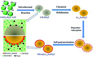
Poly-dopamine coated Li1−xFeSO4F is prepared via a self-polymerization process. The material shows larger discharge capacities, better rate capability and longer cycle stability than the pristine LiFeSO4F. The improved electrochemical properties are attributed to the highly hydrophilic and elastic properties of poly-dopamine.
Herein, we report a method that could significantly improve the electrochemical properties of LiFeSO4F via a non-aqueous polymer coating process. Fig. 1 shows a schematic diagram for the synthesis and design principles of the material. The first step is carried out by preparing tavorite LiFeSO4F using the tetraethylene glycol (TEG) assisted solvothermal method. Then, dopamine monomers are self-polymerized on the surface of Li1−xFeSO4F. The Li1−xFeSO4F material was prepared by chemical de-lithiation of the pristine LiFeSO4F in NO2BF4. The material thus obtained is called as PDA@Li1−xFeSO4F. Detailed experimental procedures are described in the ESI.† Dopamine is a nature-inspired bio-mimetic material containing several functional groups such as catechol, amine and imine. As an organic semiconductor with limited electronic conductivity, dopamine is not competitive enough to compensate the low electronic conductivity of LiFeSO4F. But, the robust wet-resistant adhesion and strong elasticity of PDA may be highly beneficial for LiFeSO4F to adsorb electrolyte efficiently and release its large volume change (ΔV/V = 10.4%) during repeated cycling.1 We show here that the PDA coated Li1−xFeSO4F exhibits much improved electrochemical properties than the pristine LiFeSO4F. To the best of our knowledge, this is the first report that improving the electrochemical performance of cathode materials by surface coating with highly hydrophilic polymers.
Fig. 2a shows the X-ray diffraction (XRD) patterns of the LiFeSO4F and PDA@Li1−xFeSO4F samples. The diffraction pattern of the pristine material fits well with that of tavorite LiFeSO4F without any detectable impurities. The lattice parameters of the material are calculated as a = 5.1855(1) Å, b = 5.5048(1) Å, c = 7.2296(1) Å, α = 106.517(1)°, β = 107.184(1)°, γ = 87.818(1)°, which are in good agreement with those reported in literatures.1,2 This batch of material was then chemically de-lithiated by NO2BF4 in acetonitrile via the reaction, LiFeSO4F + xNO2BF4 → Li1−xFeSO4F + xLiBF4 + xNO2↑ (1) in the following step, the oxidative Fe3+ ions of Li1−xFeSO4F could initiate the polymerization of dopamine, forming the PDA@Li1−xFeSO4F target material. XRD shows that the crystal structure of PDA@Li1−xFeSO4F is almost identical to that of FeSO4F which indicates that most Li ions are removed from the LiFeSO4F lattice.12 No evidence of PDA is observed because of its small amount and polymeric state in the material.
![]() | ||
Fig. 2 (a) X-ray diffraction patterns and (b) Fourier transform infrared spectra of the LiFeSO4F and PDA@Li1−xFeSO4F samples. |
Fig. 2b shows the Fourier transform infrared spectra of the LiFeSO4F and PDA@Li1−xFeSO4F samples. Both materials show a series adsorption peaks below 1400 cm−1 which are due to the SO4 tetrahedral.13 PDA@Li1−xFeSO4F shows several additive peaks. The one located at 1635 cm−1 corresponds to the CC vibration of the aromatic ring and the other broad one between 3000 and 3600 cm−1 is attributed to the overlap of the N–H and catechol-OH vibrations.14,15 The amount of PDA in PDA@Li1−xFeSO4F could be determined by thermogravimetric analysis (TGA, ESI, Fig. S1†). Comparing to the TG curve of Li1−xFeSO4F, PDA@Li1−xFeSO4F shows an extra weight loss of 1.8 wt% between 300 and 500 °C which is attributed to the carbonization of PDA. Therefore, the amount of PDA in the PDA@Li1−xFeSO4F sample is calculated as 2.87 wt%.
The morphologies of the samples are studied by scanning electron microscope (ESI, Fig. S2†). The pristine LiFeSO4F shows polyhedral shape with particle size ranging from 500 to 800 nm. The particle shape and particle size of Li1−xFeSO4F are maintained well after PDA coating.
However, the particle surface becomes much rougher comparing to the smooth surface of LiFeSO4F. Transmission electron microscope shows that the pristine LiFeSO4F possesses an overwhelmingly clean surface (Fig. 3a). In comparison, the surface of PDA@Li1−xFeSO4F shows a uniform amorphous layer with thickness ca. 10 nm which is attributed to PDA (Fig. 3b). Owing to the excellent adhesion ability of the catechol group, dopamine has a strong inclination to spontaneous deposit a conformal PDA film on virtually any substrates.16–18 X-ray energy dispersive spectroscopy (Fig. 3c) shows that not only the Fe, S, O, F elements but also N are uniformly distributed in the material, suggesting that PDA is evenly coated on the Li1−xFeSO4F sample.
![]() | ||
Fig. 3 TEM images of (a) LiFeSO4F and (b) PDA@Li1−xFeSO4F; (c) mappings of Fe, S, O, F, N elements of the PDA@Li1−xFeSO4F material. |
X-ray photoelectron spectroscopy (XPS) was used to study the surface chemistry of the materials (ESI, Fig. S3†). The N 1s peak of PDA@Li1−xFeSO4F could be fitted by two components centred at 400.2 and 402.1 eV which are assigned to the C–N and N–H bonds of PDA, respectively.19 The Fe 2p XPS of LiFeSO4F shows two peaks at 711.3 and 724.8 eV, corresponding to the 2p3/2 and 2p1/2 binding energies of Fe2+, respectively.20 As for PDA@Li1−xFeSO4F, the above XPS peaks shift to 713.9 and 727.6 eV which are close to those of Fe3+ ions.21 Mössbauer spectroscopy was further used to analysis the Fe2+/Fe3+ concentrations and their coordinative environments in the crystal lattice. As shown in Fig. 4, the Mössbauer spectrum of LiFeSO4F is composed of two sets of well fitted doublets. These doublets have almost equal area indicating that the iron ions equally occupy two crystallographic sites, i.e. Fe(1) and Fe(2). The obtained isomer shift, 1.28 mm s−1, fits well with that of high spin Fe2+. The Mössbauer spectrum of PDA@Li1−xFeSO4F indicates that about 87 at% of the Fe2+ ions are oxidized to Fe3+. The oxidation power of Fe3+, φ (Fe3+/Fe2+) = 0.771 V vs. SHE (standard hydrogen electrode), is much higher than that needed for adsorption of dopamine (φ ≈ 0.2 V vs. SHE).22 Consequently, the adsorbed dopamine monomers are self-polymerized to form a PDA film on the Li1−xFeSO4F surface. Reasonably, the pristine LiFeSO4F which do not contain Fe3+ ions could not initiate self-polymerization.
Fig. 5a shows the first charge–discharge curves of the samples evaluated at the 0.1C rate (I = 15 mA g−1). Different from the pristine LiFeSO4F, the electrochemical reactions of PDA@Li1−xFeSO4F starts with a discharge process due to lack of Li in the material. Both materials show a pair of voltage plateaus at about 3.6 V which are attributed to the LiFeSO4F/FeSO4F two-phase transition. The voltage gap between the charge/discharge plateaus gets to be smaller with PDA coating, indicating that the PDA film reduces the polarization and ohmic resistance of the battery. During the first charge process, the PDA@Li1−xFeSO4F seems to have a small additional potential plateau at ca. 4.4 V. In order to clearly understand this additional potential plateau, we study the CV curves of PDA and PDA@Li1−xFeSO4F (ESI, Fig. S4†). The CV curve of PDA@Li1−xFeSO4F shows an irreversible oxidation peak at ca. 4.2 V which is correlated with the aforementioned additional potential plateau. This current peak is consistent well with the oxidation peak of PDA. This indicates that the PDA layer of PDA@Li1−xFeSO4F is oxidized in the initial electrochemical process. The oxidized PDA layer could not be reduced again in the following electrochemical process which indicates high stability of the surface coating layer. The initial charge/discharge capacities of pristine LiFeSO4F are 119/101 mA h g−1, resulting in a coulombic efficiency of 84.9%. The irreversible capacity loss could be attributed to the formation of solid electrolyte interface (SEI) film.23 The formation of SEI film is further confirmed by TEM analysis. It is seen from Fig. 6a that a thick SEI film was formed on the surface of LiFeSO4F after the first charge. But only a thin surface film is observed for PDA@Li1−xFeSO4F (Fig. 6b) which could be attributed to the PDA layer and the SEI film. This indicates that PDA@Li1−xFeSO4F has less SEI film than that of LiFeSO4F. Fig. 5b shows the cycling performances of the samples. The discharge capacity of LiFeSO4F gradually decreases to 61 mA h g−1 after 100 cycles, corresponding to capacity retention of 60.4%. The fast capacity fading could be attributed to the relatively large volume change of 10.4% between the LiFeSO4F and FeSO4F phases resulting in loss of electrical contact after repeated lithium removal and uptake. In comparison, the initial discharge/charge capacities of PDA@Li1−xFeSO4F are recorded as 121/114 mA h g−1, resulting in a larger coulombic efficiency of 94.2%. Moreover, the material shows improved cycle stability. A much larger discharge capacity of 97 mA h g−1 is obtained after 100 cycles, corresponding to capacity retention of 85.1%. The rate dependent cycling performances of the materials are displayed in Fig. 5c. At each current rate, PDA@Li1−xFeSO4F always shows a larger discharge capacity than that of LiFeSO4F. For example, LiFeSO4F delivers a discharge capacity of 14 mA h g−1 at the 1C rate, while PDA@Li1−xFeSO4F exhibits a much larger discharge capacity of 56 mA h g−1. When the current rate returns to C/20, the discharge capacity can still return to 114 mA h g−1. The low rate capability of LiFeSO4F is most likely related to the poor electronic conductivity of the material.
![]() | ||
Fig. 6 TEM images of the LiFeSO4F (a) and PDA@Li1−xFeSO4F (b) materials after the first charge; SEM images of the LiFeSO4F (c and e) and PDA@Li1−xFeSO4F (d and f) electrodes after 50 cycles. |
Electrochemical impedance spectroscopy was used to study the effects of PDA coating on the electrochemical kinetic properties of LiFeSO4F. The impedance data were collected at 3.5 V of the first discharge. In the Nyquist plots of the materials (Fig. 5d), the well-defined semicircles in the high-to-medium frequency region are due to the SEI film and the charge transfer process, respectively.24 The slope line in the low frequency region is due to lithium diffusion in the electrode bulk. Based on this, the Nyquist plots can be simulated by the equivalent circuit given in the inset of Fig. 5d. In this equivalent circuit, Rs represents the ohmic resistance of the cell. Rf and Csl are the resistance and capacitance of the SEI film, respectively. Rct and Cdl represent the charge transfer resistance and double layer capacitance, respectively. W is the Warburg diffusion parameter. The simulated Rs, Rf and Rct values are listed in Table 1. It clearly shows that all of the electrochemical kinetic parameters of PDA@Li1−xFeSO4F are smaller than those of LiFeSO4F. Specifically, the smaller Rf of PDA@Li1−xFeSO4F is attributed to its thin SEI film as observed by TEM, and its smaller Rct indicates improved charge transfer kinetics of the electrode after PDA coating.
Rs (Ω) | Rf (Ω) | Rct (Ω) | |
---|---|---|---|
LiFeSO4F | 1.694 | 107.8 | 610.5 |
PDA@Li1−xFeSO4F | 2.22 | 84.36 | 186.5 |
As an organic semiconductor, the electronic conductivity of PDA is only 10−13 S cm−1.1,25 AC impedance spectroscopy (Fig. S5†) shows that the electronic conductivity of PDA@Li1−xFeSO4F is 1.55 × 10⁻¹¹ S which is even a little smaller than that of pristine LiFeSO4F. Therefore, it seems that the improved electrochemical performance of PDA@Li1−xFeSO4F is not ascribed to the electronic conductivity of PDA. In spite of this, the strong and robust wet-resistant adhesion of PDA could be highly beneficial for the battery performance since all the battery components are in contact with each other within a liquid environment. Inspired by the wet-resistant adhesion ability of PDA, Choi et al. prepared PDA modified polyethylene separator for lithium ion batteries. After PDA modification, the contact angle of the separator with the electrolyte was significantly decreased, indicating that the surface of the separator becomes highly hydrophilic.26 Moreover, PDA was also used as a binder for the silicon anode,27 as well as a composite additive for SnO2 to substantially improve the electrochemical properties.28 Fig. 6c–f shows the SEM images of the LiFeSO4F and PDA@Li1−xFeSO4F electrodes after 50 cycles. It is seen that the electrode film of PDA@Li1−xFeSO4F is very uniform after charge–discharge cycling. From the cross section image, it is seen that the electrode film of LiFeSO4F is cracked and tends to be peeling off from the current collector. On the contrary, the electrode film of PDA@Li1−xFeSO4F is still compact and sticks on the current collector very well. The improved electrode stability could be attributed to the wet-resistant adhesion property of PDA which improves the compatibility of the electrode. In addition, the intrinsic elasticity of PDA is beneficial in allowing the LiFeSO4F material to release its partial pressure caused by the large volume change during Li insertion/extraction. Additionally, Zhang and co-workers reported that tavorite LiFeSO4F is highly sensitive to moisture, which will rapidly decompose to FeSO4·nH2O and LiF in a humid environment.29 After PDA coating, the water contamination in the electrolyte could be consumed by this polymer layer due to the hydration reaction of PDA with water,25 thus eliminating the decomposition of LiFeSO4F. Moreover, the PDA layer isolates the direct contact of the active material and the electrolyte, thus protecting the material from any possible side reactions at the electrode/electrolyte interface resulting in a thin SEI film.
Footnote |
† Electronic supplementary information (ESI) available: Experiment details, TG curves, SEM images, XPS spectra. See DOI: 10.1039/c5ra24488a |
This journal is © The Royal Society of Chemistry 2016 |