C.
Ringwald
a and
V.
Ball
*ab
aInstitut National de la Santé et de la Recherche Médicale, Unité Mixte de Recherche 1121, 11 rue Humann, 67085 Strasbourg Cedex, France. E-mail: vball@unistra.fr
bUniversité de Strasbourg, Faculté de Chirurgie Dentaire, 8 rue Sainte Elisabeth, 67000 Strasbourg, France
First published on 5th January 2016
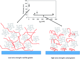
Tannic acid (TA), among other polyphenols, interacts strongly with proteins, in particular proline rich proteins, a mechanism which is at the origin of mouth astringency. Among such proline rich proteins are salivary rich proteins and collagen or gelatin. The formation of protein–polyphenol complexes is rarely, with some exceptions, of utility in materials science. However, when the complexation is controlled on surface templates, using the layer-by-layer deposition method, useful materials such as membranes for controlled separations or controlled polyphenol release can be obtained. The performance of such protein–TA containing films can only be improved through a better understanding of the parameters controlling the film deposition. Indeed it is widely believed and partially demonstrated that the protein–polyphenol interactions are driven through an interplay of hydrophobic interactions and hydrogen bonding. In this article, the deposition of (gelatin–TA)n films is investigated as a function of the ionic strength using a combination of characterization techniques: quartz crystal microbalance with dissipation monitoring (QCM-D), atomic force microscopy (AFM), Fourier transform spectroscopy in the attenuated total reflection mode (FTIR-ATR) and cyclic voltammetry (CV). It is shown that the film thickness and the amount of deposited film decreases strongly when the ionic strength is increased below about 50–100 mM but these film properties become almost ionic strength independent above 100 mM. The transition dynamics between these two growth regimes is investigated by putting the as prepared films in contact with a solution of different ionic strength.
On the other hand protein–TA complexes in particular and protein–polyphenol complexes in general are devoid of interesting applications because they reduce protein assimilation by binding otherwise useful digestive enzymes and dietary proteins.14 It is hence of the highest interest to exploit the strong TA–proline rich protein interactions, mostly hydrogen bonds and hydrophobic interactions,6 by immobilizing those complexes at solid–liquid interfaces. It has been shown that hydrogen bonds between uncharged polymers and TA allow to build-up films of controlled thickness using the step-by-step deposition15–17 of polymers like poly(N-vinylcaprolactam), poly(N-vinylpyrrolidone) or poly(ethyleneoxide) and TA.18 When gelatin, a denatured proline rich protein (obtained through denaturation of collagen from lime-cured tissues) is used, the (gelatin–TA)n films deposited on poly(acrylonitrile) ultrafiltration membranes displayed high water/ethanol permselectivity.19 As another application, (poly(N-vinylpyrrolidone)–TA)n films erode spontaneously in a pH dependent manner allowing a controlled release of TA.20 Materials based on gelatin and TA may be of high practical interest for the design of multifunctional tissue engineering matrixes associating the mechanical properties of gelatin (which gelifies at temperatures lower than about 37 °C) and the biological/chemical properties of TA. Upon oxidation of TA in the composite, quinone reactive groups will be formed offering the possibility to establish covalent bonds with lysine residues present on gelatin thus modifying the mechanical properties of the gelatin–TA composite. The stabilization of films made using the layer-by-layer method and incorporating a polyamine and TA upon oxidation with sodium periodate has already been demonstrated.21 We wish herein to investigate the interactions between a biologically relevant and cheap protein, gelatin, and TA.
The gelatin–TA composites will be deposited using the layer-by-layer deposition which allows to control the film thickness and its properties like the ionic permeability and the elasticity to yield (gelatin–TA)n films in an easy and reproducible way. In this notation of the film layering sequence, n is the number of deposition cycles. It has to be noted that such films are deposited alternating the deposition of mutually interacting macromolecular species or nanoparticles but that their final structure does not necessarily reflect the layering sequence because of possible intermixing effects.22 Depending on the strength of the interactions between the film's building blocks one can get either rigid and stratified polymer based films or intermixed 2D gels with totally different properties which will be of major influence when interactions with cells are envisioned.
In the present case of gelatin and TA, it is of particular interest is to investigate if electrostatic interactions do or not contribute to the gelatin/TA interactions allowing for the deposition of those films. It is indeed possible that TA undergoes a deprotonation in pH conditions where it is uncharged in solution i.e. below pH 7–8. To this aim the deposition of the PEI–(gelatin–TA)n film was investigated at a constant pH of 5.0 but at variable ionic strength in order to control the thickness of the electrostatic double layer. Deliberately we did not change the pH because TA has an average pKa close to 8.5 (ref. 18) and is strongly affected by oxidation in basic solutions. A set of complementary characterization methods, namely quartz crystal microbalance with dissipation monitoring (QCM-D), atomic force microscopy (AFM) in the contact mode, Fourier transform infra-red spectroscopy in the total attenuated reflection mode (FTIR-ATR) and cyclic voltammetry (CV) were employed.
Fig. 1 and 2 in the ESI file† show the deposition kinetics of the PEI–(gelatin–TA)n films at different overtones (ν = 3, 5 and 7) in the presence of 10 mM sodium acetate buffer and 50 mM acetate buffer + 150 mM NaCl respectively. It appears that the energy dissipation increases when the ionic strength decreases and that the different overtones do not perfectly overlap whatever the used ionic strength. This is a strong indication that the PEI–(gelatin–TA)n films are rather soft materials.
Some of the films deposited on silica coated quartz crystals were imaged by AFM after rinse with distilled water and drying under a stream of filtered air (Fig. 2). The average thickness of the films was obtained after imaging in a direction perpendicular to needle scratched lines in the film (Fig. 2D, see the Experimental section for details). Note that the films made from 5.5 layer pairs were homogeneous as shown by profile lines across the scratched regions (Fig. 3 of the ESI†). The film thickness was a decreasing function of the ionic strength up to about 50–100 mM where a plateau region at about 30 nm was reached for the PEI–(gelatin–TA)5–gelatin films. The root mean squared roughness of those films was also determined on 5 μm × 5 μm, 10 μm × 10 μm and 20 μm × 20 μm sized images (Fig. 4 of the ESI†). To address the homogeneity of the PEI–(gelatin–TA)n coatings we also imaged a given film prepared in the presence of 50 mM sodium acetate + x mM NaCl at different randomly chosen locations. The surface topographies, displayed in Fig. 5 of the ESI file† show a good homogeneity over different randomly chosen regions of the coatings for two films prepared in an independent manner. The same holds true for films prepared at other values of the ionic strength, the more so the higher the ionic strength (data not shown), in agreement with a decrease in the root mean squared roughness when the ionic strength increases (Fig. 4 of the ESI†).
![]() | ||
Fig. 2 (A–C) Surface topography of PEI–(gelatin–TA)5–gelatin films deposited on silica coated quartz crystals in the presence of buffers with increasing ionic strength as indicated. (D) Evolution of the film thickness as determined by AFM from height changes between the uncoated substrate and the top of the film (see Fig. 3 in the ESI file†). |
The root mean squared roughness of the PEI–(gelatin–TA)5–gelatin films is of the same order of magnitude as the film thickness determined by profile height measurements using the AFM, see Fig. 3 of the ESI.† Those measurements rely on the accurate displacement of the piezoelectric ceramic on which the sample was glued. This ceramic is regularly calibrated using an e-beam modified silicon master. It seems that even if the films are homogeneously coating the substrates (Fig. 3 in the ESI file†), their high roughness originates from the presence of large sized clusters as shown in Fig. 2 and 5 of the ESI file.† It appears surprising that the root mean squared roughness of the film (Fig. SI4† were the analysis from different independently prepared films are given) is of the same order of magnitude than the thickness values given in Fig. 2D. Note that the film thickness is determined by the height difference between the flat silicon surface and the average pixel height over the region covered by the film (see Fig. SI3†). In addition the films are covered by islands having size distributions in between 100 and 1000 nm, much larger and higher than the radius of gyration of gelatin. These islands are mainly responsible for the huge value of surface roughness. We believe these island domains to originate from a phase change of gelatin upon deposition on the film. Gelatin is in the soluble state in solution at 1 mg mL−1 but undergoes a huge increase in concentration in the adsorbed state and thus undergoes gelation. The presence of such gelatin gels can also explain the huge difficulty encountered in imaging such surfaces in the contact mode: some image artifacts remain even when the acquisition frequency is reduced to 0.5 Hz (the data displayed in Fig. 2 were obtained by scanning at 2 Hz and the data displayed in row B of Fig. SI4† by scanning at 1 Hz).
The deposition of those PEI–(gelatin–TA)n films was finally monitored by means of ATR-FTIR spectroscopy (Fig. 3) in order to investigate the composition of the films and to investigate if the local pH in the film is different or not than the environmental pH in solution. Indeed the infrared spectrum of TA is pH sensitive (Fig. 6 in the ESI file†). The absorbance change at 1640 cm−1, corresponding to the amide I band of gelatin and the absorbance at 1200 cm−1, characteristic of ether bonds of the glucose moiety of TA, (see ref. 23 for the IR spectra of TA) increase after the deposition of the corresponding molecule and do not change when the other molecule constituting the film is deposited. Globally, the same trend as in the QCM-D experiments and during the thickness determination by AFM was found, namely that the film growth, measured by the absorbance at 1640 and 1200 cm−1, was almost ionic strength independent above 50–100 mM and that the amount of deposited material was higher in the case of lower ionic strengths (after the deposition of a given number of layers) (Fig. 4). The IR spectra of a TA solution at different pH values is given in Fig. 6 of the ESI.† It is seen that the peak at 1717 cm−1, attributed to carbonyl groups in TA20 shifts progressively to 1709 cm−1 as the pH increases from 3.45 to 8.25 and constitutes hence a pH probe of the environment in which TA is located. In all the investigated PEI–(gelatin–TA)n films, this peak is located at 1717 cm−1 a value expected at pH = 5, hence the pH value at which the film deposition was performed. This means that the water in the bulk of the film is at the same pH as in the solution in equilibrium with the film.
![]() | ||
Fig. 3 (A) FTIR-ATR spectra of a PEI–gelatin (—), PEI–(gelatin–TA) (- - -), PEI–(gelatin–TA)–gelatin (![]() ![]() ![]() ![]() ![]() ![]() ![]() ![]() ![]() |
The results obtained from QCM-D, AFM measurements and FTIR spectroscopy are collected in Fig. 5. It appears that the results from all the characterization methods are coherent: the film thickness (AFM) and the amount of deposited material (QCM-D and ATR-FTIR) decreases when the total ionic strength increases up to about 50–100 mM and reach plateau values for higher ionic strengths. This result is surprising when compared with the thickness evolution of LBL films made from oppositely charged polyelectrolytes where the film thickness versus the salt concentration (or ionic strength) displays most often a bell shaped curve with an ionic strength allowing for maximal film thickness at constant pH and after a given number of deposition steps.24,25 Such a behavior is expected for films based on polyelectrolytes because the hydrodynamic diameter of the chains increases with the ionic strength (due to a screening of intramolecular electrostatic repulsions) leading to an increase in film thickness up to an ionic strength where the interactions between oppositely charged chains are also strongly screened. Above this critical (system dependent) ionic strength, the film thickness decreases again. It cannot be excluded, a priori, that the maximal thickness for PEI–(gelatin–TA)n films is reached for ionic strengths lower than 3.2 mM, the smallest value investigated herein. It was however not possible to investigate such a regime of low ionic strength because it was not possible anymore to control the pH value in this case. We will come back to the possible influence of electrostatic interactions later on, after some additional experiments.
The permeability of the PEI–(gelatin–TA)5–gelatin films for hexacyanoferrate anions (Fe(CN)64−) was also investigated to understand if the interaction of the film with this anion is ionic strength dependent. Since Fe(CN)64− is not only a redox probe but also an infrared active metal coordination complex, the investigation was performed by using FTIR-ATR spectroscopy and by cyclic voltammetry (CV). Hence, these experiments were aimed to probe the accessibility of the electrode to the redox probe through the PEI–(gelatin–TA)n–gelatin films as well as the possibility of that film to act as an anion exchanger. A representative experiment for a PEI–(gelatin–TA)5–gelatin film deposited in the presence of 10 mM sodium acetate buffer is given in Fig. 6. After only 10 min of contact with the hexacyanoferrate solution, and rinse with buffer, the infrared spectrum of the film contains a new band centered at 2040 cm−1 which is characteristic of elongation vibrations of the CN group (Fig. 6A). Hence the film incorporates some Fe(CN)64− anions. This result may seem in contradiction with the results of the CV experiment where no oxidation/reduction peak due to Fe(CN)64−/Fe(CN)63− is detected when the working electrode is coated with a PEI–(gelatin–TA)5–gelatin film (Fig. 6B). In fact CV can detect redox species only when at least a minimal number of them are in molecular contact with the surface of the working electrode whereas FTIR-ATR spectroscopy detects all the species present in the film depth sensed by the evanescent wave. The film thickness is about 80 nm in the presence of 10 mM sodium acetate (Fig. 5) which is much less than the 600–700 nm characteristic penetration depth of the evanescent wave in the aqueous solution. The apparent contradictory results from the FTIR-ATR and CV experiments may easily be conciliated by assuming that the hexacyanoferrate anions are not evenly distributed in the PEI–(gelatin–TA)5–gelatin film and accumulate at the film/solution interface but are excluded from the solid/film interface. This is in agreement with many other studies showing that the properties of films produced in a step-by-step deposition manner are dependent on the distance from the solid/film interface.26–28
The assumption that the PEI–(gelatin–TA)5–gelatin films are inhomogeneously filled with Fe(CN)64− anions was tested by performing the same experiments but at different values of the ionic strength. It is found again that Fe(CN)64− anions are detected by FTIR-ATR spectroscopy but that the cyclicvoltamogram in the presence of 1 mM K4Fe(CN)6 is superimposed with the capacitive curve, implying again that no redox probe has direct access to the electrode surface (Fig. 8 of the ESI†).
Finally, owing to the strong decrease in film thickness up to a plateau value, we investigated the dynamics of PEI–(gelatin–TA)5–gelatin films upon a change in the ionic strength. Two kinds of experiments were performed as explained in the Experimental section.
Even after prolonged exposure to a solution of lower or higher ionic strength than used during the film build up, no significant changes appear in the infrared spectra and hence in the absorbance value at 1640 cm−1 (Fig. 7), as well as at other wavenumbers (data not shown). This means that the high thickness and low thickness state reached at low and high ionic strength are almost insensitive with respect to changes in ionic strength. This is in absolute contrast with most of the films prepared using polyelectrolytes where electrostatic interactions between the oppositely charged partners contribute significantly to the cohesion of the films. In this later case the films swell-de swell and can even undergo partial29 or complete erosion30 upon a change in ionic strength. This is obviously not the case here, at least during a duration of 24 h and points to the absence in electrostatic interactions between TA and gelatin. In addition, the average pKa of TA has been found to be equal to 8.5 (ref. 18) a value significantly higher than the pH at which the present experiments were performed. The FTIR–ATR experiments also show that the carboxyl groups of TA are detected at 1717 cm−1 in the films, the same value as in solution at pH = 5.0. This shows that the TA molecules remain mostly uncharged in the film.
It is hence necessary to explain the ionic strength dependence of the film thickness and the amount of deposited material (Fig. 5) on another basis than electrostatic considerations. Three important experimental findings may be helpful at this level of the discussion.
(i) The root mean squared roughness of the PEI–(gelatin–TA)5–gelatin films decreases when the ionic strength increases (Fig. 4 in the ESI file†). This implies that the molecules that adsorb at low ionic strength are probably rigid and are not able reorganize in a smooth film. Since TA is a rigid and pretty small molecule, only gelatin may be responsible for this effect.
(ii) The adsorption kinetics of gelatin is less fast at low ionic strength and leads to a much marked frequency decrease at the end of 10 min deposition period (even if it is not finished in these conditions) than at higher ionic strength (Fig. 1). This means that gelatin needs more time to find binding partners on already present TA molecules and that more gelatin molecules are required to compensate the functionalities of TA in conditions of low ionic strength.
(iii) All the films are able to incorporate hexacyanoferrate anions (Fig. 6 and 8 of the ESI file†). This is only possible if some positive charges compensated by negative counter anions are available in the film. They must originate from the presence of gelatin with an isoelectric point of 4.9,31 very close to the pH at which the experiments were performed whereas TA is uncharged at this pH (based on IR experiments, see Fig. 6 of the ESI†).
On the basis of these findings, it is assumed that the observed ionic strength dependence of the film thickness is due to a conformational change of gelatin which may adsorb as a pretty rigid slightly charged rod at low ionic strength but as a soft coil at higher ionic strength. Our finding of a decrease in the film thickness up to a plateau value is in opposition with the experimental findings where the thickness first increases up to a maximum and eventually a plateau value21,22 with an increase in ionic strength when both species used to build up the film are charged. However, since TA is uncharged at pH 5, the TA–gelatin interactions are not of ionic nature. This may explain that a further increase in ionic strength does not induce a further reduction in film thickness (Fig. 5). This reasoning is summarized in Scheme 1. The effect of the ionic strength on the film properties may be of indirect nature: the interactions between TA and gelatin are by themselves not affected but the conformation of gelatin and hence its availability for TA is ionic strength dependent. This needs to be confirmed using structurally sensitive methods like small angle neutron scattering.
In future investigations the influence of the temperature on the deposition of gelatin–TA containing films will be investigated owing to the assumption that hydrogen bonds are highly temperature sensitive.
All the used solutions were prepared freshly and equilibrated at ambient temperature during a few hours before each new experiment. They were made from doubly distilled and deionized water having a resistivity of 18.2 MΩ cm (Milli Q+ system, Millipore). Potassium hexacyanoferrate (K4Fe(CN)6, ref. 9387 from Sigma-Aldrich) was dissolved at 1 mM in the sodium acetate + x mM NaCl buffer in order to investigate the film permeability for this redox probe. It interacts strongly with positively charged sites in thin films and hence allows to probe the presence of cationic sites which may originate from gelatin in the PEI–(gelatin–TA)n films.
The polyelectrolyte multilayer films were deposited on silica coated quartz crystal (QX301 from Q Sense AB) or on ZnSe crystals for the FTIR-ATR experiments. The SiO2 coated quartz crystals were cleaned in situ in the QCM-D flow chamber by the successive contact with a 2% (v/v) Hellmanex solution (Hellma Gmbh, Germany) during 30 min, with distilled water, a 0.1 M HCl solution, and finally with distilled water. These cleaning steps were performed just before the beginning of each new film deposition experiment and the substrate was then put in contact with flowing sodium acetate buffer + x mM in NaCl at pH = 5.0 for a duration sufficient to reach almost no drift in the resonance frequency of the quartz crystal, i.e. less than 0.1 Hz min−1. These silica coated quartz crystals were used to follow the film deposition in situ using quartz crystal microbalance with dissipation monitoring (QCM-D) as well as for surface topography determination after the deposition of the last layer using Atomic Force Microscopy (AFM). All the film deposition experiments were performed at (25 ± 1) °C in the thermostated flow chamber of the QCM-D device.
The QCM-D experiments were performed with an E1 device from QSense (Göteborg, Sweden). The gelatin, TA or buffer solutions were injected by means of a peristaltic pump at a constant flow rate of 250 μL min−1. The reduced frequency changes, Δfν/ν, as well as the dissipation changes, ΔDν, at the 3rd (ν = 3), 5th (ν = 5) and 7th (ν = 7) overtone were followed as a function of time. When the reduced frequency changes were smaller than 0.1 Hz per minute, the adsorption process of gelatin or TA was considered to be finished. The films were produced from a first priming layer of PEI which was allowed to adsorb on the silica surface for 5 min. The buffer solution, the gelatin solution, buffer, TA solution and buffer were then injected in this sequence in the flow chamber of the QCM-D device.
The surface topography of the PEI–(gelatin–TA)5–TA films was characterized by AFM (Nanoscope IV, Veeco) in the dry state and in the contact mode using MSCT cantilevers with a nominal spring constant of 0.1 N m−1. The scan rate was of 2 Hz (Fig. 2 and SI4† row A) or of 1 Hz (Fig. SI4† row B) with a 512 × 512 pixel resolution. The procedure to determine the film thickness consists in saving ten profile lines such as those displayed in Fig. SI3.† On each such profile, the average pixel height (taking the inclination of the line with respect to the horizontal line into account) is calculated on both the silicon substrate and the film surface. The difference between these two average heights yields the film thickness on that profile line. All the ten values were averaged to yield the average film thickness given in Fig. 2D and 5C with the error bars corresponding to ± one standard deviation. The RMS roughness was calculated by the Nanoscope Image Analysis Software. The calculation procedure yields to an overestimation of the film roughness because every pixel height difference with the average pixel size over an equivalent planar surface is squared. In the presence of large islands as those displayed in Fig. 2 the RMS roughness can be of the same order of magnitude as the average film thickness.
Infrared spectra were acquired in the attenuated total reflection mode using a PIKE ATR element fitted with a ZnSe crystal on a Spectrum Two spectrophotometer (Perkin Elmer). Before the film deposition, the ZnSe crystal was cleaned with 1 mg mL−1 NaClO3 and EtOH with the aid of optical paper. The spectra were obtained after averaging 16 interferograms acquired between 700 and 4000 cm−1 with a spectral resolution of 4 cm−1. The polyelectrolyte or buffer solutions were injected in the liquid cell atop the ZnSe reflexion element and adsorption was allowed for a duration identical to the optimal value determined by means of QCM-D. At the end of the film deposition, a 1 mM K4Fe(CN)6 solution in the presence of the same buffer as used during the rest of the experiment was allowed to interact with the film during 10 min. The surface was rinsed with the sodium acetate + x mM NaCl buffer and a new spectrum was acquired in order to determine if the polyelectrolyte multilayer film is able to incorporate Fe(CN)64− anions. The infra-red spectrum of Fe(CN)64− anions is characterized by an intense peak centered at about 2030–2040 cm−1.34
The response of PEI–(gelatin–TA)5–gelatin films to changes in ionic strength was also investigated by means of FTIR-ATR spectroscopy. To that aim two different kinds of experiments were performed:
(i) The film was deposited in the presence of 50 mM sodium acetate buffer + 500 mM NaCl (532 mM in ionic strength) and was subsequently exposed to 10 mM sodium acetate buffer (6.4 mM in ionic strength).
(ii) The film was deposited in the presence of 10 mM sodium acetate buffer and was subsequently exposed to 50 mM sodium acetate buffer + 500 mM NaCl.
After the buffer change, infrared spectra were regularly collected during 24 h.
The cyclic voltammetry experiments were performed with a CHI 604B potentiostat (CH Instruments, Austin, Texas) in a three electrode configuration using an amorphous carbon working electrode (ref. 104, CH Instruments, 2 mm in diameter), an Ag/AgCl (ref. 111, CH Instruments) and a Pt wire (ref. 115, CH Instruments) as the working electrode, the reference and the auxiliary electrode respectively. The amorphous carbon electrode was polished with alumina powders 1 and 0.1 μm in diameter (Escil, France) and sonicated 2 times for 1 min each in distilled water just before the beginning of a coating experiment. The cyclic voltammetry experiments were performed at a scan rate of 100 mV s−1 between −0.2 and 0.8 V vs. Ag/AgCl. The electrode polishing was considered satisfactory when the potential difference between the anodic and the cathodic peak current of the Fe(CN)63−/Fe(CN)64− redox couple was lower than 80 mV, the theoretical value for a one electron reversible redox process being of 59 mV at 25 °C.
Footnote |
† Electronic supplementary information (ESI) available. See DOI: 10.1039/c5ra24337h |
This journal is © The Royal Society of Chemistry 2016 |