A ratiometric, fluorescent BODIPY-based probe for transition and heavy metal ions

Wenwu Qin*a, Wei Doua, Volker Leenb, Wim Dehaenb, Mark Van der Auweraerb and Noël Boens*b
aKey Laboratory of Nonferrous Metal Chemistry and Resources Utilization of Gansu Province and State Key Laboratory of Applied Organic Chemistry, College of Chemistry and Chemical Engineering, Lanzhou University, Lanzhou 730000, China. E-mail: qinww@lzu.edu.cn; Noel.Boens@chem.kuleuven.be
bDepartment of Chemistry, KU Leuven (Katholieke Universiteit Leuven), Celestijnenlaan 200f, 3001 Leuven, Belgium

Received 10th November 2015 , Accepted 8th January 2016

First published on 13th January 2016


Abstract

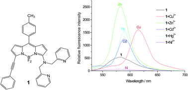
A novel metal ion-sensitive fluorescent probe – 4,4-difluoro-8-(4-methylphenyl)-5-(phenylethynyl)-3-[bis(pyridin-2-ylmethyl)amino]-4-bora-3a,4a-diaza-s-indacene – based on the BODIPY platform with di(2-picolyl)amine as chelator has been synthesized and spectroscopically and photophysically characterized. The generalized treatment of the solvent effect shows that solvent dipolarity is primarily responsible for the observed shifts of the absorption and fluorescence emission maxima. Complex formation with various metal ions is investigated in acetonitrile solution by means of spectrophotometric and fluorometric titrations. The BODIPY indicator forms 1[thin space (1/6-em)]:[thin space (1/6-em)]1 complexes with several transition metal (Ni2+, Cu2+, Zn2+) and heavy metal (Cd2+, Hg2+) ions, producing large bathochromic shifts in the absorption and fluorescence spectra and, except for Ni2+, cation-induced fluorescence amplifications. The dissociation constants of the metal ion complexes range from 4 μM for Hg2+ to 48 μM for Zn2+.


Introduction

Fluorescent probes for the measurement of concentrations of analytically and biologically important ions are essential tools in numerous fields of modern medicine and materials science.1,2 The design and synthesis of fluorescent indicators with high selectivity and sensitivity for metal ions remain an exciting field of supramolecular chemistry. Of major importance are fluoroionophores targeting transition and heavy metal ions (such as Ni2+, Cu2+, Zn2+, Cd2+, Hg2+, …). Ratiometric fluorescent probes which exhibit shifts in the absorption and/or fluorescence spectra upon coordination of the analyte of interest are preferred over probes with on/off switching of fluorescence. Indeed, measuring the ratio of the fluorescence signals at two suitably chosen wavelengths avoids the influence of many extraneous artifacts in fluorescence changes at a single wavelength. Therefore, the design, synthesis and spectroscopic characterization of ratiometric fluorescent chemosensors have emerged as an important area of supramolecular chemistry.

Here we describe a new ratiometric, fluorescent probe for transition and heavy metal ions based on the 4,4-difluoro-4-bora-3a,4a-diaza-s-indacene (BODIPY or boron dipyrrin or boron dipyrromethene)3,4 platform. Lately there is intense research interest in the synthesis and photophysical/spectroscopic properties of BODIPY-based fluorescent dyes and their application in selective and sensitive fluorescent chemosensors.4c,d As a result, there are now thousands of different BODIPY dyes described in the literature with exciting structural variations. BODIPY is an outstanding fluorophore characterized by valuable properties including bright fluorescence [due to the combination of a high fluorescence quantum yield Φ with a large molar absorption coefficient ε(λ)] with absorption and fluorescence emission bands in the visible range, robustness towards chemicals and light, and generally a negligible intersystem-crossing.4 Moreover, the BODIPY core can be postfunctionalized easily (at the pyrrole carbons, the meso-carbon and the boron atom), leading to dyes with custom-made, fine-tuned spectroscopic properties for use in bioscience and material research.5

The transition and heavy metal ions of which the fluorometric/spectrophotometric detection is investigated are Cd2+, Hg2+, Ni2+, Cu2+ and Zn2+. As chelator for these ions, we chose bis(pyridin-2-ylmethyl)amine [commonly known as di(2-picolyl)amine, DPA], linked to the BODIPY core at the 3-position. Di(2-picolyl)amine is a chelator of several metal ions, including Mn2+, Fe2+, Co2+, Ni2+, Cu2+, Zn2+, Ag+, Cd2+, Hg2+ and Pb2+.6 Reported values of the dissociation constants Kd of the 1[thin space (1/6-em)]:[thin space (1/6-em)]1 complex [Zn(DPA)]2+ are 158 nM (ref. 7) and 70 nM.8

The literature of BODIPY-based fluorescent indicators, among them those for transition and heavy metal ions, has been reviewed recently by the KU Leuven authors.4c In addition, there are excellent reviews on fluorescent chemosensors for zinc ions based on different fluorophore platforms and various receptor units, including DPA and related N,N-bis(2-pyridylmethyl)ethane-1,2-diamine.9 Of significant importance for this study are reports on fluoroionophores built on the BODIPY platform with DPA as ion receptor. A BODIPY-DPA Zn2+ probe with 30-fold fluorescence enhancement was reported by Nagano et al. (Chart 1).10 A fluorescent sensor for Zn2+ (Kd = 1 nM), based on photoinduced electron transfer (PET), utilizing BODIPY linked at the meso-position to the DPA chelator (Chart 1), which displays a significant fluorescence enhancement upon Zn2+ binding, was described by Peng and coworkers.11 The same research group also reported a selective sensor for imaging Cd2+ in living cells, using the BODIPY scaffold (Kd is ca. 60 μM), but with the DPA chelator attached at the 3-position through a p-styryl spacer (Chart 1).12 A very similar BODIPY-styryl linked DPA chelator (Chart 1) was described by Akkaya and coworkers, but it was reported as being selective for Zn2+ (Kd = 20 μM), though Hg2+ and Cd2+ also showed some response.13 Although the ratiometric probes of Peng12 and Akkaya13 are very alike (see Chart 1), they display a different selectivity (Cd2+ vs. Zn2+). Usually discrimination between Cd2+ and Zn2+ is very difficult because they are stereoelectronic isosteres. For Ni2+ detection, only one account on a BODIPY-based fluorescent sensor can be found in the literature.14 This fluorescence turn-on (PET) probe (Kd = 0.2 mM) shows a ca. 25-fold fluorescence increase upon Ni2+ binding with no shifts in absorption and emission maxima. Since paramagnetic Cu2+ is a notorious fluorescence quencher, few ratiometric fluorescent chemosensors for Cu2+ are available in the literature.4c,15 A colorimetric and near-IR fluorescent turn-on BODIPY-based probe with DPA as a chelator with high selectivity for Cu2+ among several transition metal ions has been reported by the KU Leuven authors (Chart 1).16a Similar BODIPY-DPA derived colorimetric and NIR fluorescent chemosensors for Cu2+ (Chart 1) have been described by Yin and coworkers.16b,c BODIPY-DPA functionalized hydroxyapatite (HA) nanoparticles formed 1[thin space (1/6-em)]:[thin space (1/6-em)]1 complexes with Cd2+ or Zn2+ with large cation-induced fluorescence amplifications,17 whereas BODIPY-functionalized silica nanoparticles exhibited high specificity for Cu2+ over other transition metal ions in aqueous-organic media and resulted in notable fluorescence quenching, as reported by Qin et al.18 Fluorescent probes for lead, cadmium and mercury19 ions have been reviewed recently.20


image file: c5ra23751c-c1.tif
Chart 1 BODIPY-DPA linked probes described in the literature.10–13,16

In this work, we synthesized the chemosensor 1 with BODIPY as fluorophore coupled at the 3-position to the DPA chelator and to a phenylethynyl subunit at the 5-position. The phenylethynyl moiety at 5-position causes a red shift of the spectra in relation to unsubstituted and common BODIPY dyes.5,21 The solvent-dependent spectroscopic/photophysical characteristics of 1 were investigated by steady-state UV-vis spectrophotometry and fluorometry. Coordination of 1 to various metal ions (Na+, K+, Mg2+, Ca2+, Ni2+, Cu2+, Zn2+, Cd2+, Hg2+) was spectroscopically studied and discussed. The measured ground-state dissociation constants Kd for the formed complexes, the absorption and fluorescence emission maximal wavelengths in the absence and presence of ions, the fluorescence quantum yields Φ of the apo and ion-bound species of 1 are presented and discussed.

Results and discussion

Synthesis

Fluorescent probe 4,4-difluoro-8-(4-methylphenyl)-5-(phenylethynyl)-3-[bis(pyridin-2-ylmethyl)amino]-4-bora-3a,4a-diaza-s-indacene (1) was synthesized by nucleophilic substitution of 3-chloro-4,4-difluoro-8-(4-methylphenyl)-5-(phenylethynyl)-4-bora-3a,4a-diaza-s-indacene (2) with bis(pyridin-2-ylmethyl)amine (DPA, 3) (Scheme 1). Compound 2 was synthesized according to our published procedure.22
image file: c5ra23751c-s1.tif
Scheme 1 Synthesis of compound 1.

UV-vis spectroscopy

A selection of UV-vis absorption and fluorescence emission spectra of 1 dissolved in a series of solvents is depicted in Fig. 1. The spectroscopic/photophysical properties of 1 are compiled in Table 1. The maximum of the main absorption band [λabs(max)] of 1, attributed to the 0–0 vibrational band of a strong S1 ← S0 transition, is located between 525 nm (in acetonitrile) and 561 nm (in cyclohexane), corresponding to a energy difference of ca. 1200 cm−1. A shoulder in the 510–525 nm range at the short wavelength side of the spectrum is assigned to the 0–1 vibrational band of the same transition. This vibrational fine structure is undetectable in the more polar solvents (i.e. DMSO, acetonitrile, DMF and methanol, see Fig. 1a). The typical, narrow BODIPY-like main absorption band is not observed for 1. The most narrow absorption bands are detected in cyclohexane, toluene, dibutyl ether, pentan-1-ol and octan-1-ol. The full width at half height of the maximum of the absorption band (fwhmabs) increases from ca. 2700 cm−1 in cyclohexane to ca. 3900 cm−1 in butanenitrile. The broadening of the absorption spectra in the more polar solvents indicates a change in the size or orientation of the permanent dipole moment upon excitation, which could correspond to a partial charge-transfer character for the lowest excited state in the ground-state geometry. To make this observation compatible with the blue shift of the absorption spectra upon increasing solvent dipolarity, one has to assume that the size of the permanent dipole moment probably decreases upon excitation or that excitation changes rather the orientation than the size of the permanent dipole moment. A closer inspection of the data in Table 1 suggests that a red shift related to an increased solvent polarizability and the resulting increased dispersion and van der Waals interactions is accompanied by a blue shift upon increasing the dipolarity of the solvent.
image file: c5ra23751c-f1.tif
Fig. 1 (a) Normalized absorption spectra of 1 in a selection of solvents. (b) Corresponding normalized fluorescence emission spectra (excitation at 530 nm, except for acetonitrile, DMF and DMSO with excitation at 510 nm).
Table 1 Spectroscopic and photophysical properties of 1 in several solvents
#a Solvent λabs(max) [nm] λem(max) [nm] λex(max) [nm] Δ[small nu, Greek, macron] [cm−1] fwhmabs [cm−1] fwhmem [cm−1] Φb
a The solvents are numbered according to increasing dielectric constant.b Φ values were determined vs. cresyl violet as reference (Φρ = 0.55). Excitation wavelength λex was 530 nm, except for DMF, acetonitrile and DMSO with λex = 510 nm.c THF = tetrahydrofuran, DMF = N,N-dimethylformamide, DMSO = dimethyl sulfoxide.
1 Cyclohexane 561 589 561 847 2720 1300 0.18 ± 0.03
2 Toluene 554 594 555 1216 2720 1340 0.180 ± 0.002
3 Dibutyl ether 553 587 556 1047 2770 1310 0.134 ± 0.005
4 Diethyl ether 550 590 552 1233 3000 1390 0.106 ± 0.002
5 Chloroform 552 590 553 1167 3450 1320 0.24 ± 0.03
6 Ethyl acetate 539 584 543 1430 3180 1450 0.12 ± 0.01
7 THFc 542 585 545 1356 3100 1430 0.156 ± 0.006
8 1-Octanol 554 587 554 1015 2910 1330 0.497 ± 0.004
9 1-Pentanol 552 586 552 1051 2930 1390 0.30 ± 0.01
10 1-Butanol 550 586 552 1117 3040 1530 0.38 ± 0.01
11 2-Propanol 548 586 550 1183 2980 1390 0.313 ± 0.006
12 Acetone 530 582 534 1686 3490 1750 0.103 ± 0.008
13 Butanenitrile 529 582 532 1721 3900 1580 0.131 ± 0.006
14 Ethanol 542 583 543 1298 3220 1410 0.165 ± 0.008
15 Methanol 538 582 541 1405 3760 1360 0.115 ± 0.004
16 DMFc 527 577 530 1644 3620 1660 0.158 ± 0.001
17 Acetonitrile 525 575 527 1656 3670 1710 0.108 ± 0.006
18 DMSOc 526 581 530 1800 3660 1580 0.26 ± 0.02


The structureless fluorescence emission of 1 also is solvent dependent (Fig. 1b). The maximum of the structureless emission, λem(max), shifts from 575 nm in acetonitrile to 594 nm in toluene. This red shift (ca. 560 cm−1) is accompanied by a decrease of the emission bandwidth fwhmem (from ca. 1700 cm−1 in acetonitrile to ca. 1300 cm−1 in toluene). This suggests an increase of the permanent dipole moment or a change of the orientation of the permanent dipole moment during the return to the Franck–Condon ground state.

It is useful to determine the origin of the solvent-dependent spectral changes because these deliver information on the nature of the ground and excited states. The most recent, comprehensive treatment of the solvent effect (based on a set of four empirical, complementary, mutually independent solvent scales, i.e., dipolarity, polarizability, acidity and basicity of the medium) has been proposed by Catalán.23 In this method, the polarizability and dipolarity of a particular solvent are characterized by the parameters SP and SdP, respectively, whereas acidity and basicity are described by the scales SA and SB, respectively. The {SA, SB, SP, SdP} parameters for a large number of solvents can be found in ref. 23. Mathematically, the solvent effect on the physicochemical observable y can be expressed by the multilinear eqn (1):

 
y = y0 + aSASA + bSBSB + cSPSP + dSdPSdP (1)
where y0 denotes the physicochemical property of interest in the gas phase; aSA, bSB, cSP and dSdP are regression coefficients that describe the sensitivity of the property y to the various solvent–solute interaction mechanisms; {SA, SB, SP, SdP} are independent solvent parameters (indices) accounting for the various types of solvent–solute interactions.

The spectroscopic observables y analyzed in this paper are the absorption maxima [small nu, Greek, macron]abs [= 1/λabs(max)] and the fluorescence emission maxima [small nu, Greek, macron]em [= 1/λem(max)], both expressed in cm−1. The use of {SA, SB, SP, SdP} (eqn (1)) gives high-quality fits of [small nu, Greek, macron]abs of 1 (for the solvents listed in Table 1), using the correlation coefficient r as goodness-of-fit criterion (r = 0.939, eqn (2a) and , ESI). Good-quality fits are also obtained for the multilinear analysis of [small nu, Greek, macron]em according to eqn (1) (r = 0.827, eqn (2b) and ESI). The extra benefit of the generalized (Catalán) treatment of the solvent effect is that it allows one to separate the relative contributions of dipolarity, polarizability, acidity and basicity of the medium. Therefore, we utilized the new methodology to resolve which solvent property/properties is/are responsible for the observed shifts of [small nu, Greek, macron]abs and [small nu, Greek, macron]em. The relative importance of each of the Catalán solvent scales was studied by omitting in turn one, two or three solvent scales from the regression analysis (for details, see ESI). These analyses clearly identify solvent dipolarity (SdP) as the most critical parameter that accounts for the observed shifts of [small nu, Greek, macron]abs and [small nu, Greek, macron]em. Moreover, only for SdP are the estimated dSdP coefficients significantly larger than their associated standard errors.

 
[small nu, Greek, macron]abs = (18.2 ± 0.5) × 103 + (−841 ± 249) SA + (−54 ± 170) SB + (−625 ± 649) SP + (1.3 ± 0.1) × 103 SdP (2a)
 
[small nu, Greek, macron]em = (17.2 ± 0.3) × 103 + (−178 ± 142) SA + (31 ± 97) SB + (−474 ± 371) SP + (3.9 ± 0.8) × 102 SdP (2b)

Complex formation between 1 and various metal ions in acetonitrile

To investigate the chelating ability of 1, UV-vis spectrophotometric and fluorometric titrations were carried out in acetonitrile solution as a function of metal ion concentration. Complex formation was examined for 1 with alkali (Na+ and K+), alkaline-earth (Mg2+ and Ca2+), transition metal (Ni2+, Cu2+, Zn2+) and heavy metal (Cd2+ and Hg2+) ions. The results are summarized in Table 2.
Table 2 Spectroscopic and photophysical characteristics of 1 in the absence and presence of transition metal (Ni2+, Cu2+, Zn2+) and heavy metal (Cd2+, Hg2+) ions in acetonitrile. Kd is the average value determined from all the (direct and ratiometric) analyses of fluorometric and spectrophotometric titrations
Complex λabs(max) [nm] λem(max) [nm] Isosbestic points [nm] Δ[small nu, Greek, macron] [cm−1] Kd [μM] fwhmabs [cm−1] fwhmem [cm−1] Φ
1 525 575   1656   3670 1710 0.108[thin space (1/6-em)]±[thin space (1/6-em)]0.006
1–Ni2+ 565 580 540, 443 458 13 ± 2 1120 1800 0.020[thin space (1/6-em)]±[thin space (1/6-em)]0.004
1–Cu2+ 594 616 547, 463 601 9 ± 3 950 0.40 ± 0.03
1–Zn2+ 565 584 540, 434 576 48 ± 11 1100 900 0.29 ± 0.05
1–Cd2+ 575 590 540, 438 442 9 ± 2 950 920 0.23 ± 0.02
1–Hg2+ 575 590 537, 438 442 4 ± 1 2000 840 0.27 ± 0.02


Upon addition of the alkali ions Na+ and K+ and the alkaline-earth ions Mg2+ and Ca2+ to a solution of 1 in acetonitrile, no change in the UV-vis absorption and fluorescence spectra could be detected. Obviously, the interaction between 1 and these ions is too weak to cause any change of either the UV-vis absorption spectra or the vis fluorescence spectra.

Conversely, the transition metal (Ni2+, Cu2+, Zn2+) and heavy metal (Cd2+, Hg2+) ions produced spectral changes of 1 (Fig. 2). For example, the lowest-energy absorption band of 1 shifts bathochromically by ca. 40 nm, from 525 nm in an ion-free environment to 565 nm upon addition of Zn2+ to the acetonitrile solution (Fig. 3a). The relative contributions of the 565/525 nm signals change with varying [Zn2+] and the vis absorption spectra show isosbestic points at 434 and 540 nm. Similar changes were observed in the fluorescence excitation spectra (Fig. S1, ESI). The maximum of the fluorescence emission band shifts bathochromically from 575 nm in ion-free acetonitrile to 584 nm in the presence of Zn2+ and is accompanied by an increase in intensity (Fig. 3b). A pseudo-isoemissive point can be observed at 564 nm, which can be used to simplify analysis of ratiometric fluorometric titrations. The fluorescence quantum yield Φ of 1 increases from 0.11 in the absence of Zn2+ to 0.29 for the 1–Zn2+ complex. These red shifts can be rationalized as follows. Upon complexation of Zn2+, the lone pair of N of DPA connected to C3 of BODIPY will become less available for delocalization over the BODIPY moiety. One should note also that, upon complexation of Zn2+, the features of the absorption spectra become closer to that of unsubstituted and common BODIPYs showing a narrow band and a hint of vibrational fine structure even in polar solvents.5,24 This can be attributed again to the blocking of the delocalization of the lone pair of N of DPA in 3-position over the rest of the conjugated system. The same effect will decrease the dipole moments (and their differences between ground and excited state) in the ground and excited state. This also accounts for the strong decrease of the Stokes shift from 1660 cm−1 in 1 to 580 cm−1 in 1–Zn2+ and of both fwhmabs and fwhmem of 1–Zn2+ (Table 2). Often BODIPYs with explicit electron-withdrawing or electron-donating substituents are characterized by low Φ values, especially in polar solvents.25 Upon complexation by Zn2+, the electron-donating effect of N in 3-position is significantly decreased which leads to an increase of Φ from 0.11 to 0.29.


image file: c5ra23751c-f2.tif
Fig. 2 (a) Absorption and (b) fluorescence emission spectra of apo 1 and of 1 in the presence of the transition metal ions Cu2+ (60 μM), Zn2+ (600 μM), Ni2+ (170 μM) and the heavy metal ions Cd2+ (235 μM) and Hg2+ (50 μM) in acetonitrile.

image file: c5ra23751c-f3.tif
Fig. 3 Compound 1 as a function of [Zn2+] in acetonitrile solution. (a) Absorption spectra. (b) Fluorescence emission spectra (excitation at 510 nm). The full line in the inset shows the best fit to the ratiometric emission (eqn (4) with n = 1) titration data at λem1/λem2 = 582 nm/564 nm (isoemissive point) as a function of [Zn2+].

Analysis of the UV-vis spectrophotometric and vis fluorometric titration data in the presence of varying concentrations of Zn2+ allows one to extract the ground-state dissociation constant Kd and the stoichiometry of the 1–Zn2+ complex. The values of Kd and the stoichiometry (n) of binding of Zn2+ by the DPA ligand of 1 were determined by nonlinear fitting eqn (3) (direct fluorometric titration)26 and eqn (4) (ratiometric fluorometric titration)26 to the fluorescence excitation or emission spectral data F (eqn (3)) and ratios R (eqn (4)), measured as a function of metal ion concentration, i.e., [X] = [Zn2+]. Nonlinear least-squares analyses of the corresponding spectrophotometric titrations (direct and ratiometric) as a function of metal ion were also performed.27 In all titrations, no metal ion buffer was used to control the free metal ion concentration. It was assumed that the free metal ion concentration [X] could be approximated by its analytical concentration.

 
image file: c5ra23751c-t1.tif(3)

In eqn (3), F stands for the fluorescence signal at free ion concentration [X], whereas Fmin and Fmax denote the fluorescence signals at minimal and maximal [X], respectively, and n is the number of cations bound per probe molecule (i.e., stoichiometry of binding). Because the fits of eqn (3) to the fluorescence data F with n, Kd, Fmin and Fmax as freely adjustable parameters always gave values of n close to 1 (indicative of a 1[thin space (1/6-em)]:[thin space (1/6-em)]1 complex), n = 1 was used in the final curve fittings, from which the estimated Kd values are reported here. The plot of F against −log[X] has a sigmoidal shape. This plot starts at Fmin at very low [X] and changes most in the X-concentration range from 0.1 × Kd to 10 × Kd to asymptotically reach Fmax at high [X]. Outside this critical X-concentration range, F does not change much. Therefore, it important that the dissociation constant Kd is well matched with the X-concentration range of interest.

 
image file: c5ra23751c-t2.tif(4)

In the excitation ratiometric method, one measures R = F(λem,λex1)/F(λem,λex2) at a common emission wavelength, λem, and two different excitation wavelengths, λex1 and λex2. Rmin is the ratio of the fluorescence intensities at two distinct excitation wavelengths and one emission wavelength of the apo form of the indicator (minimum [X]). Rmax represents the ratio of the fluorescence intensities of the bound form of the indicator (maximum [X]). R denotes the ratio of the fluorescence intensities corresponding to intermediate [X] and ξ = Fmin(λem,λex2)/Fmax(λem,λex2). Choosing the wavelength of the pseudo-isoemissive point as λex2 makes the analysis simpler. In this case we have ξ = 1 and the expression for R (eqn (4)) simplifies to that of F (eqn (3)). Fitting eqn (4) to excitation ratiometric values R as a function of [X] yields values for Kdξ(λem,λex2), Rmin, Rmax and n. Because ξ(λem,λex2) – the ratio of the fluorescence signal of the apo form of the indicator over that of the bound form at the indicated wavelengths – is experimentally accessible, a value for Kd can be recovered from ratiometric excitation fluorescence data.

In the emission ratiometric method, one determines R = F(λem1,λex)/F(λem2,λex) at the indicated wavelengths as a function of cation concentration [X]. In this case, ξ is defined as ξ = Fmin(λem2,λex)/Fmax(λem2,λex). ξ = 1 when λex2 is chosen as the wavelength corresponding to the pseudo-isoemissive point and, in that case, the expression for R (eqn (4)) simplifies to that of F (eqn (3)). Fitting eqn (4) to the emission ratiometric fluorescence data R as a function of [X] yields values for Kdξ(λem2,λex), Rmin, Rmax and n. Because ξ(λem2,λex) can be determined from the fluorescence signals of the apo and bound forms of the indicator at the indicated wavelengths, a value Kd for the complex can be obtained.

From the analyses of all the direct and ratiometric spectrophotometric and fluorometric titrations of 1 with Zn2+ ions it is clear that a 1[thin space (1/6-em)]:[thin space (1/6-em)]1 ligand/cation stoichiometry is found with an average Kd value for the 1–Zn2+ complex of 48 ± 11 μM.

The vis absorption and fluorescence (excitation/emission) spectral changes observed when Cd2+ is added to 1 in acetonitrile solution are comparable to those for Zn2+ (Fig. S2, ESI), although the bathochromic shifts upon complex formation are somewhat larger [λabs(max) = 575 nm, λem(max) = 590 nm] and Φ of the 1–Cd2+ complex is slightly lower than that of 1–Zn2+ (Table 2). In the emission spectra as a function of [Cd2+] pseudo-isoemissive points can be found at 571 and 550 nm. An average Kd value of 9 ± 2 μM was obtained for the 1–Cd2+ complex. This smaller Kd value (compared to Kd of the 1–Zn2+ complex) suggests a stronger bond between Cd2+ and the N ligands of DPA. Hence, the lone pair of N connected to C3 of the BODIPY framework will be less available for delocalization over the conjugated system, leading to somewhat larger red spectral shifts than those observed for Zn2+.

The response of the absorption and fluorescence (excitation/emission) spectra of 1 in acetonitrile upon addition of Hg2+ (Fig. 4) is nearly the same as that found for 1 in the presence of Cd2+, but Φ of 1–Hg2+ is somewhat higher than Φ of 1–Cd2+ (Table 2). This Φ value is unexpected taking into account the possible heavy-atom quenching by Hg2+. The emission spectra as a function of [Hg2+] show two pseudo-isoemissive points, at 569 and 544 nm. The average Kd value of the 1–Hg2+ complex was found to be 4 ± 1 μM.


image file: c5ra23751c-f4.tif
Fig. 4 Compound 1 in acetonitrile solution as a function of [Hg2+]. (a) Fluorescence excitation spectra (emission observed at 620 nm). (b) Fluorescence emission spectra (excitation at 510 nm). The full line in the insets of (a) and (b) shows the best fit to (a) the direct fluorometric excitation (eqn (3) with n = 1) titration data at λex = 575 nm, λem = 620 nm and (b) the ratiometric emission (eqn (4) with n = 1) at λem1/λem2 = 590 nm/569 nm (isoemissive point) titration data as a function of [Hg2+].

Upon addition of Ni2+ to a solution of 1 in acetonitrile, the vis absorption spectra respond analogously to the addition of Zn2+: a ca. 40 nm red shift (to 565 nm) is found in addition to the appearance of isosbestic points at 443 and 540 nm. In contrast, addition of Ni2+ causes strong fluorescence quenching: Φ of 1–Ni2+ is only 0.02 compared to 0.11 for apo 1. Quenching of fluorescence upon Ni2+ binding arises from a PET from the BODIPY fluorophore to the metal center. Ni2+ has – in contrast to the close-shell ions Zn2+, Cd2+ and Hg2+ – several low-lying dd-type excited states and is moreover paramagnetic. The influence of Ni2+ on the spectral maxima and the features of the absorption and emission spectra can be clarified in the same way as for Zn2+, Cd2+ and Hg2+. The absorption and fluorescence emission spectra of 1 in acetonitrile as a function of [Ni2+] are given in Fig. S3 (ESI). The average Kd value of the 1–Ni2+ complex amounted to 13 ± 2 μM.

Compared to the transition and heavy metal ions Ni2+, Zn2+, Cd2+ and Hg2+, addition of Cu2+ triggers a large fluorescence enhancement (Φ = 0.40 for 1–Cu2+) and the largest red shifts [λabs(max) = 594 nm, λem(max) = 616 nm] (Fig. 2 and 5). The average Kd value of 1–Cu2+ complex was 9 ± 3 μM. The Stokes shift of the 1–Cu2+ complex (600 cm−1) is larger than those of the 1–Cd2+ and 1–Hg2+ complexes (440 cm−1) and close to that of the 1–Zn2+-complex (580 cm−1). Also the large increase in Φ is quite unexpected because Cu2+ is paramagnetic and has low-lying dd-states. Both properties are analogous to Ni2+, which strongly quenches the BODIPY fluorescence. Although the absorption spectrum of 1 undergoes a red shift upon complex formation with Cu2+ (in analogy to what occurs upon complex formation with the other divalent ions), the features of the absorption spectrum of the 1–Cu2+ complex differ in several aspects from those of the other complexes. First, the apparent ratio of the 0–1 to the 0–0 band of the S1 ← S0 transition decreases from 0.9 for Cu2+ to 0.3–0.4 for the other ions. Second, the ratio of the maximum absorbance of the S2 ← S0 to that of the S1 ← S0 transition decreases from 0.9 for Cu2+ to 0.15–0.25 for the other ions. Finally, the long wavelength edge of the 0–0 band of the S1 ← S0 transition is considerably shallower. Conversely, the features of the fluorescence spectrum of 1–Cu2+, although bathochromically shifted, resembles quite well those of the other complexes. This makes it unlikely that in 1–Cu2+ the nature of the excited state differs strongly from that in the complexes of 1 with the other metal ions. In those complexes, the excited state resembles that of a BODIPY fluorophore without strong electron-donating or electron-withdrawing substituents. Taking into account that Cu2+ complexes are known to absorb strongly between 550 and 650 nm, the unexpected features of BODIPY in the presence of Cu2+ is perhaps attributable to an overlap of the absorption spectrum of BODIPY and that due to dd-transitions of Cu2+ in 1–Cu2+. The relative short wavelength of the absorption spectrum of Cu2+ in 1–Cu2+ will lead to a poor overlap with the emission spectrum of 1–Cu2+. Hence energy transfer to the dd-type excited states of Cu2+ will be rather slow which may account for the (unexpected) absence of quenching of the BODIPY fluorescence by Cu2+ in the 1–Cu2+ complex.


image file: c5ra23751c-f5.tif
Fig. 5 Compound 1 in acetonitrile solution as a function of [Cu2+]. (a) Absorption spectra. (b) Fluorescence emission spectra (excitation at 510 nm). The full line in the inset of (b) shows the best fit to the direct fluorometric emission titration data (eqn (3) with n = 1) at λem = 580 nm as a function of [Cu2+].

High selectivity towards the analyte of interest is an important property of any chemosensor. BODIPY indicator 1 forms 1[thin space (1/6-em)]:[thin space (1/6-em)]1 complexes with several transition metal (Ni2+, Cu2+, Zn2+) and heavy metal (Cd2+, Hg2+) ions. The dissociation constants Kd of these formed complexes (Table 2) provide numerical criteria for evaluating the selectivity of 1 for a particular ion. As shown above, the determination of Kd of each metal ion complex requires the laborious execution and analysis of fluorometric titrations.

In the literature, competitive, steady-state fluorescence measurements are frequently used as a quick alternative for assessing the selectivity of a fluorescent indicator for a certain ion. These simple tests are typically carried out as follows. First, one has to establish through fluorometric titration that the probe is sensitive to a specific ion, let's say X. The analysis of the titration data (eqn (3)) yields a value of Kd of the complex with X. To determine if the probe is selective for X, one then measures the steady-state fluorescence signal F0 (either the whole, integrated fluorescence spectrum or at a single emission wavelength) of the probe in the presence of X at a certain concentration. Subsequently, the competing ion Y at a specific concentration is added to this solution and the fluorescence signal F is measured. The outcome of these competition experiments is often displayed in the form of bar graphs, as presented in Fig. S5 (ESI), and shows the change/constancy of the steady-state fluorescence upon addition of the competing ion Y to a solution of the probe in the presence of ion X (X = Cu2+ or Cd2+ in Fig. S5, ESI). If no change is found upon addition of Y, it is commonly assumed that Y does not compete with X for the probe and hence that the probe is selective for X. We shall show that such fast, easy competition experiments often lead to erroneous conclusions either because the experiments are carried out defectively or for fundamental reasons. For an in-depth investigation of the fluorescence signal F arising from a photophysical system consisting of a probe in the presence of competing ions X and Y, a correct mathematical description of F is needed. Although such analysis is beyond the scope of this paper, it is still possible, without going into mathematical detail, to discuss the present photophysical system (Fig. S5, ESI) consisting of 1 in the presence of metal ions X and Y. It can be demonstrated mathematically that the fluorescence F of 1 in the presence of X and Y is dependent on several parameters, namely (i) the Kd values of the complexes of 1 (1–X and 1–Y) with the competing ions X and Y, (ii) the stationary fluorescence Fmin of the apo form of 1, (iii) the fluorescence F1–X and F1–Y of the pure complexes 1–X and 1–Y, respectively and (iv) the concentrations [X] and [Y] of the unbound (free) ions X and Y, respectively.28 F describes a 3D data surface as a function of the independent variables [X] and [Y]. Using the equation of F with the above (i–iv) parameters, one can predict mathematically how addition of ion Y to a solution of 1 in the presence of X will affect its steady-state fluorescence F. The dependence of the steady-state fluorescence on the addition of Y at a specific concentration to a solution of 1 in the presence of certain concentration of X is commonly measured by competition experiments (such as those shown in Fig. S5, ESI). However, in such experiments only two data points of the full 3D data surface are determined: F0 and F in Fig. S5 (ESI) are even fused into the unique data point F/F0. F0 stands for the fluorescence signal of 1 in the presence of 10 μM X (= Cu2+ or Cd2+) and F corresponds to 1 in the presence of 10 μM X (= Cu2+ or Cd2+) plus a 50 μM solution of other metal ions Y. These two data points and a priori the single data point F/F0 are insufficient to determine the selectivity of indicator 1 vs. X or Y. Addition of the competitive ion (50 μM) to a solution of 1 in the presence of 10 μM Cu2+ (or 10 μM Cd2+) shows no significant variation in the fluorescence intensity (Fig. S5, ESI). This indicates that the fluorescence of 1–Cu2+ (or 1–Cd2+) is not influenced by other coexisting metal ions Y, at least not at the concentrations used for the original ion X (Cu2+ or Cd2+) and the competing metal ions Y.

The near-invariability of the fluorescence intensities F0 and F (i.e., F/F0 ≈ 1.0) cannot be taken as unambiguous proof that 1 is not selective for Y, for the following reasons. It is crucial to start the experiment beginning with the pure complex 1–X; F0 should therefore correspond to the fluorescence signal F1–X of the pure complex 1–X, which is not the case here. At 10 μM Cu2+ (or 10 μM Cd2+) (Fig. S5, ESI), the fluorescence intensity of the 1–Cu2+ (or 1–Cd2+) complex is not reached. This is only the case when the free concentration [Cu2+] (or [Cd2+]) is equal to at least 10 × Kd (or preferably > 102 × Kd) of the corresponding 1–Cu2+ (or 1–Cd2+) complex (Kd = 9 μM) (see eqn (3)).27 At [Cu2+] (or [Cd2+]) = 10 μM and with Kd = 9 mM, we are approximately at the mid-point of the titration. Hence, the fluorescent indicator appears both in its free form 1 and its complexed form 1–X (= 1–Cu2+ or 1–Cd2+). At low [X] ([Cu2+] or [Cd2+]), addition of competing metal ion Y to a mixture containing both 1 and 1–X (1–Cu2+ or 1–Cd2+) may form the complex 1–Y directly from 1 or/and by displacement of X (Cu2+ or Cd2+) from the 1–X (1–Cu2+ or 1–Cd2+) complex. Although under those conditions there might be a change of fluorescence, it is impossible to estimate from these two data points the Kd values of the complex 1–Y and/or F1–Y. Even if only 1–X (1–Cu2+ or 1–Cd2+ in Fig. S5, ESI) is present before addition of the competing metal ion Y, constancy of fluorescence upon addition of Y may simply be attributable to the concentration [Y] used that is too low for influencing the fluorescence. Then, a large excess of [Y] might reveal fluorescence change or invariability in relation to the initial experimental condition where only 1–X is present. The fluorescence will vary from F1–X (only 1–X present) at low [Y] ([Y] → 0) to F1–Y (only 1–Y present) for [Y] → ∞. However, it is still impossible to estimate a value for Kd of the complex 1–Y. Theoretically, constancy of the fluorescence as a function of [Y] may also be the result of F1–X and F1–Y being equal (F1–X = F1–Y). In this case, the fluorescence remains constant, irrespective of the concentration [Y]. Finally, the competition between X and Y is a dynamic process: it is possible that decomplexation of the original 1–X complex and/or subsequent formation of the new 1–Y complex is too slow to occur on the timescale used. To summarize, there are several reasons for the invariability of the fluorescence signal upon addition of Y (too low [Y] used, F1–X = F1–Y, decomplexation of 1–X and/or succeeding formation of 1–Y is too slow, or the probe is simply not sensitive to Y), so that no reliable decision about the selectivity of the probe can be reached. To conclude, these quick competitive tests are not capable of providing a clear answer to the question of selectivity; only Kd values estimated from fluorometric titrations can do.

Besides selectivity, another important parameter defining fluorescent indicators is sensitivity. Chemosensor 1 forms 1[thin space (1/6-em)]:[thin space (1/6-em)]1 complexes with various transition metal (Ni2+, Cu2+, Zn2+) and heavy metal (Cd2+, Hg2+) ions. An excellent linear correlation (r = 0.994, n = 7) between the steady-state fluorescence F and the Zn2+ concentration (log[Zn2+]) was obtained over the range 0–186 μM Zn2+ (Fig. S6, ESI). The detection limit (DL) was estimated based on the following equation: DL = 3σ/k, where σ is the standard deviation of 10 blank samples (containing only probe 1 without any metal ions) and k is the slope of the calibration curve (fluorescence intensity plotted against [metal ion] or log[metal ion]).29 The detection limit was calculated to be as low as 1.0 μM for Zn2+. The sensitivity of the probe towards each metal ion is compiled in Table 3 and is shown to be 1.0 μM or lower.

Table 3 The sensitivity of probe 1 towards each metal ion
Metal ion Linear range/μM Fa DL/μM r
a Equation used for the fitting of F as a function of ion concentration. The Zn2+ and Cd2+ concentrations are expressed in M (mol L−1), whereas [Ni2+], [Cu2+] and [Hg2+] are in μM.
Ni2+ 0–25.7 F = 534 − 15.3 × [Ni2+] 0.1 0.990
Cu2+ 1.5–26 F = 81.6 + 53.7 × [Cu2+] 0.04 0.992
Zn2+ 0–186 F = 5591 + 879 × log[Zn2+] 1.0 0.994
Cd2+ 1.3–235.5 F = 876.7 + 122.2 × log[Cd2+] 0.5 0.994
Hg2+ 0–5.3 F = 389 + 142.7 × [Hg2+] 0.01 0.996


Conclusions

We have synthesized the visible light excitable, fluorescent BODIPY-based sensor 1, which shows solvent-dependent spectroscopic/photophysical properties. The generalized treatment of the solvent effect shows that solvent dipolarity is primarily responsible for the observed shifts of the absorption and fluorescence emission maxima. Fluorescent indicator 1 forms 1[thin space (1/6-em)]:[thin space (1/6-em)]1 complexes with several transition metal ions (Ni2+, Cu2+, Zn2+) and heavy metal ions (Cd2+, Hg2+), but not with alkali-metal ions (Na+, K+) and earth-alkaline-metal ions (Ca2+, Mg2+). Kd values of the metal ion complexes range from 4 μM for Hg2+ to 48 μM for Zn2+. The new compound is an example of a very sensitive fluorescent probe for several metal ions displaying large absorption and fluorescence changes in an analytically interesting wavelength region.

Experimental

The materials used, the steady-state UV-vis absorption and fluorescence spectroscopy, the determination of Kd through direct and ratiometric fluorometric titration are described in the ESI.

Synthesis of 4,4-difluoro-8-(4-methylphenyl)-5-(phenylethynyl)-3-[bis(pyridin-2-ylmethyl)amino]-4-bora-3a,4a-diaza-s-indacene (1)

3-Chloro-4,4-difluoro-8-(4-methylphenyl)-5-(phenylethynyl)-4-bora-3a,4a-diaza-s-indacene (2) was synthesized following a literature procedure.22 To a solution of 2 (104 mg, 0.25 mmol) in acetonitrile (50 mL) under argon atmosphere was added bis(pyridin-2-ylmethyl)amine 3 (60 mg, 0.30 mmol, 1.2 equiv.). The reaction mixture was stirred at room temperature for 16 h. Afterwards the resulting solution was poured in water and extracted with dichloromethane (2 × 100 mL). The combined organic layer was washed with brine, dried over MgSO4, filtered and evaporated to dryness under reduced pressure. The crude solid was purified by column chromatography on silica gel using ethyl acetate as eluent to yield 129 mg (89% yield) of a red solid. M.p. 176 °C. 1H NMR (CDCl3) δ 8.55 (d, 2H, J = 4.1 Hz), 7.66 (td, 2H, J = 7.9 Hz, J = 1.5 Hz), 7.48 (dd, 2H, J = 7.9 Hz, J = 1.5 Hz), 7.44 (d, 2H, J = 7.9 Hz), 7.34 (d, 2H, J = 7.9 Hz), 7.28 (m, 3H), 7.24 (d, 2H, J = 7.9 Hz), 7.20 (dd, 2H, J = 5.3 Hz, J = 1.8 Hz), 6.82 (d, 1H, J = 4.9 Hz), 6.59 (d, 1H, J = 4.1 Hz), 6.36 (d, 1H, J = 3.8 Hz), 6.28 (d, 1H, J = 5.2 Hz), 5.29 (s, 4H), 2.42 (s, 3H, tolyl). 13C NMR (CDCl3) δ 164.2, 156.6, 149.6, 139.2, 137.1, 136.3, 135.4, 133.7, 132.2, 132.0, 131.6, 130.6, 128.2, 128.0, 126.0, 125.6, 123.8, 122.7, 122.1, 120.3, 119.5, 115.3, 100.1, 95.6, 84.0, 58.1, 22.8. IR (cm−1) 3055 (m), 3014 (m), 2920 and 2852 (CH2, s), 2202 (C[double bond, length as m-dash]C, w). LRMS (EI, 70 eV) m/z 579. HRMS: calcd for C36H28BF2N5 579.2406, found 579.23967.

1H and 13C NMR spectra were recorded at room temperature on a Bruker 600 instrument operating at a frequency of 600 MHz for 1H and 150 MHz for 13C. 1H NMR spectra were referenced to tetramethylsilane (0.00 ppm) as an internal standard. Chemical shift multiplicities are reported as s = singlet, d = doublet and m = multiplet. 13C spectra were referenced to the CDCl3 (77.67 ppm) signal. Mass spectra were recorded on a Hewlett Packard 5989A mass spectrometer (EI mode and CI mode). High-resolution mass data were obtained with a KRATOS MS50TC instrument. The IR spectrum was recorded on a Bruker FT-IR spectrometer Alpha-P with Diamond ATR. Melting points were taken on a Reichert Thermovar and are uncorrected.

Acknowledgements

WD and MVdA are thankful to BELSPO (Belgium) for funding through IAP 06/27 and IAP7/05. WQ thanks the Natural Science Foundation of China (no. 21271094).

References

  1. Chemosensors of Ion and Molecule Recognition, ed. J.-P. Desvergne and A. W. Czarnik, Kluwer, Dordrecht, The Netherlands, 1997 Search PubMed.
  2. New Trends in Fluorescence Spectroscopy. Applications to Chemical and Life Sciences, ed. B. Valeur and J.-C. Brochon, Springer, Berlin, 2002 Search PubMed.
  3. A. Treibs and F.-H. Kreuzer, Liebigs Ann. Chem., 1968, 718, 208–223 CrossRef CAS.
  4. (a) A. Loudet and K. Burgess, Chem. Rev., 2007, 107, 4891–4932 CrossRef CAS; (b) G. Ulrich, R. Ziessel and A. Harriman, Angew. Chem., Int. Ed., 2008, 47, 1184–1201 CrossRef CAS; (c) N. Boens, V. Leen and W. Dehaen, Chem. Soc. Rev., 2012, 41, 1130–1172 RSC; (d) T. Kowada, H. Maeda and K. Kikuchi, Chem. Soc. Rev., 2015, 44, 4953–4972 RSC; (e) S. Yin, V. Leen, C. Jackers, M. van der Auweraer, M. Smet, N. Boens and W. Dehaen, Dyes Pigm., 2011, 88, 372–377 CrossRef CAS; (f) S. Yin, V. Leen, C. Jackers, D. Beljonne, B. van Averbeke, M. van der Auweraer, N. Boens and W. Dehaen, Chem.–Eur. J., 2011, 17, 13247–13257 CrossRef CAS.
  5. N. Boens, B. Verbelen and W. Dehaen, Eur. J. Org. Chem., 2015, 6577–6595 CrossRef CAS.
  6. K. Kyose, H. Kojima, Y. Urano and T. Nagano, J. Am. Chem. Soc., 2006, 128, 6548–6549 CrossRef.
  7. D. W. Gruenwedel, Inorg. Chem., 1968, 7, 495–501 CrossRef CAS.
  8. G. K. Walkup, S. C. Burdette, S. J. Lippard and R. Y. Tsien, J. Am. Chem. Soc., 2000, 122, 5644–5645 CrossRef CAS.
  9. (a) K. Kikuchi, K. Komatsu and T. Nagano, Curr. Opin. Chem. Biol., 2004, 8, 182–191 CrossRef CAS; (b) P. Jiang and Z. Guo, Coord. Chem. Rev., 2004, 248, 205–229 CrossRef CAS; (c) N. C. Lim, H. C. Freake and C. Brückner, Chem.–Eur. J., 2005, 11, 38–49 CrossRef; (d) P. Carol, S. Sreetjith and S. Ajayaghosh, Chem.–Asian J., 2007, 2, 338–348 CrossRef CAS.
  10. H. Koutaka, J.-i. Kosuge, N. Fukasaku, T. Hirano, K. Kikuchi, Y. Urano, H. Kojima and T. Nagano, Chem. Pharm. Bull., 2004, 52, 700–703 CrossRef CAS.
  11. Y. Wu, X. Peng, B. Guo, J. Fan, Z. Zhang, J. Wang, A. Cui and Y. Gao, Org. Biomol. Chem., 2005, 3, 1387–1392 CAS.
  12. X. Peng, J. Du, J. Fan, J. Wang, Y. Wu, J. Zhao, S. Sun and T. Xu, J. Am. Chem. Soc., 2007, 129, 1500–1501 CrossRef CAS PubMed.
  13. S. Atilgan, T. Ozdemir and E. U. Akkaya, Org. Lett., 2008, 10, 4065–4067 CrossRef CAS.
  14. S. C. Dodani, Q. He and C. J. Chang, J. Am. Chem. Soc., 2009, 131, 18020–18021 CrossRef CAS PubMed.
  15. J. A. Cotruvo Jr, A. T. Aron, K. M. Ramos-Torres and C. J. Chang, Chem. Soc. Rev., 2015, 44, 4400–4414 RSC.
  16. (a) S. Yin, V. Leen, S. van Snick, N. Boens and W. Dehaen, Chem. Commun., 2010, 46, 6329–6331 RSC; (b) S. Yin, W. Yuan, J. Huang, D. Xie, B. Liu, K. Jiang and H. Qiu, Spectrochim. Acta, Part A, 2012, 96, 82–88 CrossRef CAS; (c) J. Huang, X. Ma, B. Liu, L. Cai, Q. Li, Y. Zhang, K. Jiang and S. Yin, J. Lumin., 2013, 141, 130–136 CrossRef CAS.
  17. X. Jia, X. Yu, G. Zhang, W. Liu and W. Qin, J. Coord. Chem., 2013, 66, 662–670 CrossRef CAS.
  18. X. Yu, X. Jia, X. Yang, W. Liu and W. Qin, RSC Adv., 2014, 4, 23571–23579 RSC.
  19. E. M. Nolan and S. J. Lippard, Chem. Rev., 2008, 108, 3443–3480 CrossRef CAS.
  20. H. N. Kim, W. X. Ren, J. S. Kim and Y. Yoon, Chem. Soc. Rev., 2012, 41, 3210–3244 RSC.
  21. W. Qin, T. Rohand, W. Dehaen, J. N. Clifford, K. Driessen, D. Beljonne, B. van Averbeke, M. van der Auweraer and N. Boens, J. Phys. Chem. A, 2007, 111, 8588–8597 CrossRef CAS.
  22. T. Rohand, W. Qin, N. Boens and W. Dehaen, Eur. J. Org. Chem., 2006, 4658–4663 CrossRef CAS.
  23. J. Catalán, J. Phys. Chem. B, 2009, 113, 5951–5960 CrossRef PubMed.
  24. L. Jiao, C. Yu, J. Wang, E. A. Briggs, N. A. Besley, D. Robinson, M. J. Ruedas-Rama, A. Orte, L. Crovetto, E. M. Talavera, J. M. Alvarez-Pez, M. van der Auweraer and N. Boens, RSC Adv., 2015, 5, 89375–89388 RSC.
  25. (a) W. Qin, M. Baruah, M. Sliwa, M. van der Auweraer, D. Beljonne, B. van Averbeke and N. Boens, J. Phys. Chem. A, 2008, 112, 6104–6114 CrossRef CAS PubMed; (b) W. Qin, V. Leen, T. Rohand, W. Dehaen, P. Dedecker, M. van der Auweraer, K. Robeyns, L. van Meervelt, D. Beljonne, B. van Averbeke, J. N. Clifford, K. Driesen, K. Binnemans and N. Boens, J. Phys. Chem. A, 2009, 113, 439–447 CrossRef CAS PubMed; (c) I. Esnal, J. Bañuelos, I. López Arbeloa, A. Costela, I. Garcia-Moreno, M. Garzón, A. R. Agarrabeitia and M. J. Ortiz, RSC Adv., 2013, 3, 1547–1556 RSC.
  26. E. Cielen, A. Tahri, A. Ver Heyen, G. J. Hoornaert, F. C. de Schryver and N. Boens, J. Chem. Soc., Perkin Trans. 2, 1998, 1573–1580 RSC.
  27. A. Kowalczyk, N. Boens, V. van den Bergh and F. C. de Schryver, J. Phys. Chem., 1994, 98, 8585–8590 CrossRef CAS.
  28. N. Boens, unpublished results.
  29. W. Qin, W. Liu and M. Tan, Anal. Chim. Acta, 2002, 468, 287–292 CrossRef CAS.

Footnote

Electronic supplementary information (ESI) available: Experimental (materials, steady-state UV-vis absorption and fluorescence spectroscopy, determination of Kd through direct and ratiometric fluorometric titration). The Catalán analyses of the solvent-dependent absorption and fluorescence emission maxima. Absorption, fluorescence excitation and emission spectra of 1 in acetonitrile as a function of [Zn2+], [Cd2+] and [Ni2+]. Cyclic voltammetry. Fluorescence experiments with competing ions. Sensitivity of 1 towards Zn2+, Cu2+ and Ni2+. 1H NMR and 13C NMR spectra of 1 in CDCl3. See DOI: 10.1039/c5ra23751c

This journal is © The Royal Society of Chemistry 2016