A lanthanide metal–organic framework (MOF-76) for adsorbing dyes and fluorescence detecting aromatic pollutants

Xiao Lian and Bing Yan*
Shanghai Key Lab of Chemical Assessment and Sustainability, Department of Chemistry, Tongji University, Siping Road 1239, Shanghai 200092, China. E-mail: byan@tongji.edu.cn

Received 10th November 2015 , Accepted 18th January 2016

First published on 22nd January 2016


Abstract

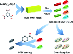
A series of nano-sized lanthanide metal–organic frameworks (Ln-MOFs) which are isostructural with parent MOF-76(Ln) (Ln = Eu, Tb, Sm, Dy) are synthesized under solvothermal conditions. The morphology and particle size of the Ln-MOFs demonstrated by scanning electron microscopy can be changed by increasing the quantities of sodium acetate whose capping effect provides more coordinate sites for lanthanide ions. Furthermore, we explore the adsorption properties of nanosized MOF-76(Ln) for organic dyes, indicating that nanosized MOF-76(Ln) can adsorb some cationic dyes like methylene blue, etc. In addition, MOF-76(Eu) and MOF-76(Tb) are selected for fluorescence sensing for monoaromatic hydrocarbons (BTEX). Such system serves as an acetophenone detection platform through a fluorescence quench process. This work highlights the practical application of MOF-76(Ln) as sensors, and it paves the way for detecting BTEX in solution or in vapor phase.


Introduction

Metal–organic frameworks (MOFs), also called porous coordination polymers (PCPs), have been rapidly growing as a new type of multifunctional materials in the last two decades.1 Such materials can directly self-assemble from one or several kinds of metal ions and organic linkers.2 The tunable nature of MOFs through rational design endows new materials various applications, such as gas separation and storage,3 luminescence,4 heterogeneous catalysis,5 chemical sensing,6 proton conducting,7 drug delivery and biomedical imaging, etc.8 Perhaps one of the most widely studied properties of MOF materials is the existence of porosity. The porous nature of MOFs promotes favorable host–molecule interactions because of their high surface areas and tunable pore sizes. Therefore, some MOFs can be used for the adsorption of pollutants in the atmosphere and aqueous solutions.9

Compared with transition metal–organic frameworks, lanthanide MOFs (Ln-MOFs) hold outstanding photoluminescent properties due to the unique electronic transition arising from 4f electrons of lanthanide ions.10 In fact, a variety of luminescent Ln-MOFs and organic–inorganic hybrid materials have been realized for their diverse applications on chemical sensing,6c,7,11 light-emitting devices,12 biological imaging13 and biomedicine, etc.8b,14

Organic dyes and monoaromatic hydrocarbons (BTEX), are two kinds of significant pollutants in our environment.15 Organic dye pollutant have become one of the main sources of pollution in the world. This problem has caused great harm to the environment and drinking water.16 At presence, the most effective way to solve this problem is to adsorb dye molecules by porous materials. Benzene, toluene, ethylbenzene, acetophenone and mixture of xylenes (BTEX) are monoaromatic hydrocarbon compounds. BTEX is an important pollutant from anthropogenic activities and BTEX in the urban atmosphere has the serious influence to the region and local distance. Water contamination caused by monoaromatic compounds is a very serious problem as these compounds are toxic and often be classified as carcinogens for humans.15 So the accurate prediction and detection of these pollutants are necessary.17

Herein, a series of lanthanide MOFs are synthesized via solvothermal method and used for dye adsorption and luminescence sensing (Fig. 1).18 We find that both the shape and size of MOF-76(Ln) have been changed from pillar-like rods to broad bean with the addition of sodium acetate. The nanosized MOF-76(Ln) with a large specific surface area and porosity is used for dye adsorption experiment. Furthermore, MOF-76(Ln) has excellent photoluminescent properties. Especially for the MOF-76(Eu) and MOF-76(Tb) reveal sharp and strong emission in the visible light region.19 We find that MOF-76(Eu) and MOF-76(Tb) exhibits high selectivity and sensitivity of their luminescent intensity for benzene homologues solutions and BTEX vapors. In addition, the relevant mechanism of these phenomena also has received the preliminary attention and investigation.


image file: c5ra23681a-f1.tif
Fig. 1 Schematic diagram of dye adsorption and sensing process of MOF-76(Ln).

Experimental section

Reagents and chemicals

All chemicals and solvents were commercially available and analytical grade. Lanthanide nitrates Ln(NO3)3·6H2O (Ln = Eu, Tb, Sm, Dy, Nd, Yb) were prepared by dissolving their respective oxides in concentrated nitric acid with heating and stirring to promote the reaction. All the other reagents (1,3,5-benzenetricarboxylic acid (H3BTC), N,N-dimethylformamide (DMF), sodium acetate, benzene, toluene, ethylbenzene, o-xylene, m-xylene, p-xylene, Cl-benzene, aniline, anisole, benzonitrile, m-cresol and acetophenone) were purchased from Aldrich and used without further purification.

Physical characterization

The powder X-ray powder diffraction patterns (PXRD) were collected using a Bruker Focus D8 at 40 kV, 40 mA for Cu-Kα with a scan speed of 0.10 s per step and a step size of 0.02°; the data were collected within 2θ range of 5–40°. Scanning electron microscopy (SEM) and energy dispersive analysis by X-rays (EDX) were performed on a Hitachi S-4800 field emission scanning electron microscope operating at 15 kV. Thermogravimetric analysis (TGA) was carried out at a heating rate of 5 K min−1 under nitrogen protection by simultaneous thermal analysis STA409PC. Fourier transform infrared spectra (FT-IR) were recorded with KBr slices from 4000–400 cm−1 using a Nexus 912 AO446 infrared spectrophotometer. Nitrogen adsorption–desorption isotherms were measured at liquid nitrogen temperature using a TriStar 3000 analyzer. Samples were out gassed for 3 h at 473 K before measurements. Surface areas were calculated by the Brunauer–Emmett–Teller (BET) method. UV-vis absorption spectra were carried on Agilent 8453 spectrometer and the ultraviolet diffuse-reflectance spectra of the powdered samples were recorded by a B&W Tek BWS003 spectrophotometer.

Luminescent measurements

Luminescence spectroscopy and lifetime measurements were carried out on an Edinburgh Analytical Instrument FLS920 using a microsecond pulse lamp as excitation source. The quantitative value of lifetime was calculated by linear fitting. The outer luminescent quantum efficiency was determined using an integrating sphere (150 mm diameter, BaSO4 coating) from Edinburgh FLS920 phosphorimeter.

Synthesis of MOF-76(Ln) (1)

MOF-76 was synthesized according to Duan et al. work.18 In a typical synthesis process, Ln(NO3)3·6H2O (0.56 mmol) and H3BTC (0.042 g, 0.2 mmol) were dissolved in a mixture solution which contained DMF (16 mL) and H2O (8 mL) at room temperature. The mixture was stirred further in a polytetrafluoroethylene (PTFE)-lined steel autoclave, and was subsequently heated to 353 K for 24 h. The white flocculent product was collected by centrifugation, washed several times with ethanol and dried in air before characterization.

Synthesis of nanosized MOF-76(Ln) (2 and 3)

A mixture of Ln(NO3)3·6H2O (0.56 mmol), H3BTC (0.042 g, 0.2 mmol) and CH3COONa (0.033 g, 0.4 mmol) was dissolved in DMF (16 mL) and H2O (8 mL) at room temperature. The mixture was heated further in a polytetrafluoroethylene (PTFE)-lined steel autoclave for the hydrothermal reaction for 24 h at 353 K. The solid products 2 were collected via centrifugation. Finally, the products were washed with methanol for several times, and then dried in air. Smaller products 3 were obtained by a homologous synthesis process, but sodium acetate should be added into 0.8 mmol.

Dye adsorption experiments

Freshly prepared compound 3 (50 mg) was transferred to the ethanol solutions of methyl orange (MO), methylene blue (MB), rhodamine B (RhB), crystal violet (CV), semixylenol orange (SO), and basic red 2 (BR-2) (5 mL, 20 mg L−1), respectively. After two days of impregnation, the materials were washed with ethanol and dried in a vacuum.

Luminescence-sensing experiments

MOF-76(Eu) and MOF-76(Tb) were detected for the sensing of aromatic compounds in solutions. MOF-76(Eu) and MOF-76(Tb) powders (6.0 mg) were simply immersed in DMF, benzene, toluene, ethylbenzene, o-xylene, m-xylene, p-xylene, Cl-benzene, anisole, benzonitrile, m-cresol, ethyl benzoate, diphenyl ether and acetophenone, respectively. The mixtures were then sonicated for 3 min before luminescent measurements.

Powder samples of MOF-76(Tb) were used for sensing BTEX volatiles. For each experiment, 100 mg of MOF-76(Tb) was dispersed in 2 mL ethanol, and then the mixture was smeared on a glass and dried in air. The glass was put in a small bottle which contains about 3 mL of solvent and exposed to various solvent vapors. This setup was placed in an airtight environment at 298 K for 1 h before luminescent measurements.

Results and discussion

Physical characterization of 1 and nano sized materials 2 and 3

A series of MOF-76(Ln) (Ln = Eu, Tb, Sm, Dy, Yb and Nd) are prepared, whose structure and coordination environment of Ln3+ are shown in the Scheme S1. According to the reported literature,18,20 in the structures of MOF-76(Ln), the central Ln3+ ions are coordinated by six oxygen atoms from the carboxylate groups of H3BTC ligands and further formation of a tetragonal porous framework of P4322 space group. It has one-dimensional channels of about 6.6 Å × 6.6 Å along the [001] direction.

The identical powder X-ray diffraction (PXRD) patterns (Fig. 2 and S1) of the as-synthesized MOF-76(Eu, Tb, Sm, Dy) materials demonstrate they are isostructural, even though different lanthanide ions are employed in the synthesis process. The morphology of the as-synthesized materials is characterized by SEM. As illustrated in Fig. 3a and b, the crystals of the MOF-76(Ln) (Fig. 3a and b) are uniform and exist as pillar-like rods. The widths and the lengths of materials are 7.85 μm and 120 μm, respectively. The composition of MOF-76(Tb) is subsequently analysed by energy dispersive analysis by X-rays (EDX) spectroscopy (Fig. S2).


image file: c5ra23681a-f2.tif
Fig. 2 PXRD patterns of MOF-76(Ln) with different dimensions.

image file: c5ra23681a-f3.tif
Fig. 3 SEM images of the bulk MOF-76(Ln) 1 with pillar-like rod shapes (a and b), size reduced MOF-76(Ln) 2 (c and d) and nanosized MOF-76(Ln) 3 with broad bean shaped (e and f).

To obtain nanosized MOF-76(Ln), the synthesis of them are performed under the same conditions except for adding sodium acetate as capping reagents. The method we used to reduce the size of the MOF-76(Ln) is adding capping reagents with the same chemical functionality as the H3BTC. Typically, sodium acetate is used as a capping reagent to control the size and morphology of the resulting crystals. The PXRD patterns of the nanosized MOF-76(Ln) 2 and 3 are identical with the normal sample 1 (Fig. 2), which indicate that the phase purity of nano materials has not changed. The morphologies of the nanosized materials 2 and 3 are shown in Fig. 3c–f. The nanosized material 2 is also cylindrical crystals, but its dimension (width and length) has been reduced to about 500 nm and 1950 nm, respectively (Fig. 3c and d). A smaller sized material 3 is obtained when the amount of sodium acetate attains to 0.8 mmol. The material 3 is a broad bean shaped crystals (crystal) with a width of about 360 nm and a length of about 725 nm (Fig. 3e and f). It is clearly that with the addition of sodium acetate, both the shape and size of MOF-76(Ln) have been changed uncommonly. The mechanism for reducing the size of MOF-76(Ln) is considered as the role of sodium acetate. The modulating effect of the sodium acetate on the coordinating interactions between the lanthanide ions and organic linkers will have an influence on the react process. In the initial stage of the synthesis reaction, lanthanide cations are coordinated with carboxylic groups, which are not only from the organic linker 1,3,5-benzenetricarboxylic acid but also from the added reagent sodium acetate. Therefore, the crystal growth is handicapped in an early stage and has been allowed to form more nuclei. In a word, the competitive coordination between the linkers and sodium acetate regulate the rates of nuclei formation and crystal growth.21 In our experiments, for example, when sodium acetate participates in the reaction, the formation of crystal nuclei of MOF-76(Ln) is accelerated and the rate of crystal growth is slowed down, thus leading to the changes of the materials' morphology.

The thermogravimetric analysis (TGA) curve is illustrated in Fig. S4. On the whole, there are four thermal weight loss stages shown in the curve. The first two states of weightlessness represent the evaporation of solvents locating in the channel of MOF-76(Ln). The inflection points at 93.2 °C and 177.4 °C are corresponded with the boiling points of solvents, water and DMF. The third weight loss (the inflection point is 315.5 °C) is attributed to the preliminary thermal decomposition of the ligands. The sum of lost weight owing to these three stages is 19.9 wt%, and the largest weight loss appears in last stage. From 471.2 to 532.1 °C, the thermogravimetric rate has changed from 21.1 wt% to 54.0 wt%. This abrupt change is the result of the collapse of framework under high temperature. The thermogravimetric analysis result demonstrates that MOF-76(Ln) possesses excellent thermal stability for further research and application.

The Fourier transform infrared spectra (FT-IR) exhibit the information of the chemical bonds of MOF-76(Ln). As shown in Fig. S5, a broad band at about 3400 cm−1 is ascribed to the stretching vibration of the O–H bond. These remaining bonds are not coordinated with lanthanide ions in the synthesis process. The stretching vibrations of C[double bond, length as m-dash]O and C–O bonds bring about two sharp peaks that located at 1614 cm−1 and 1383 cm−1, respectively. As a result of the vibration of the chemical bonds on benzene ring, two groups of multiple peaks have appeared in 1400–1650 cm−1 frequency region and fingerprint region. The former group of multiple peaks, which is corresponded to the stretching vibrations of the C[double bond, length as m-dash]C bonds on the benzene ring, is situated at 1574 cm−1, 1535 cm−1 and 1441 cm−1, respectively. And the fingerprint peaks (771 cm−1, 706 cm−1 and 567 cm−1) are owing to the deformation vibrations of the C–H bonds of the benzene ring.

The dye absorption properties

Fig. 4 provides the N2 adsorption–desorption isotherms of materials 1, 2 and 3. These isotherms display typical type IV curves with H1-type hysteresis loops at high relative pressure, which are commonly seen in microporous materials. The atypical hysteresis of these N2 adsorption isotherms may be due to the steric hindrance of the free non-coordinated carboxyl groups in the channels, which reduces the access of N2 molecules. The nanosized MOF-76(Ln) exhibits similar behavior with 1 in N2 adsorption–desorption experiment. Furthermore, nanosized materials show larger specific surface areas than that of the ordinary sample. The BET surface areas of 1, 2 and 3 are 262 m2 g−1, 375 m2 g−1 and 391 m2 g−1, respectively, and show a gradual increase with decrease of materials' particle size.
image file: c5ra23681a-f4.tif
Fig. 4 The N2 adsorption–desorption isotherms of materials 1, 2 and 3.

Inspired by the adsorption of these as-synthetic materials, we have tried to evaluate the adsorption ability towards different dyes.22 The material 3 is immersed in various solutions of methyl orange (MO), methylene blue (MB), rhodamine B (RhB), crystal violet (CV), semixylenol orange (SO), and basic red 2 (BR-2), respectively. Subsequently, these suspensions are stirred at room temperature for about 48 h. The result indicates that materials 3 represent different adsorption behaviors and this difference can be found just by naked eye (Fig. 5). Samples which soaked in the MB, RhB, CV and BR-2 are dyed blue, red, violet and pink, respectively. But the other two samples soaked in the MO and the SO are remained colorless. That means the cationic dyes, such as MB, RhB, CV and BR-2, can be adsorbed over a period of time with the colorless crystals gradually becoming colored. However, the anionic dyes like MO and SO could not be adsorbed. We speculate that this selective adsorption of different dyes is due to the interaction of the dye molecules and framework of 3.23 Some carboxyl groups, which are coordinated to lanthanide ions, are present in framework in the form of carboxylic anion. The electrostatic interaction makes dye molecules can combine with the framework. As is shown in Fig. S6, the successful adsorption of cationic dyes does not influence the crystalline structure integrity of 3 after a dye adsorption experiment. This result indicates that MOF-76(Ln) can be developed for a potential adsorbent to remove the cationic dyes in the aquatic environment.


image file: c5ra23681a-f5.tif
Fig. 5 The schematic diagram and photographs of dye adsorption experiment. The original MOF-76(Ln) powders were immersed in different dye solutions include methyl orange (MO), methylene blue (MB), rhodamine B (RhB), crystal violet (CV), semixylenol orange (SO), and basic red 2 (BR-2) for 48 h, respectively.

Photoluminescence properties of MOF-76(Ln)

The excitation and emission spectra of MOF-76(Ln) are displayed in Fig. 6. The luminescence of MOF-76(Ln) is mainly attributed to the excited energy levels of organic ligand with a subsequent energy transfer to the resonant excited levels of lanthanide ions, which then exhibits a sharp and strong light emission. In the luminescence experiment, the emission spectra of MOF-76(Ln) obtained using an appropriate wavelength display their characteristic emission peaks. The emission spectra of MOF-76(Eu) in Fig. 6a show that all the transitions of Eu3+ from the emitting 5D0 level to the ground-state manifold (590 nm, 5D07F1; 616 nm, 5D07F2; 652 nm, 5D07F3; and 701 nm, 5D07F4). It is obviously observed that a very intense 5D07F2 transition at 616 nm is the dominant peak in the emission spectra. The 5D07F2 transition is a typical electric dipole transition and is very sensitive to the local symmetry of europium ions. In the luminescence spectrum of MOF-76(Tb) (Fig. 6b), the typical narrow-band emissions of Tb3+ appear at 492, 545, 584 and 622 nm, corresponding to the 5D47FJ (J = 6–3) transitions. For MOF-76(Sm), the characteristic emission peaks of Sm3+ ions (563, 598, 644 and 707 nm, Fig. S7) are originated from their lowest-emitting states of the 4G5/2 to 6H5/2, 6H7/2, 6H9/2, 6H11/2 levels. Upon excited at 296 nm (Fig. S8), MOF-76(Dy) exhibits emission lines of Dy3+ at 484 and 576 nm, which are assigned to 4F9/26F15/2 and 4F9/26F13/2 transitions. Furthermore, it is clearly shown that all the excitation spectra (the black line) are dominated by broad absorption bands located in the ultraviolet region centered at about 296 nm, suggesting that the resulting materials can absorb the ultraviolet light efficiently and then sensitize the emission of lanthanides by energy transfer. The CIE (Commission International de L'Eclairage) chromaticity diagram of these materials is illustrated in Fig. S9.
image file: c5ra23681a-f6.tif
Fig. 6 Photoluminescent spectra of (a) MOF-76(Eu) and (b) MOF-76(Tb).

For further investigation of the luminescence properties, the luminescence lifetime decay curves and outer luminescent quantum efficiency of MOF-76(Ln) are measured, and the resulting luminescent data are given in Table S1. MOF-76(Eu) and MOF-76(Tb) show much higher quantum efficiencies than MOF-76(Sm) and MOF-76(Dy). This is probably due to the fact that the energy match between H3BTC and Eu3+, Tb3+ is more effective than that between H3BTC and Sm3+, Dy3+.

Sensing for BTEX molecules

To examine the potential of the MOF-76(Eu) for the sensing of BTEX molecules, it is immersed in different organic solvents (DMF, benzene, toluene, ethylbenzene, o-xylene, m-xylene, p-xylene, Cl-benzene, anisole, benzonitrile, m-cresol, ethyl benzoate, diphenyl ether and acetophenone) for luminescence studies. The mixtures are then treated with ultrasonic for 5 min followed by photoluminescence measurement. As shown in Fig. 7 and S10, the intensities of MOF-76(Eu) at 614 nm are strongly influenced by the solvent, especially in the case of acetophenone, which exhibits the conspicuous significant quenching effects. The different effects on the emission between acetophenone and other solvents are clearly observed, indicating that MOF-76(Eu) can be considered as a promising luminescent probe for acetophenone. Such sensing properties of MOF-76(Eu) for acetophenone are of interest, and have been examined in more detail.
image file: c5ra23681a-f7.tif
Fig. 7 The 5D07F2 transition intensity ratios from the emission spectra of MOF-76(Eu) introduced into various pure BTEX solvents when excited at 297 nm.

MOF-76(Eu) powder is immersed in DMF as the standard suspension, and the acetophenone content is successively increased to detect the luminescent response. Fig. 8 shows the emission spectra of MOF-76(Eu) dispersed in acetophenone–DMF mixed solvent with different volume ratio (Vacetophenone/VDMF). The luminescence intensity of MOF-76(Eu) suspension decreases progressively with the increase of the proportion of acetophenone, and almost disappears when acetophenone content is 5.0 vol%.


image file: c5ra23681a-f8.tif
Fig. 8 The emission spectra of MOF-76(Eu) dispersed in different content acetophenone–DMF mixed solvent.

Analogous materials MOF-76(Tb) are also used for sensing benzene homologous molecules. The initial luminescent intensity of MOF-76(Tb) is much stronger than that of MOF-76(Eu). As shown in the Fig. 9 and S11, MOF-76(Tb) exhibit similar behavior with MOF-76(Eu), the luminescence intensity is responding to acetophenone and reduces rapidly with the content of acetophenone increasing. However, it is notable that the fluorescence of MOF-76(Tb) is disappeared just as an acetophenone content of 2.0 vol% (Fig. 9c). We fit the linear relationship between the fluorescence intensity (545 nm) of MOF-76(Tb) vs. the content of acetophenone. The detecting threshold of acetophenone is also calculated according to the fitting curve (Fig. S12). The description of the linear equation is Y = 6941(−ln[thin space (1/6-em)]ω) + 27[thin space (1/6-em)]054 (R = 0.98267, ω is the volume content of acetophenone), and the limit of detection (LOD) can be calculated from the equation which describes as LOD = 3Sb/S. S is the slope of the fitting calibration curve; Sb is the standard deviation for replicating detections of blank solutions.24 According to this method, the value of LOD is equal to 0.02 vol%.


image file: c5ra23681a-f9.tif
Fig. 9 (a) Photograph of the various mixtures consisting of MOF-76(Tb) and different BTEX solvents; (b) the 5D47F5 transition intensity ratios from the emission spectra of MOF-76(Tb) introduced into various pure BTEX solvents when excited at 296 nm; (c) emission spectra of MOF-76(Tb) dispersed in different content acetophenone–DMF mixed solvent.

In order to investigate the effect of MOF-76(Tb) sensor for detecting BTEX volatiles, the luminescence response of MOF-76(Tb) powders upon exposure to vapors of various BTEX for 1 h is measured, and the results are shown in Fig. S13. We can observe a remarkable attenuation in luminescence intensity for the vapor of acetophenone. This indicates that our nanosized MOF-76(Tb) shows a high capacity to discriminate acetophenone molecule among other BTEX volatiles. However, the selectivity of BTEX vapors sensing experiment is much poorer than which for sensing BTEX solvents. For this experiment, in fact, the strength of the quenching effect is influenced by the evaporation rates of various solvents. For this reason, the weak volatility of acetophenone is a limiting factor for high selectivity.

The mechanism of this quenching phenomenon is also investigated.25 PXRD is employed to study the structural data of the original and treated materials. The powder XRD patterns of the solvents-MOF-76(Tb) are similar to that of the original one, suggesting that the basic frameworks remain in the materials (Fig. S14). Subsequently the luminescence lifetimes of BTEX-MOF-76(Tb) suspensions are measured (Table S2). The results reveal that lifetime remained almost unchanged in various BTEX solvents, which suggests that there are no interactions between BTEX molecules and the Tb3+ ions. In order to further elucidate the detection mechanism of the sensors, the UV-vis absorption spectra of these solvents as well as ultraviolet diffuse reflectance spectra of 1,3,5-benzenetricarboxylic acid are measured. The strong absorption band in Fig. S15 indicates that 1,3,5-benzenetricarboxylic acid ligands absorb the ultraviolet light centered at 362 nm. In the solvents we used, there are no significant adsorption bands in this region except acetophenone. As shown in Fig. S16, the strong absorption band of acetophenone is located from 250 to 400 nm, which is largely overlapped by the absorption band of 1,3,5-benzenetricarboxylic acid. Therefore, the quenching behavior for acetophenone is related to the interactions between acetophenone and the ligand H3BTC.

According to the absorption spectra and luminescence spectra, the interaction among ligand and acetophenone is a major reason in this luminescence quenching effect of benzene homologous molecules. Upon excitation, there is a competition between the absorption of acetophenone and the excitation of MOF-76(Ln), resulting in a decrease in PL intensity, even quenching. This quenching mechanism is consistent with some previous literature.26

Conclusion

In summary, a series of lanthanide metal–organic frameworks MOF-76(Ln) are synthesized via conventional solvothermal method. It is found that both the shape and size of MOF-76(Ln) change from pillar-like rods to broad bean with the addition of sodium acetate. We infer the additive sodium acetate can increase the coordinate sites for lanthanide ions so as to accelerate the formation of crystal nucleus of MOF-76(Ln) and slow down the rate of crystal growth. The nanosized MOF-76(Ln) with a large specific surface area and porosity can adsorb some cationic dyes such as MB, RhB, CV and BR-2. Furthermore, MOF-76(Eu) and MOF-76(Tb) with the strong luminescence intensity are selected for sensing BTEX molecules and the latter exhibits high selectivity and sensitivity of their luminescent intensity for benzene homologues, even in BTEX vapors. The results indicate that MOF-76(Ln) materials have the potential for fluorescent sensor to detect benzene homologues as a kind of important organic pollutants and toxicant to our environment.

Acknowledgements

This work is supported by the National Natural Science Foundation of China (21571142), Developing Science Funds of Tongji University and Science & Technology Commission of Shanghai Municipality (14DZ2261100).

Notes and references

  1. (a) H. Furukawa, K. E. Cordova, M. O'Keeffe and O. M. Yaghi, Science, 2013, 341, 1230444 CrossRef PubMed; (b) N. Stock and S. Biswas, Chem. Rev., 2012, 112, 933 CrossRef CAS PubMed; (c) S. Li and F. Huo, Nanoscale, 2015, 7, 7482 RSC.
  2. (a) F.-X. Coudert and A. H. Fuchs, Coord. Chem. Rev., 2015, 307, 211 CrossRef; (b) T. D. Bennett and A. K. Cheetham, Acc. Chem. Res., 2014, 47, 1555 CrossRef CAS PubMed.
  3. (a) S. Furukawa, J. Reboul, S. Diring, K. Sumida and S. Kitagawa, Chem. Soc. Rev., 2014, 43, 5700 RSC; (b) A. U. Czaja, N. Trukhan and U. Muller, Chem. Soc. Rev., 2009, 38, 1284 RSC; (c) B. Li, H.-M. Wen, H. Wang, H. Wu, T. Yildirim, W. Zhou and B. Chen, Energy Environ. Sci., 2015, 8, 2504 RSC; (d) V. Finsy, L. Ma, L. Alaerts, D. E. De Vos, G. V. Baron and J. F. M. Denayer, Microporous Mesoporous Mater., 2009, 120, 221 CrossRef CAS; (e) D. Britt, H. Furukawa, B. Wang, T. G. Glover and O. M. Yaghi, Proc. Natl. Acad. Sci. U. S. A., 2009, 106, 20637 CrossRef CAS PubMed.
  4. M. D. Allendorf, C. A. Bauer, R. K. Bhakta and R. J. Houk, Chem. Soc. Rev., 2009, 38, 1330 RSC.
  5. (a) R. Li, J. Hu, M. Deng, H. Wang, X. Wang, Y. Hu, H. L. Jiang, J. Jiang, Q. Zhang, Y. Xie and Y. Xiong, Adv. Mater., 2014, 26, 4783 CrossRef CAS PubMed; (b) J. Gascon, U. Aktay, M. Hernandezalonso, G. Vanklink and F. Kapteijn, J. Catal., 2009, 261, 75 CrossRef CAS.
  6. (a) B. Chen, Y. Yang, F. Zapata, G. Lin, G. Qian and E. B. Lobkovsky, Adv. Mater., 2007, 19, 1693 CrossRef CAS; (b) L. E. Kreno, K. Leong, O. K. Farha, M. Allendorf, R. P. Van Duyne and J. T. Hupp, Chem. Rev., 2012, 112, 1105 CrossRef CAS PubMed; (c) Z. Hu, B. J. Deibert and J. Li, Chem. Soc. Rev., 2014, 43, 5815 RSC; (d) J. F. Callan, A. P. de Silva and D. C. Magri, Tetrahedron, 2005, 61, 8551 CrossRef CAS.
  7. L. V. Meyer, F. Schonfeld and K. Muller-Buschbaum, Chem. Commun., 2014, 50, 8093 RSC.
  8. (a) D. Cunha, M. Ben Yahia, S. Hall, S. R. Miller, H. Chevreau, E. Elkaïm, G. Maurin, P. Horcajada and C. Serre, Chem. Mater., 2013, 25, 2767 CrossRef CAS; (b) K. Deng, Z. Hou, X. Li, C. Li, Y. Zhang, X. Deng, Z. Cheng and J. Lin, Sci. Rep., 2015, 5, 7851 CrossRef CAS PubMed; (c) J. L. Vivero-Escoto, R. C. Huxford-Phillips and W. Lin, Chem. Soc. Rev., 2012, 41, 2673 RSC.
  9. (a) K.-Y. A. Lin, S.-Y. Chen and A. P. Jochems, Mater. Chem. Phys., 2015, 160, 168 CrossRef CAS; (b) M. T. Luebbers, T. Wu, L. Shen and R. I. Masel, Langmuir, 2010, 26, 11319 CrossRef CAS PubMed.
  10. (a) J. Rocha, L. D. Carlos, F. A. Paz and D. Ananias, Chem. Soc. Rev., 2011, 40, 926 RSC; (b) Y. Cui, Y. Yue, G. Qian and B. Chen, Chem. Rev., 2012, 112, 1126 CrossRef CAS PubMed; (c) Y. Cui, B. Chen and G. Qian, Coord. Chem. Rev., 2014, 273–274, 76 CrossRef CAS; (d) J. Feng and H. J. Zhang, Chem. Soc. Rev., 2013, 42, 387 RSC; (e) B. Yan, RSC Adv., 2012, 2, 9304 RSC.
  11. (a) J.-N. Hao and B. Yan, J. Mater. Chem. C, 2014, 2, 6758 RSC; (b) Y. Lu and B. Yan, Chem. Commun., 2014, 50, 13323 RSC; (c) Y. L. Hou, H. Xu, R. R. Cheng and B. Zhao, Chem. Commun., 2015, 51, 6769 RSC.
  12. (a) Y. Lu and B. Yan, Chem. Commun., 2014, 50, 15443 RSC; (b) Y. Zhou and B. Yan, Inorg. Chem., 2014, 53, 3456 CrossRef CAS PubMed.
  13. (a) X. Q. Ge, L. N. Sun, S. Dang, J. L. Liu, Y. X. Xu, Z. W. Wei, L. Y. Shi and H. J. Zhang, Microchim. Acta, 2015, 182, 1653 CrossRef CAS; (b) Y. Liu, L. Sun, J. Liu, Y. X. Peng, X. Ge, L. Shi and W. Huang, Dalton Trans., 2015, 44, 237 RSC.
  14. J. N. Hao and B. Yan, Chem. Commun., 2015, 51, 14509 RSC.
  15. M. Farhadian, D. Duchez, C. Vachelard and C. Larroche, Water Res., 2008, 42, 1325 CrossRef CAS PubMed.
  16. C. I. Pearce, J. R. Lloyd and J. T. Guthrie, Dyes Pigm., 2003, 58, 179 CrossRef CAS.
  17. (a) Y. Takashima, V. M. Martinez, S. Furukawa, M. Kondo, S. Shimomura, H. Uehara, M. Nakahama, K. Sugimoto and S. Kitagawa, Nat. Commun., 2011, 2, 168 CrossRef PubMed; (b) X. G. Liu, H. Wang, B. Chen, Y. Zou, Z. G. Gu, Z. Zhao and L. Shen, Chem. Commun., 2015, 51, 1677 RSC; (c) R. Volkamer, J. L. Jimenez, F. San Martini, K. Dzepina, Q. Zhang, D. Salcedo, L. T. Molina, D. R. Worsnop and M. J. Molina, Geophys. Res. Lett., 2006, 33 Search PubMed.
  18. T.-W. Duan and B. Yan, J. Mater. Chem. C, 2014, 2, 5098 RSC.
  19. (a) K. Liu, H. You, Y. Zheng, G. Jia, Y. Song, Y. Huang, M. Yang, J. Jia, N. Guo and H. Zhang, J. Mater. Chem., 2010, 20, 3272 RSC; (b) W. Yang, Z. Q. Bai, W. Q. Shi, L. Y. Yuan, T. Tian, Z. F. Chai, H. Wang and Z. M. Sun, Chem. Commun., 2013, 49, 10415 RSC.
  20. W. Yang, J. Feng, S. Song and H. Zhang, ChemPhysChem, 2012, 13, 2734 CrossRef CAS PubMed.
  21. H. Guo, Y. Zhu, S. Qiu, J. A. Lercher and H. Zhang, Adv. Mater., 2010, 22, 4190 CrossRef CAS PubMed.
  22. A. Ahmed, M. Forster, J. Jin, P. Myers and H. Zhang, ACS Appl. Mater. Interfaces, 2015, 7, 18054 CAS.
  23. S. N. Zhao, X. Z. Song, M. Zhu, X. Meng, L. L. Wu, J. Feng, S. Y. Song and H. J. Zhang, Chem. –Eur. J., 2015, 21, 9748 CrossRef CAS PubMed.
  24. (a) R. J. Gui, X. Q. An and W. X. Huang, Anal. Chim. Acta, 2013, 767, 134 CrossRef CAS PubMed; (b) G. Y. Gao, W. J. Qu, B. B. Shi, Q. Lin, H. Yao, Y. M. Zhang, J. Chang, Y. Cai and T. B. Wei, Sens. Actuators, B, 2015, 213, 501 CrossRef CAS; (c) L. L. Yang, X. M. Liu, K. Liu, H. Liu, F. Y. Zhao, W. J. Ruan, Y. Li, Z. Chang and X. H. Bu, Talanta, 2014, 128, 278 CrossRef CAS PubMed.
  25. A. Douvali, A. C. Tsipis, S. V. Eliseeva, S. Petoud, G. S. Papaefstathiou, C. D. Malliakas, I. Papadas, G. S. Armatas, I. Margiolaki, M. G. Kanatzidis, T. Lazarides and M. J. Manos, Angew. Chem., Int. Ed., 2015, 54, 1651 CrossRef CAS PubMed.
  26. (a) Y. Xiao, L. Wang, Y. Cui, B. Chen, F. Zapata and G. Qian, J. Alloys Compd., 2009, 484, 601 CrossRef CAS; (b) Z. Hao, X. Song, M. Zhu, X. Meng, S. Zhao, S. Su, W. Yang, S. Song and H. Zhang, J. Mater. Chem. A, 2013, 1, 11043 RSC; (c) X.-Z. Song, S.-Y. Song, S.-N. Zhao, Z.-M. Hao, M. Zhu, X. Meng, L.-L. Wu and H.-J. Zhang, Adv. Funct. Mater., 2014, 24, 4034 CrossRef CAS.

Footnote

Electronic supplementary information (ESI) available. See DOI: 10.1039/c5ra23681a

This journal is © The Royal Society of Chemistry 2016