A depropargylation-triggered spontaneous cyclization based fluorescent “turn-on” chemodosimeter for the detection of palladium ions and its application in live-cell imaging

Yu Chen a, Minmin Zhangb, Yifeng Han*a and Jie Wei*b
aDepartment of Chemistry, The Key Laboratory of Advanced Textile Materials and Manufacturing Technology, Zhejiang Sci-Tech University, Hangzhou, 310018, China. E-mail: zstuchem@gmail.com
bDepartment of Physiology, Medical College of Nanchang University, Bayi Road 461, Nanchang, Jiangxi 330006, China. E-mail: jwei@ncu.edu.cn

Received 9th November 2015 , Accepted 12th January 2016

First published on 15th January 2016


Abstract

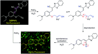
A novel depropargylation-triggered spontaneous cyclization reaction based fluorescent turn-on chemodosimeter for the detection of palladium ions has been rationally designed and developed. Based on the specific reactivity of the palladium promoted hydrolysis reaction, the probe exhibited a high selectivity and sensitivity for palladium ions. Furthermore, the probe was successfully used for fluorescence imaging of Pd2+ in living cells.


Palladium is widely distributed in the environment due to its use in alloys, jewellery, dental crowns, fuel cells, chemical catalysts and especially in automobile catalytic converters.1 Palladium is not biodegradable, and hence can be concentrated through the food chain. Excess palladium accumulation may result in degradation of DNA and cell mitochondria, allergic reactions, and also enzyme inhibition.2 Therefore, the determination of palladium in environmental and biological samples is crucial both to the monitoring of environmental pollution and to the diagnosis of clinical disorders.

Whereas conventional techniques used for quantification of palladium species, such as atomic absorption spectroscopy, inductively coupled plasma atomic emission spectroscopy, and solid-phase microextraction high-performance liquid chromatography, usually suffer from the expensive and sophisticated instrumentation, and/or complicated sample preparation, and are therefore not suitable for real-time and in situ analysis.3 In comparison with those conventional methods for palladium species, fluorescent probe techniques display apparent advantages because of their ease of application in solution as well as their high sensitivity to and selectivity for trace analytes with spatial and temporal resolution.4

Over the past several years, considerable efforts have been made to develop fluorescent probe for palladium ions based on the coordination of Pd2+ to heteroatom-based ligands, Pd2+ catalyzed ring-opening reaction, Pd2+ catalyzed oxidative cyclization reaction, and palladium catalyzed depropargylation and deallylation reaction (Fig. 1).5 However, many of them still have limitations such as interference from other coexisting metal ions, long response time, and need additional reagents. Therefore, for practical applications, it is still strongly desirable to develop novel fluorescent probes with high sensitivity, and quick response for real-time detection of palladium ions.


image file: c5ra23645b-f1.tif
Fig. 1 Some reported palladium probes.

Herein, the aim of this work is to develop a new fluorescent probe with novel response mechanism for palladium ions. As shown in Scheme 1, probe SPd1 was developed with coumarin precursor derivative and a terminal propargyl ether moiety. It is well known that the terminal propargyl ether can be cleaved by palladium-catalyzed hydrolysis reaction to generate the corresponding free hydroxyl group.5d,5m We envisioned that the fluorescent intensity of the SPd1 is greatly reduced due to the effect of intramolecular rotation. However, the deprotection of the propargyl ether group of SPd1 by palladium ions promoted hydrolysis reaction would releases the hydroxy intermediate, which wills readily spontaneous cyclize to form a highly fluorescent coumarin derivative 4 (Scheme 1). To the best of our knowledge, this is the first example of depropargylation-triggered spontaneous cyclization based fluorescent chemodosimeter for the detection of palladium ions. Furthermore, SPd1 can be successfully applied for Pd2+ imaging in living cells.


image file: c5ra23645b-s1.tif
Scheme 1 The “deprotection–cyclization” strategy for the design of SPd1.

As shown in Scheme 2, SPd1 can be readily prepared in two convenient steps under facile conditions with high yield starting with commercially available 4-(diethylamino)-2-hydroxybenzaldehyde. The product (SPd1) was well characterized by 1H, 13C NMR, and HR-MS (ESI).


image file: c5ra23645b-s2.tif
Scheme 2 Synthesis of SPd1: (a) 3-bromopropyne/K2CO3, acetone, reflux, 12 h, 93%; (b) Et3N, EtOH, rt, 12 h, 65%; (c) PdCl2, THF–H2O (1[thin space (1/6-em)]:[thin space (1/6-em)]1), rt, 3 h, 72%.

We firstly assessed the UV-vis spectroscopic properties of SPd1 in PBS buffer solution (10 mM, pH 7.4, containing 50% EtOH). SPd1 (20.0 μM) displayed a moderate UV-vis absorption around 474 nm. Upon incremental addition of Pd2+ (0–10.0 equiv.), the peak at 474 nm slightly decreased, and the absorption at 350 and 555 nm increased instantly with two clear isosbestic points at 452 and 502 nm, respectively, indicating that compound 4 was formed in the present of Pd2+ (Fig. 2). Furthermore, a good linear relationship was observed between the changes in the absorbance at 452 and 520 nm with Pd2+ in the range of 0–10.0 equiv. (Fig. S1, ESI).


image file: c5ra23645b-f2.tif
Fig. 2 Absorption spectra of SPd1 (20.0 μM) in PBS buffer solution (10 mM, pH 7.4, containing 50% EtOH) in the presence of different concentrations of Pd2+ (0–10.0 equiv.).

The emission spectra of SPd1 and its fluorescent titration with Pd2+ were recorded in PBS buffer solution (10 mM, pH 7.4, containing 50% EtOH) (Fig. S2, ESI). As expected, SPd1 alone is almost non-fluorescent (λex = 510 nm, Φ = 0.004, Table S1, ESI) due to the effect of intramolecular rotation (Scheme 1). However, upon progressive addition of Pd2+, the emission band at 542 nm rapidly increased (Fig. 3), which was attributed to the cleavage of propargyl ether group by palladium ions-promoted hydrolysis followed by spontaneous cyclization reaction to form the highly fluorescent coumarin derivative 4 (Scheme 1). Moreover, the fluorescence titration curve revealed that the fluorescence intensity at 542 nm increased linearly with increasing concentration of Pd2+ (R2 = 0.99213) (Fig. S3 and S4, ESI) and further smoothly increased until a maximum was reached up to 100.0 μM Pd2+ (λex = 510 nm, Φ = 0.023, Table S1, ESI). Based on these results, the detection limit of SPd1 for Pd2+ was calculated to be 9.3 × 10⁻⁸ M.6 Owing to the specific reactivity of palladium ions-promoted hydrolysis reaction, SPd1 displayed a high sensitivity toward Pd2+.


image file: c5ra23645b-f3.tif
Fig. 3 Fluorescence spectra of SPd1 (10.0 μM) in PBS buffer solution (10 mM, pH 7.4, containing 50% EtOH) in the presence of different concentrations of Pd2+ (0–150.0 μM) (λex = 510 nm). Inset: cuvette images of probe SPd1 before and after addition of PdCl2 taken under a hand held UV-lamp (λex = 365 nm).

The plausible mechanism of the palladium ions induced fluorescence response is shown in Scheme 1. Efforts were then made to explore the nature of the palladium ions induced response. To this end, a comparison of fluorescent spectra between the SPd1–Pd2+ system and compound 4 was made to confirm the generation of 4 after treatment with Pd2+ (Fig. S5, ESI). The 1H NMR spectra of the isolated product of the SPd1–Pd2+ solution were also measured to support the depropargylation-triggered spontaneous cyclization of SPd1 (see ESI).

Subsequently, the time-dependence of fluorescence was also evaluated in the presence of Pd2+ in PBS buffer solution (10 mM, pH 7.4, containing 50% EtOH) (Fig. 4, ESI). The result shows that the fluorescence of the tested solutions remarkably increased to the maximum value within 70 minutes. Accordingly, the observed rate constant (kobs) for the formation of compound 4 has been calculated to be 2.4 × 10⁻² min−1 (Fig. S6, ESI).7


image file: c5ra23645b-f4.tif
Fig. 4 Time-dependent fluorescence intensity changes of SPd1 (10.0 μM) upon addition of Pd2+ (10.0 equiv.) in PBS buffer solution (10 mM, pH 7.4, containing 50% EtOH) (λex = 510 nm).

Further, the fluorescence titration of SPd1 with various metal ions was conducted to examine the selectivity (Fig. 5, and S7, ESI). Much to our delight, the examined alkali, alkaline-earth metal ions, transition metal ions, and even Hg2+ showed nominal changes to the fluorescence spectra of SPd1. It should be mentioned that SPd1 still responded to palladium ions sensitively even in the presence of other relevant competing ions (Fig. 5, and S8, ESI). Therefore, these results suggest that SPd1 displays high selectivity toward palladium ions in neutral aqueous solution.


image file: c5ra23645b-f5.tif
Fig. 5 Fluorescence responses of SPd1 to various metal ions (including Na+, K+, Ag+, Co2+, Mn2+, Al3+, Cd2+, Cr3+, Cs+, Cu2+, Fe2+, Fe3+, Hg2+, Mg2+, Zn2+, Pb2+, and Pd2+). Black bars represent the addition of 10.0 equiv. of the appropriate metal ions to a 10.0 μM solution of SPd1 (in PBS buffer solution, 10 mM, pH 7.4, containing 50% EtOH). Red bars represent the addition of 10.0 equiv. of Pd2+ to the solutions containing SPd1 (10.0 μM) and the appropriated metal ions (10.0 equiv.) (λex = 510 nm).

Moreover, the palladium ions sensing ability of SPd1 at a wide range of pH values was investigated. As depicted in Fig. S9, ESI, SPd1 alone was inert to pH in the range of 5.5–9.8. On the other hand, it readily reacted with palladium ions within the biologically relevant pH range (6.5–8.5). These results indicate that SPd1 could be used in living cells without interference from pH effects.

Due to the favorable properties of SPd1 in vitro, the potential utility of SPd1 in living cells was studied. HeLa cells were incubated with 10.0 μM of SPd1 for 30 min at 37 °C and exhibited only weak fluorescence (Fig. 6b). The cells were then treated with PdCl2 (30.0 μM) for 30 min at 37 °C, which resulted in a dramatic increase of intracellular green fluorescence (Fig. 6d). These obvious changes indicated that SPd1 was cell membrane permeable and capable of image Pd2+ in living cells.


image file: c5ra23645b-f6.tif
Fig. 6 Fluorescence image of HeLa cells incubated with SPd1 (10.0 μM) for 0.5 h, and then washed quickly with PBS for imaging (b). The cells were then treated with PdCl2 (30.0 μM) for 0.5 h which resulted in a dramatic increase in intracellular green fluorescence (d). (a) and (c) Bright-field images of live cells in (b) and (d).

In conclusion, we have rationally developed a novel and simple depropargylation-triggered spontaneous cyclization based fluorescent chemodosimeter for the detection of palladium ions. The probe displayed a specific fluorescence response towards palladium ions under mild conditions with a low detection limit. Furthermore, fluorescence imaging of Pd2+ in living cells indicated that this probe might be favorable for biological applications.

Acknowledgements

This work was supported by the National Natural Science Foundation of China (20902082), the Project Grants 521 Talents Cultivation of Zhejiang Sci-Tech University, and the Program for Innovative Research Team of Zhejiang Sci-Tech University (13060052-Y).

Notes and references

  1. (a) G. Zeni and R. C. Larock, Chem. Rev., 2004, 104, 2285–2310 CrossRef CAS PubMed; (b) T. W. Lyons and M. S. Sanford, Chem. Rev., 2010, 110, 1147–1169 CrossRef CAS PubMed; (c) K. Ravindra, L. Bencs and R. V. Grieken, Sci. Total Environ., 2004, 318, 1–43 CrossRef CAS PubMed; (d) S. Rauch, H. F. Hemond, C. Barbante, M. Owari, G. M. Morrison, B. Peucker-Ehrenbrink and U. Wass, Environ. Sci. Technol., 2005, 39, 8156–8162 CrossRef CAS PubMed; (e) F. Zereini, C. Wiseman and W. Puttmann, Environ. Sci. Technol., 2007, 41, 451–456 CrossRef CAS PubMed.
  2. (a) A. Hensten-Pettersen, Adv. Dent. Res., 1992, 6, 38–43 CAS; (b) J. C. Wataha and C. T. Hanks, J. Oral Rehabil., 1996, 23, 309–320 CrossRef CAS PubMed; (c) J. Kielhorn, C. Melber, D. Keller and I. Mangelsdorf, Int. J. Hyg. Environ. Health, 2002, 205, 417–432 CrossRef CAS PubMed; (d) International Programme on Chemical Safety, Palladium; Environmental Health Criteria Series 226, World Health Organization, Geneva, 2002 Search PubMed.
  3. (a) B. Dimitrova, K. Benkhedda, E. Ivanova and F. Adams, J. Anal. At. Spectrom., 2004, 19, 1394–1396 RSC; (b) C. Locatelli, D. Melucci and G. Torsi, Anal. Bioanal. Chem., 2005, 382, 1567–1573 CrossRef CAS PubMed; (c) K. Van Meel, A. Smekens, M. Behets, P. Kazandjian and R. Van Grieken, Anal. Chem., 2007, 79, 6383–6389 CrossRef CAS PubMed; (d) L. Duan, Y. Xu and X. Qian, Chem. Commun., 2008, 6339–6341 RSC.
  4. (a) M. Y. Berezin and S. Achilefu, Chem. Rev., 2010, 110, 2641–2684 CrossRef CAS PubMed; (b) K. P. Carter, A. M. Young and A. E. Palmer, Chem. Rev., 2014, 114, 4564–4601 CrossRef CAS PubMed; (c) E. M. Nolan and S. J. Lippard, Chem. Rev., 2008, 108, 3443–3480 CrossRef CAS PubMed; (d) X. Chen, T. Pradhan, F. Wang, J. S. Kim and J. Yoon, Chem. Rev., 2012, 112, 1910–1956 CrossRef CAS PubMed; (e) V. S. Lin, W. Chen, M. Xian and C. J. Chang, Chem. Soc. Rev., 2015, 44, 4596–4618 RSC; (f) H. Zhu, J. Fan, B. Wang and X. Peng, Chem. Soc. Rev., 2015, 44, 4337–4366 RSC; (g) R. T. K. Kwok, C. W. T. Leung, J. W. Y. Lam and B. Z. Tang, Chem. Soc. Rev., 2015, 44, 4228–4238 RSC; (h) J. Yin, Y. Hu and J. Yoon, Chem. Soc. Rev., 2015, 44, 4619–4644 RSC; (i) M. Kaur and D. H. Choi, Chem. Soc. Rev., 2015, 44, 58–77 RSC; (j) Z. Guo, S. Park, J. Yoon and I. Shin, Chem. Soc. Rev., 2014, 43, 16–29 RSC; (k) L. Yuan, W. Lin, K. Zheng, L. He and W. Huang, Chem. Soc. Rev., 2013, 42, 622–661 RSC; (l) J. Fan, M. Hu, P. Zhan and X. Peng, Chem. Soc. Rev., 2013, 42, 29–43 RSC; (m) J. Du, M. Hu, J. Fan and X. Peng, Chem. Soc. Rev., 2012, 41, 4511–4535 RSC; (n) K. Wu, Y. Gao, Z. Yu, F. Yu, J. Jiang, J. Guo and Y. Han, Anal. Methods, 2014, 6, 3560–3563 RSC; (o) X. Li, Y. Gong, K. Wu, S. H. Liang, J. Cao, B. Yang, Y. Hu and Y. Han, RSC Adv., 2014, 4, 36106–36109 RSC; (p) X. Li, C. Yang, K. Wu, Y. Hu, Y. Han and S. H. Liang, Theranostics, 2014, 4, 1233–1238 CrossRef CAS PubMed; (q) J. Liu, K. Wu, S. Li, T. Song, Y. Han and X. Li, Dalton Trans., 2013, 42, 3854–3859 RSC; (r) J. Liu, K. Wu, X. Li, Y. Han and M. Xia, RSC Adv., 2013, 3, 8924–8928 RSC; (s) X. Li, S. Zhang, J. Cao, N. Xie, T. Liu, B. Yang, Q. He and Y. Hu, Chem. Commun., 2013, 49, 8656–8658 RSC.
  5. (a) A. L. Garner and K. Koide, J. Am. Chem. Soc., 2008, 130, 16472–16473 CrossRef CAS PubMed; (b) A. L. Garner, F. Song and K. Koide, J. Am. Chem. Soc., 2009, 131, 5163–5171 CrossRef CAS PubMed; (c) M. E. Jun and K. H. Ahn, Org. Lett., 2010, 12, 2790–2793 CrossRef CAS PubMed; (d) M. Santra, S.-K. Ko, I. Shin and K. H. Ahn, Chem. Commun., 2010, 46, 3964–3966 RSC; (e) H. Li, J. Fan, J. Du, K. Guo, S. Sun, X. Liu and X. Peng, Chem. Commun., 2010, 46, 1079–1081 RSC; (f) S. Goswami, D. Sen, N. K. Das, H.-K. Fun and C. K. Quah, Chem. Commun., 2011, 47, 9101–9103 RSC; (g) J. Jiang, H. Jiang, W. Liu, X. Tang, X. Zhou, W. Liu and R. Liu, Org. Lett., 2011, 13, 4922–4925 CrossRef CAS PubMed; (h) W. Chen, B. D. Wright and Y. Pang, Chem. Commun., 2012, 48, 3824–3826 RSC; (i) H. Li, J. Fan and X. Peng, Chem. Soc. Rev., 2013, 42, 7943–7962 RSC; (j) J. Wang, F. Song, J. Wang and X. Peng, Analyst, 2013, 138, 3667–3672 RSC; (k) M. Kumar, N. Kumar and V. Bhalla, RSC Adv., 2013, 3, 1097–1102 RSC; (l) J.-T. Hou, K. Li, K.-K. Yu, M.-Z. Ao, X. Wang and X.-Q. Yu, Analyst, 2013, 138, 6632–6638 RSC; (m) H. Chen, W. Lin and L. Yuan, Org. Biomol. Chem., 2013, 11, 1938–1941 RSC; (n) R. Balamurugan, C.-C. Chien, K.-M. Wu, Y.-H. Chiu and J.-H. Liu, Analyst, 2013, 138, 1564–1569 RSC; (o) D. Keum, S. Kim and Y. Kim, Chem. Commun., 2014, 50, 1268–1270 RSC; (p) P. Kaur, N. Kaur, M. Kaur, V. Dhuna, J. Singh and K. Singh, RSC Adv., 2014, 4, 16104–16108 RSC; (q) S. Sun, B. Qiao, N. Jiang, J. Wang, S. Zhang and X. Peng, Org. Lett., 2014, 16, 1132–1135 CrossRef CAS PubMed; (r) X. Wang, Z. Guo, S. Zhu, H. Tian and W. Zhu, Chem. Commun., 2014, 50, 13525–13528 RSC; (s) W. Liu, J. Jiang, C. Chen, X. Tang, J. Shi, P. Zhang, K. Zhang, Z. Li, W. Dou, L. Yang and W. Liu, Inorg. Chem., 2014, 53, 12590–12594 CrossRef CAS PubMed; (t) A. K. Mahapatra, S. K. Manna, K. Maiti, S. Mondal, R. Maji, D. Mandal, S. Mandal, M. R. Uddin, S. Goswami, C. K. Quah and H.-K. Fun, Analyst, 2015, 140, 1229–1236 RSC; (u) M. Wang, X. Liu, H. Lu, H. Wang and Z. Qin, ACS Appl. Mater. Interfaces, 2015, 7, 1284–1289 CrossRef CAS PubMed; (v) U. Reddy, G. F. Ali, N. Taye, S. Chattopadhyay and A. Das, Chem. Commun., 2015, 51, 3649–3652 RSC; (w) J. Cui, D.-P. Li, S.-L. Shen, J.-T. Liu and B.-X. Zhao, RSC Adv., 2015, 5, 3875–3880 RSC; (x) L. Zhou, Q. Wang, X.-B. Zhang and W. Tan, Anal. Chem., 2015, 87, 4503–4507 CrossRef CAS PubMed.
  6. (a) C. Yang, Y. Chen, K. Wu, T. Wei, J. Wang, S. Zhang and Y. Han, Anal. Methods, 2015, 7, 3327–3330 RSC; (b) T. Wei, J. Wang, Y. Chen and Y. Han, RSC Adv., 2015, 5, 57141–57146 RSC; (c) B. P. Joshi, C. R. Lohani and K. H. Lee, Org. Biomol. Chem., 2010, 8, 3220–3226 RSC.
  7. J. Jo, H. Y. Lee, W. Liu, A. Olasz, C.-H. Chen and D. Lee, J. Am. Chem. Soc., 2012, 134, 16000–16007 CrossRef CAS PubMed.

Footnotes

Electronic supplementary information (ESI) available: Experimental details, characterization of the compounds, and additional spectroscopic data. See DOI: 10.1039/c5ra23645b
These authors contributed equally.

This journal is © The Royal Society of Chemistry 2016