Carbon nano onions cross the blood brain barrier

Bholanath Pakhiraad, Mitrajit Ghoshb, Afreen Allamc and Sabyasachi Sarkar*a
aNano Science and Synthetic Leaf Laboratory, Centre for Healthcare Science and Technology, Indian Institute of Engineering Science and Technology, Shibpur, Botanic Garden-711103, Howrah, West Bengal, India. E-mail: abya@iitk.ac.in
bInstitute for Stroke and Dementia Research (ISD) and Munich Cluster for System, Neurology (Synergy), University of Munich, Medical Centre, Munich-81377, Germany
cCromoz. Inc. 2 Davis Drive, Research Triangle Park, NC-27709, USA
dDept. of Chemistry, Sister Nibedita Govt. General Degree College for Girls, Hastings House, Alipore, Kolkata 700027, India

Received 8th November 2015 , Accepted 16th March 2016

First published on 17th March 2016


Abstract

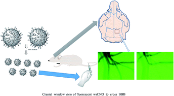
We show the crossing of small sized water soluble fluorescent carbon nano onion (wsCNO) through the blood brain barrier (BBB) in the Cerebral Autosomal-Dominant Arteriopathy with Subcortical Infarcts and Leukoencephalopathy (CADASIL) murine model as well as in glioblastoma multiforme (GBM) induced mice. It is readily excreted from the body after a few days suggesting its possible use as cargo in drug delivery.


The formation of onion like nano carbon along with fullerenes and carbon nanotubes was first recognized by Iijima.1 Ugarte and co-workers further elaborated its formation by electron beam irradiation on multi wall nano carbon and by high heat treatment of pure carbon soot.2–4 Ultra disperse diamond under thermal annealing also showed the transformation into carbon nano onions.2–6 The carbon nano onions so created require exotic conditions not routinely available in any wet laboratory of a chemist. Furthermore these carbon nano onions like other nano carbons or graphite are hydrophobic and cannot be used as a probe under aqueous environment. To explore the utility of carbon nano onions under wet conditions we used a simple approach to pyrolyze cheap carbon sources like wood wool.7–9 Such carbonization is well known and used in the preparation of incandescent carbon filament for light bulb by Swan and Edison10 long ago. Derivatization of these carbon nano onions under oxidative treatment introduce several hydrophilic hydroxyl and carboxylic acid groups which led to surface passivation leading to their photo luminescent properties. These became soluble in water and were used in vivo imaging of the entire life cycle of Drosophila melanogaster.7 We also explored their utility in plant kingdom.8 Based on such usefulness we now explore the utility of water soluble carbon nano onion to probe brain in the light of the existence of blood brain barrier. To attempt such an experiment we checked the size distribution of wsCNO as produced earlier.7 Our intention this time to use smaller sized11,12 wsCNO. Fragmentation to smaller size of nano carbon materials even in solution phase is possible. For this a simple process to fragment the earlier availed wsCNO7 has been developed. Nitric acid treatment of the available wsCNO in water bath to dryness and repeating this acid treatment followed by vacuum drying over solid NaOH yielded the smaller size distribution of wsCNO (Fig. 1). The XRD data of such treated wsCNO (Fig. SI-1) clearly show the retention of its graphite nature even under repetitive nitric acid treatment. Representative FESEM (Field-Emission Scanning Electron Microscope) and HRTEM (High-Resolution Transmission Electron Microscopy) microscopic images of these wsCNO are shown in Fig. 1a and b respectively. However, three times nitric acid treatment resulted a crop of wsCNO and a DLS (Dynamic Light Scattering) study of these in PBS (Phosphate-Buffered Saline) buffer showed the presence of remarkably smaller fractions. Roughly 35% of the total particles belong to a size under 1 nm and the rest 65% belong to a size around 15 nm (Fig. 1c). The corresponding zeta potential show two peaks at −9.18 mV and −28.6 mV respectively (Fig. SI-7). This relative ratio is similar to what we observed in DLS distribution (Fig. 1c). The smaller fraction with less negative zeta potential value may be presumed to be almost neutral and the larger particles are distinctly in the negative domain and so these may not adversely affect the biological system as normally encountered with the particles with positive zeta potential values. Thus a method has been developed to create the size of wsCNO as per the choice using oxidative treatment of nitric acid to cut the larger wsCNO into smaller in sizes. We are investigating the size dependence of wsCNO per nitric acid treatment to optimize the procedure to make tailor made size of the wsCNO.
image file: c5ra23534k-f1.tif
Fig. 1 Fragmentation of wsCNO by nitric acid: (a) FESEM and (b) HRTEM (c) DLS analysis in PBS buffer after 3rd time nitric acid treatment showing two different size distribution, one below 1 nm (∼35%) and the other around 15 nm (∼65%).

The perivascular cells in a brain play a crucial role in the functionality of the selective permeable space between the blood circulatory system and central nervous system. This space, known as the blood brain barrier (BBB)13,14 which is composed of endothelial cells, pericytes and astrocytes. The BBB protects the functionality of the brain and central nervous system (CNS). Pericyte cells create the BBB with tight junctions to protect vesicle trafficking through the endothelial cells and inhibit the effects of CNS immune cells. In addition, pericytes as contractile cells also contribute to controlling the flow within blood vessels as well as between blood vessels and the brain.

CADASIL is a major contributor of vascular dementia in humans. Primarily, it is known to be caused by Notch3 mutation.15–17 Recently, ultra-structural changes in pericytes have been shown in CADASIL.18,19 It was also documented recently that pericytes initiates pathogenesis in murine model of CADASIL and that decrease in pericyte coverage leads to BBB opening in CADASIL mice.20 Therefore, we assessed BBB integrity in a murine model of CADASIL (R169C; Tg88by over expression of mutated transgene)15–17 by the passage of smaller sized water soluble carbon nano onion (wsCNO) exploiting its role as nano-platform for imaging. The easy permeability of such wsCNO in the blood serum albumin shows its amphiphilic behavior. We were interested to know the fate of these wsCNO in brain.

We report herein that these wsCNO smoothly cross not only through the BBB into the brain of CADASIL mice but also through the GBM induced mice.

Using tail vain injection of wsCNO we imaged the brain (see Experimental) in vivo with the progress of time (Fig. 2). Images were taken by constant monitoring for at least 30 minutes after injection of wsCNO after taking few baseline images (see movie clipping, ESI). The gradual enhancement in fluorescence in the vessels and also in the background clearly demonstrates the passage of wsCNO, whereas there is no such enhancement in fluorescence in control animals as shown in Fig. 2a–c.


image file: c5ra23534k-f2.tif
Fig. 2 In vivo image of brain of control mouse: (a) first few second, (b) 14th second and (c) after 35th second and mouse injected with wsCNO: (d) first few second, (e) 14th second and (f) 35th second (d–f are frozen shots from the movie file showing the passage of fluorescent wsCNO) (see ESI, movie file).

The mice were then sacrificed and their brain slice were imaged by fluorescence microscopy using two colour channels to demonstrate the presence of wsCNO (fluorescent in red) when the brain vessels are labelled with lectin by intravenous injection (fluorescent in green) to demarcate the fluorescence from wsCNO in brain vessels of cortex as shown in Fig. 3a. Interestingly the wsCNO injected mice when subjected to the wait period of three days and then sacrificed to image the brain slice; the clearance of the red fluorescence (due to wsCNO) is observed (Fig. 3b). To extend this observation we now used GBM induced mice to check if the passage of wsCNO to the brain tumour could be made with the possible reaching of these to neurons.


image file: c5ra23534k-f3.tif
Fig. 3 Labelling of floating section of brain cortex (100 μm) under fluorescent microscope with two colour channels (red, wsCNO and green, auto-fluorescence lectin stained vessels): (a) mouse sacrificed after an hour after wsCNO treatment, (b) mouse sacrificed after 3 days to show the clearance of wsCNO.

Using standard protocols21 and labelling NeuN antibody stain and GFP+ (tumour) (see Experimental) we observed the passage of wsCNO to the tumour and also to the neuronal sites (Fig. 4).


image file: c5ra23534k-f4.tif
Fig. 4 Post wsCNO injection (after 4 h). GFP+ tumor tissue, from left: CD31 antibody stain labelled blood vessels (blue), GFP (green), wsCNO (red), NeuN antibody stain labelled neurons (white), and merged images showing the proximity of wsCNO and neurons.

The wsCNO do not accumulate in the brain but readily goes out under normal condition (Fig. 3b). As observed earlier7 with the passage of time the distribution of fluorescent wsCNO gradually diminished in the exposed mice wherein the fluorescent wsCNO is continuously released in their excreta. In the present study the fluorescence from the sixth day clearance has been almost similar to that observed from the initial day of the untreated mice as monitored by fluorescence spectroscopy using 380, 488 and 560 nm excitation lines ((Fig. SI-4–6)). This property of wsCNO is unique as these are water soluble and do not deposit inside for a long period in contrast to other dispersed but insoluble nano species. The nano-onions with the related very low and negative zeta potential value (Fig. SI-7) are internalized and cross the BBB impediment most possibly through paracellular spaces as in CADASIL mice, the BBB tight-junction proteins are also markedly down regulated.20

Conclusion

Thus we are delighted to report that wsCNO have crossed through the blood brain barrier (BBB) and entered the brain without causing any perfusion. This raises immense possibilities for drug delivery in the brain. At this stage it would be prudent to exploit the basic structure and different sizes of wsCNO which should carry and de-load drug molecule of interest like Trojan horse and can readily be removed from the site after the delivery job done. A process of “close and open sesame” has already been disclosed using small sized graphene22 to hold and release molecules of interests and similar effort with wsCNO has been initiated.

Experimental procedure

All mice were bred at the Zentrum für Neuropathologie und Prionforschung animal facility (Munich, Germany). All experiments were approved by the Government of Upper Bavaria (protocol no. 220/30) regarding the use of laboratory animals or approved by the UNC, USA, Institutional Animal Care and Use Committee.

The immunofluorescence staining and in vivo experiments were performed using 7 month-old FVB/N mice (n = 6 mice per group, for a total of 12 mice). The following lines of mice used for this study: wild-type (non-transgenic); and TgNotch3R169C mice, which over express rat Notch3 with the R169C mutation. The TgNotch3R169C line is an established mouse model for CADASIL.5

1 mg mL−1 of wsCNO7 in water was made and 10.0 μL per g of the body weight of mice was administered by intravenous tail vein injection. We used 6 to 8 months old transgenic FVBN mice (R169C; Tg88by over expression of mutated transgene) (23 to 26 g) that was obtained from either Charles River (Kisslegg, Germany) or Jackson Laboratories (Bicester, UK). The animals had free access to tap water and pellet food. Mice within one experiment were housed individually throughout the experiment.

The surgical procedure was performed as previously described.23,24 In brief, animals were anesthetized by an intra-peritoneal injection of medetomidine (0.5 mg kg−1, Domitor®), fentanyl (0.05 mg kg−1), and midazolam (5 mg kg−1, Dormicum®). Following induction, the mice were endotracheally intubated and ventilated using a volume-controlled ventilator. Body temperature was maintained at 37 ± 0.1 °C with a feedback-controlled heating pad. Body temperature and end-tidal CO2 were monitored continuously. Subsequently, the animals were immobilized in a stereotactic frame, and one square (2 mm × 2 mm) cranial window was prepared over the fronto-parietal cortex of right hemisphere. The window was prepared under continuous cooling with saline, the dura mater was carefully removed, and a custom-made cover glass (Schott Displayglas, Jena, Germany) was inserted and affixed with dental cement (Cyano Veneer, Hager & Werken, Duisburg, Germany). For maintenance of physiological conditions, the exposed dura mater was continuously irrigated with warm isotonic saline solution (0.9% NaCl at 37 °C). Intra vital microscopy was performed as previously described.25 The cerebral micro vessels were then investigated in this area. The animals were placed on a computer-controlled microscope stage for repeated analyses of the same vessels. Visualization of the micro vessel was performed using an upright epifluorescence microscope AxioscopeVario (Zeiss) with COLIBRI for detection of fluorescent wsCNO in FITC channel. The vessels were visualized with a saltwater immersion objective.

Analysis of the pial microvasculature was made in the following way. After two baseline recordings of selected cerebral arterioles and venules in the window, the animals were injected with wsCNO (n = 6 mice per group) by tail vein injection both in transgenic and age-matched control mice. The previously observed vessels were constantly being monitored up to 30 min after injection. At the end of each experiment, the animals were sacrificed by transcardiac perfusion with 4% PFA. Image and video acquisition was done using a Zeiss AxioCamMRm monochrome camera equipped with the microscope and COLIBRI illumination system. The system was controlled with the Zeiss AxioVision software tools. The video acquisition was made using Fast acquisition sub tool in the multidimensional imaging tool of the software in the FITC channel.

In tumor induced experiments adult C57B6 mice (6 months old) were injected into the brain with a small number of mouse GBM cells (“orthotopic allograft”). After tumor establishment (two weeks), a GFP plasmid was expressed stably in the cells using retroviral infection. 1.0 mg mL−1 of wsCNO in water with 10 μL per g body weight of each of the mice (also with control mice of the same age group) with 3 time period exposure: 4-12-24 hours were injected into the blood stream via the tail vein. The antibody stains and other details are used following standard protocols.21

Mice were anesthetized and arterially perfused with lectin, after which they were sacrificed by transcardial perfusion with 0.9% sodium chloride (NaCl) and 4% paraformaldehyde (PFA). The brains were removed and post-fixed overnight in PFA. Coronal sections of the cerebral cortex (50 μm thick) were prepared using a VS1200 vibratome (Leica). The free-floating sections were collected either in phosphate-buffered saline (PBS) for immediate use or in a cryoprotectant solution (for later use) and imaged as slice under confocal microscope as shown in Fig. 4. All tissue sections were imaged using a Zeiss Axiovert 200M inverted fluorescence microscope and a Leica TCS SP5 II confocal microscope.

Fluorescence spectra were recorded using a Photon Technology International (PTI) Quanta Master™ 300 for Scanning Electron Microscopy (SEM), a SUPRA 40VP Field-emission SEM (Carl Zeiss NTS GmbH, Oberkochen, Germany) equipped with an energy-dispersive X-ray (EDX) unit, in high-vacuum mode operated at 10 kV was used. TEM images were taken using a FEI, TECHNAI-T-20 machine operated at the voltage of 200 kV. The powder X-ray diffraction data were collected on a Bruker D8 Advance X-ray diffractometer using Cu Kα radiation (l 1/4 1.5418 A) generated at 40 kV and 40 mA. Dynamic light scattering (DLS) measurements were carried out using Malvern NANO ZS 90 in PBS buffer (0.01 M).

Acknowledgements

BP acknowledges CSIR, New Delhi for a SRF-NET. MG thanks Prof. Dr N. Plesnila, Laboratory of Experimental Stroke Research, Institute for Stroke and Dementia Research, Munich 81377, for providing imaging facilities of CADASIL in murine model. AA thanks Dr Ralf S. Schmid, Lineberger Comprehensive Cancer Center, Chapel Hill, NC 27599-7250 for providing the data of GBM induced mice with wsCNO. We thank Subrata Ghosh for helping with DLS experiment. S. S. and A. A. thank Cromoz Inc. USA for funding the project. S. S. also thanks the SERB-DST, New Delhi for continuous funding.

Notes and references

  1. S. Iijima, J. Cryst. Growth, 1980, 6, 675 CrossRef.
  2. W. A. D. Heer and D. Ugarte, Chem. Phys. Lett., 1993, 207, 480 CrossRef.
  3. D. Ugarte, Nature, 1992, 359, 707 CrossRef CAS PubMed.
  4. D. Ugarte, Chem. Phys. Lett., 1993, 207, 473 CrossRef CAS.
  5. V. L. Kuznetsov, A. L. Chuvilin, Y. V. Butenko, I. Y. Mal'kov and V. M. Titov, Chem. Phys. Lett., 1994, 222, 343–348 CrossRef CAS.
  6. S. Tomita, T. Sakurai, H. Ohta, M. Fujii and S. Hayashi, J. Chem. Phys., 2001, 114, 7477–7482 CrossRef CAS.
  7. M. Ghosh, S. K. Sonkar, M. Saxena and S. Sarkar, Small, 2011, 7, 3170–3177 CrossRef CAS PubMed.
  8. S. K. Sonkar, M. Roy, D. G. Babar and S. Sarkar, Nanoscale, 2012, 4, 7670–7675 RSC.
  9. S. K. Sonkar, M. Ghosh, M. Roy, A. Begum and S. Sarkar, Mater. Express, 2012, 2, 105–114 CrossRef CAS.
  10. I. McNeil, in EncyclopedEncyclopedia of the History of Technology, ed. I. McNeil, Routledge, London, 1990, pp. 366–369 Search PubMed.
  11. L. Zhang, J. Liang, Y. Huang, Y. Ma, Y. Wang and Y. Chen, Carbon, 2009, 47, 3365–3368 CrossRef CAS.
  12. Z. Li, W. Zhang, Y. Luo, J. Yang and J. G. Hou, J. Am. Chem. Soc., 2009, 131, 6320–6321 CrossRef CAS PubMed.
  13. D. M. Hermann and A. ElAli, Sci. Signaling, 2012, 5, re4 CrossRef PubMed.
  14. B. Obermeier, R. Daneman and R. M. Ransohoff, Nat. Med., 2013, 19, 1584–1596 CrossRef CAS PubMed.
  15. H. Chabriat, A. Joutel, M. Dichgans, E. Tournier-Lasserve and M.-G. Bousser, Lancet Neurol., 2009, 8, 643–653 CrossRef PubMed.
  16. M. Dichgans, M. Mayer, I. Uttner, R. Bruning, J. Muller-Hocker, G. Rungger, M. Ebke, T. Klockgether and T. Gasser, Ann. Neurol., 1998, 44, 731–739 CrossRef CAS PubMed.
  17. A. Joutel, C. Corpechot, A. Ducros, K. Vahedi, H. Chabriat, P. Mouton, S. Alamowitch, V. Domenga, M. Cecillion, E. Marechal, J. Maciazek, C. Vayssiere, C. Cruaud, E.-A. Cabanis, M. M. Ruchoux, J. Weissenbach, J. F. Bach, M. G. Bousser and E. Tournier-Lasserve, Nature, 1996, 383, 707–710 CrossRef CAS PubMed.
  18. X. Gu, X.-Y. Liu, A. Fagan, M. E. Gonzalez-Toledo and L.-R. Zhao, Ultrastruct. Pathol., 2012, 36, 48–55 CrossRef PubMed.
  19. D. Dziewulska and E. Lewandowska, Neuropathology, 2012, 32, 515–521 CrossRef PubMed.
  20. M. Ghosh, M. Balbi, F. Hellal, M. Dichgans, U. Lindauer and N. Plesnila, Ann. Neurol., 2015, 78, 887–900 CrossRef CAS PubMed.
  21. R. S. McNeill, R. S. Schmid, R. E. Bash, M. Vitucci, K. K. White, A. M. Werneke, B. H. Constance, B. Huff and C. R. Miller, J. Visualized Exp., 2014, 90, e51763 Search PubMed.
  22. B. Pakhira, S. Ghosh, S. Maity, D. N. Sangeetha, A. Laha, A. Allam and S. Sarkar, RSC Adv., 2015, 5, 89076–89082 RSC.
  23. S. M. Schwarzmaier, S.-W. Kim, R. Trabold and N. Plesnila, J. Neurotrauma, 2009, 27, 121–130 CrossRef PubMed.
  24. S. C. Thal and N. Plesnila, J. Neurosci. Methods, 2007, 159, 261–267 CrossRef PubMed.
  25. S. M. Schwarzmaier, R. Zimmermann, N. B. McGarry, R. Trabold, S.-W. Kim and N. Plesnila, J. Neuroinflammation, 2013, 10, 32 CrossRef CAS PubMed.

Footnote

Electronic supplementary information (ESI) available: SI 1, SI 2, SI 3 and movie file available of vivo image of brain under intravenous (tail vein) injection of wsCNO. See DOI: 10.1039/c5ra23534k

This journal is © The Royal Society of Chemistry 2016
Click here to see how this site uses Cookies. View our privacy policy here.