DOI:
10.1039/C5RA23451D
(Paper)
RSC Adv., 2016,
6, 9717-9724
Synthesis of {110}-faceted rutile TiO2 nanocrystals from tetratitanate nanoribbons for improving dye-sensitized solar cell performance
Received
6th November 2015
, Accepted 4th January 2016
First published on 15th January 2016
Abstract
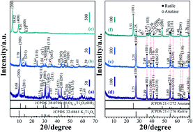
{110}-faceted rutile TiO2 nanocrystals with nanorods and nanoflower morphologies were synthesized through simple hydrothermal treatment of a tetratitanate nanoribbons precursor. The nanostructures and the formation reaction mechanism of the rutile TiO2 nanocrystals from the layered tetratitanate nanoribbons were investigated using X-ray diffraction (XRD), field emission scanning electron microscopy (FE-SEM), transmission electron microscope (TEM) and selected-area electron diffraction (SAED). The transformation from the layered phase into the rutile TiO2 may experience several stages during the hydrothermal reaction including dissolution of nanoribbons, recrystallization of [Ti(OH)6]2− octahedral fragments, and part dissolution and aggregation of the rutile TiO2 nanorods. Furthermore, the dye-sensitized solar cell (DSSC) performance of the synthesized rutile nanocrystals was also characterized, which showed a superior photovoltaic performance, compared to the benchmark P25 TiO2.
Introduction
Design and synthesis of titanium dioxide (TiO2) nanostructures with different morphologies and exposed specific facets have attracted considerable attention over the past decade, not only because of their considerable applications in photocatalysis,1,2 dye-sensitized solar cells (DSSCs),3,4 gas sensors,5 lithium-ion batteries,6 etc.; but also because of their thermal and chemical stability, relatively high photocatalytic activity, nontoxicity, and cost effectiveness.7 In nature, TiO2 mainly exists in four crystalline phases: anatase (tetragonal, space group I41/amd), rutile (tetragonal, space group P42/mmm), brookite (orthorhombic, space group Pbca), and TiO2 (B) (monoclinic, space group C2/m).8 Among the four crystalline forms, anatase TiO2 with specific active facets exposed has attracted enormous attention for its superior photocatalytic properties and excellent photovoltaic performances.9–11 The average surface energies of anatase TiO2 increase in the order of {101} facet (0.44 J m−2) < {100} facet (0.53 J m−2) < { 001} facet (0.90 J m−2).12 Since the crystal facets of rutile ({011} and {110} facets) and anatase ({001} and {011} facets) TiO2 particles and their roles in photocatalytic reactions were first reported by Ohno et al.,13 the morphologies and crystals facet-controlled synthesis of TiO2 have been extensively investigated to improve their photocatalytic properties and photovoltaic performances. For instance, anatase TiO2 nanocrystals with exposed {010} facets and controllable morphologies were firstly synthesized by hydrothermal treatment of the [Ti1.73O4]1.07− nanosheets. They exhibited higher photocatalytic activity than the normal spherical nanocrystals without a specific facet on the surface.14 After that, anatase TiO2 single crystals with a large percentage of {001} facets were synthesized by using hydrofluoric acid solution as a capping agent.15 Using organic morphology controlling agents, rhombic-shaped anatase TiO2 nanocrystals and tetragonal faceted-nanorods with exposed {010} facets were prepared, and they exhibited conspicuous photocatalytic activity and improved photovoltaic conversion efficiency, respectively.16,17 Recently, the anatase nanocrystals with a large percentage of {010} facets and [111]-facets were prepared by hydrothermal treatment of titanate nanosheets precursors. They exhibited higher photocatalytic than the commercial P25 sample.18,19
Compared with anatase TiO2, relatively few researches have been reported on rutile TiO2. Feng et al. firstly reported the fabrication of dye-sensitized solar cells from vertically oriented single crystalline rutile TiO2 nanowires arrays on transparent conducting fluorine-doped tin oxide (FTO) glass substrates.20 After that, Sun et al. reported that rutile nanowires grown on a titanate nanosheet film performed better in dye-sensitized solar cells than those on bare FTO conducting glass in terms of all cell parameters.21 Liu et al. reported a facile one-pot hydrothermal synthesis of pure rutile TiO2 photoanode with 100% exposed pyramid-shaped {111} surfaces on a FTO conducting substrate.22 Recently, hierarchical double-layered TiO2 flower-rod structure composed of three-dimensional (3D) TiO2 nanoflowers and one-dimensional (1D) nanorods on FTO conducting glass were synthesized by hydrothermal method.23,24 As shown above, the rutile crystals are usually prepared in FTO conducting glass substrates, which are relatively difficult in the process of experimental operation.
Herein, we report a facile precursor route to prepare rutile TiO2 nanorods and nanoflowers with exposed {110} facets using a layered tetratitanate as a start material. Through ion-exchange, exfoliation, and hydrothermal treatment, the layered tetratitanate precursor was converted into the desired rutile TiO2 material. The possible transformation mechanism of the rutile TiO2 nanocrystals and the photovoltaic performance were investigated. And the photovoltaic performance was compared with the benchmark P25 TiO2 (commercial TiO2 power, ∼87% anatase and ∼13% rutile).
Methods
Regents and materials
Potassium tetratitanate (K2Ti4O9) was obtained from Otsuka Chemical Co., Ltd. Tetramethylammonium hydroxide solution (TMAOH, 15%) and hydrochloric acid (HCl, 35.0–37.0%) were purchased from Wako Pure Chemical Industries, Ltd. cis-Di(thiocyanate)bis(2,2′-bipyridyl-4,4′-dicarboxylate)-ruthenium(II)bis-tetrabutylammonium (N719) was purchased from Sigma-Aldrich. Degussa P25 was obtained from Nippon Aerosil, Ltd. All the chemicals and reagents were of analytical grade and were used as received without further purification. Deionized water was obtained from Milli-Q ultra-pure system (Yamata Scientific Co., Ltd).
Preparation of layered tetratitanate nanoribbon colloidal solutions
The layered tetratitanate nanoribbon colloidal solutions were prepared from layered tetratitanate K2Ti4O9 (KTO). The KTO sample (10.0 g) was acid treated with 1 mol L−1 HCl solution (1 L) for 3 days under stirring conditions to exchange K+ ions with H+ to obtain a protonic tetratitanate H2Ti4O9·0.25H2O (HTO), or written as (H2O)0.25Ti4O7(OH)2.25 The acid solution was replaced daily with a fresh one in order to remove K+ completely from the compounds. The HTO layered tetratitanate was washed with copious Milli-Q water to remove excess acid and dried using a freeze drier. 3.0 g of the HTO sample was hydrothermally treated in a 12.5% TMAOH solution (30 mL) at 100 °C for 24 h under stirring to intercalate TMA+ ions into the interlayer and to obtain TMA+-intercalated HTO sample. And further, the obtained sample was dispersed in 300 mL of Milli-Q water at room temperature by stirring for 24 h to exfoliate the TMA+-intercalated HTO sample to its nanoribbons. After removal of the non-exfoliated particles by filtration, the tetratitanate nanoribbon colloidal solution was obtained.
Preparation of TiO2 nanorods–nanoflower from layered tetratitanate nanoribbon colloidal solution
TiO2 nanoparticles were prepared by hydrothermal treatment of the tetratitanate nanoribbon colloidal solution after adjusting pH value to 1.0 with a 3 mol L−1 HCl solution. 40 mL of the pH adjusted colloidal solution was sealed in a Teflon-lined stainless steel autoclave with a capacity of 70 mL at 120 °C and 140 °C for 24 h, respectively. After the hydrothermal treatment, the sample was separated from the solution by centrifugation, followed by washing several times with Milli-Q water, and finally dried using a freeze drier. The obtained TiO2 sample was designated as pH1.0–Tx, where x is the desired temperature of the hydrothermal treatment.
Fabrication of dye-sensitized solar cell
A paste of rutile TiO2 nanorods–nanoflowers was prepared by mixing of 0.5 g of the TiO2 nanocrystals sample, 2.5 g of ethanol, 2.0 g of α-terpineol, 1.4 g of 10 wt% solution of Wako-ethyl-cellulose 10 (8–14 mPas), and 1.1 g of 10 wt% solutions of Wako-ethyl-cellulose 45 (45–65 mPas). The mixture was dispersed by ultrasonic treatment for 5 minutes, followed by ball-milling for 3 days at room temperature. After ball-milling, a sticky paste was obtained by using a rotary evaporator to remove the ethanol. The DSSCs were fabricated by the following steps: (i) preparation of photoelectrodes, (ii) dye adsorption of photoelectrodes, and (iii) assembly. For prepare the photoelectrodes, the fluorine-doped tin oxide (FTO) conducting glass plates (12.5 × 12.5 mm) were cleaned for 5 min with Milli-Q water and alcohol, respectively, using an ultrasonic bath, in order. The FTO glass plate was immersed in 0.1 M titanium tetraisopropoxide (TTIP) solution for several seconds, and then sintered at 480 °C in muffle furnace for 60 min to coat the FTO glass surface with a dense TiO2 thin film. The prepared TiO2 paste was coated on the TTIP-treated FTO glass plate by a doctor-blade technique. The films thickness was controlled by thickness of the adhesive tape used as the spacer. After being coated with the paste on the FTO glass plate, the doctor-bladed FTO glass plate was calcined at 315 °C for 15 min. Do such working several times to obtain a thickness of TiO2 films around 14 μm, and then calcined it at 450 °C for 30 min. We also prepared similar films from the commercially available Degussa P25 TiO2 for comparison. The TiO2 films were treated with 0.1 M TTIP solution as described above, and then calcined at 480 °C for 60 min to obtain TiO2 porous electrode. For the dye adsorption, the sintered TiO2 porous electrode was soaked in 3 × 10⁻⁴ mol L−1 N719 dye solution in a 1
:
1 acetonitrile (super dehydrated)-tert-butyl alcohol for 24 h at room temperature and washed with absolute ethanol for removing the un-adsorbed dyes. The DSSC was comprised of the dye-sensitized TiO2 electrode and a Pt-counter electrode with an electrolyte solution between the electrodes. The electrolyte consisting of 0.60 mol L−1 1-butyl-3-methylimidazolium iodide, 0.03 mol L−1 iodine, 0.10 mol L−1 guanidine thiocyanate, and 0.50 mol L−1 4-tert-butylpyridine in acetonitrile/valeronitrile (85
:
15, v/v), was injected into the cell through the edges between the two electrodes.
Characterization
The powder XRD measurements were performed on each sample by using a SHIMADZU XRD-6100 diffractometer with Cu Kα radiation, λ = 0.15406 nm, operated at 30 mA current and with a voltage of 40 kV. Field emission scanning electron microscopy (FE-SEM) was performed using a HITACHI S-90X machine at an operating voltage of 15 kV and current of 10 mA. Transmission electron microscope (TEM), high-resolution TEM (HR-TEM) and selected-area electron diffraction (SAED) were done with the JEOL (JEM-3010) machine at an operating voltage of 200 kV. The Brunauer–Emmett–Teller (BET) surface areas of the samples were determined by a TriStar II 3020 volumetric adsorption analyzer at −196 °C after the samples were degassed at 120 °C for 5 h under high vacuum before the measurements. The photocurrent–voltage characteristic curves for the DSSCs were measured using a Hokuto-Denko BAS100B electrochemical analyzer under irradiation with simulated sunlight of AM 1.5 (100 mW cm−2), using a sunlight simulator (YSS-E40, Yamashita Denso) and a 0.25 cm2 mask. The thicknesses of the TiO2 films were measured using a SURFCOM 480A surface-shape determiner.
Results and discussion
Structure and morphology
The precursor, monoclinic KTO, is confirmed by a powder XRD pattern (JCPDS no. 32-0861, a = 18.25, b = 3.79, c = 12.01 Å, and β = 106.4°), as shown in Fig. 1(a). After the K+/H+ ion–exchange reaction, the basal spacing was changed from 8.68 Å for KTO to 7.78 Å for HTO (JCPDS no. 38-0700, a = 16.20, b = 3.74, c = 12.09 Å, and β = 105.7°), indicating the HTO sample maintains the same essential layered structure (Fig. 1(b)). To exfoliate the layered structure HTO into its nanoribbons, the HTO sample was hydrothermally treated in a TMAOH solution at 100 °C to intercalate TMA+ ions into the interlayer. After the hydrothermal treatment, the TMA+-intercalated tetratitanate sample still maintains the same layered structure, which shows the main diffraction peaks of (200), (400), and (600) with d-values of 18.02, 9.01, and 6.10 Å, respectively (Fig. 1(c)). The basal spacing was changed from 7.78 Å for HTO to 18.02 Å for TMA+-intercalated tetratitanate, indicating that the TMA+ ions were successfully intercalated into the interlayer space by H+/TMA+ ion-exchange. In view of the height of TMA+ ion being 5.30 Å,26 and the thickness of crystallographic nanoribbon being 5.70 Å,27,28 the large basal spacing implies that the TMA+-intercalated sample in its wet state contains double layers of the TMA+ ions (2 × 5.30 + 5.70 = 16.30 Å) in the interlayer. The interlayer gallery height is a little higher than double layers of the TMA+ ions might be caused by the presence of interlayer water.29 When the TMA+-intercalated tetratitanate is dispersed in a certain amount of Milli-Q water, the layered structure is exfoliated into nanoribbons, resulting in a tetratitanate nanoribbon colloidal solution.
 |
| Fig. 1 XRD patterns of (a) K2Ti4O9 (KTO), (b) H2Ti4O9·0.25H2O (HTO), (c) TMA+-intercalated tetratitanate sample, (d) pH1.0–T120, (e) pH1.0–T140, and (f) P25 samples. | |
As shown in Fig. 2(a), the HTO has a layer structure, and one layer consists of linear groups of four [TiO6] octahedrons with overlapping edges.30 Fig. 2(b–d) show the typical FE-SEM images of KTO, HTO, and the exfoliated tetratitanate nanoribbons sample, respectively. The KTO sample exhibits a fibrous morphology, with a size of ∼120–250 nm in width and several micrometers in length [Fig. 2(b)]. After the ion–exchange reaction, the HTO sample retaining the profile of the KTO [Fig. 2(c)], and the axis-direction of the HTO corresponds to the b-axis direction of the crystal [Fig. 2(e and f)]. The HR-TEM image [Fig. 2(g)] shows that the lattice spacing of 3.47 Å (lattice fringes perpendicular to the length of the nanoribbon) and 2.72 Å (lattice fringes parallel to the length of the nanoribbon), corresponding to the (010) and (004) facets of the HTO, respectively, revealing that the nanoribbon direction is in the [010]-direction.
 |
| Fig. 2 (a) Crystal structure of H2Ti4O9·0.25H2O (HTO), FE-SEM images of (b) K2Ti4O9 (KTO) sample, (c) H2Ti4O9·0.25H2O (HTO) sample, and (d) the exfoliated tetratitanate nanoribbons sample, TEM image and SAED pattern of (e and f) H2Ti4O9·0.25H2O (HTO) sample, and HR-TEM image of (g) an exfoliated tetratitanate nanoribbon sample. | |
The XRD patterns of the samples prepared by hydrothermal treatment of the tetratitanate nanoribbon colloidal solution at pH 1.0 at 120 and 140 °C, are shown Fig. 1(d and e), respectively. The positions of the diffraction peaks indicate that both the samples are rutile type of TiO2 (JCPDS no. 21-1276, tetragonal system, a = b = 4.59 Å, c = 2.96 Å). And no anatase diffraction peaks are observed. As shown in Fig. 1(d and e), the diffraction peaks of rutile TiO2 became stronger and sharper with the increase of temperature, implying an increase of crystallinity. As shown in Fig. 1(f), the commercial P25 TiO2 sample containing 87% anatase phase and 13% rutile phase, calculated from the integrated intensities of anatase (101), and rutile (110) peaks according to literature.31,32
Typically, FE-SEM images (Fig. 3(a–d)) show that the synthesized rutile TiO2 samples are consisted of uniformed nanorods (with a size of ∼1 μm in length and ∼120 nm in width). Partial dissolution and reaggregation of these nanorods lead to some nanoflowers structure. Actually, each nanorod is consisting of hundreds of highly ordered ultrafine rutile nanowires (with a size of ∼8 nm in width), which is confirmed by HR-TEM image shown in Fig. 3(e and f). The lattice fringe has d-spacing values of 3.25 and 2.95 Å, corresponding to (110) and (001) planes of rutile TiO2, respectively, and the angle between the two planes is 90°, which is identical to the theoretical value between the (110) and (001). Fig. 3(f) also reveals that the rodlike crystal facets are parallel to {110} facets, and the elongation of the crystal is parallel to <001> direction. That is, the long axis of the rodlike crystal corresponds to c-axis direction of the rutile structure and exposed crystal facets are {110} facets.33
 |
| Fig. 3 FE-SEM images of (a and b) pH1.0–T120, and (c and d) pH1.0–T140 sample; TEM and HR-TEM images of (e and f) pH1.0–T120 sample. | |
Transformation reaction mechanism from tetratitanate nanoribbons to nanorods–nanoflowers of rutile TiO2
On the basis of the above results, we proposed a possible reaction mechanism for the formation of nanorods–nanoflowers of rutile TiO2 from tetratitanate nanoribbons in the hydrothermal reaction process, which is illustrated in Fig. 4. In this process, first the TMA+-intercalated layered tetratitanate structure is exfoliated into elementary host [Ti4O9]2− nanoribbons. The cations of TMA+ and H3O+ locate on the surface of [Ti4O9]2− nanoribbons to balance the negative charge. The formation of the nanorods–nanoflowers of rutile TiO2 architecture from the [Ti4O9]2− nanoribbons may experience several stages during the hydrothermal reaction including dissolution of nanoribbons, recrystallization of [Ti(OH)6]2− octahedral fragments, and part dissolution and aggregation of the nanorods. Initially the [Ti4O9]2− nanoribbons are split to small, primarily [Ti(OH)6]2− octahedral monomers along the corner-shared and edge-shared oxygen atoms in high acidic conditions. Subsequently, the rutile TiO2 nanorods are formed by sharing equatorial edges of the [Ti(OH)6]2− octahedral monomers. Finally, the rutile TiO2 nanorods occurred partly dissolution and aggregated to nanoflowers. The transformation of [Ti4O9]2− nanoribbons to rutile TiO2 can be described as follows. |
[Ti4O9]2− + 15H2O = 4[Ti(OH)6]2− + 6H+
| (1) |
|
[Ti(OH)6]2− + 2H+ = TiO2 + 4H2O
| (2) |
 |
| Fig. 4 Transformation reaction mechanism from TMA+-intercalated layered tetratitanate to rutile TiO2 nanorods–nanoflowers structure. | |
The acid condition can slow down the dissolution reaction of [Ti4O9]2− by providing free H+ as present in reaction (1), which is favorable to form the oriented 1D TiO2 nanorod by dehydration reaction (i.e., oxolation) between OH ligands in [Ti(OH)6]2− octahedral monomers (2). Meanwhile, Cl− provided by HCl can preferentially adsorb on rutile TiO2 (110) plane and postpone the growth rate of (110) plane, which is facilitative for the anisotropic growth of rutile nanorods along [001] direction.23 With the proceeding of the hydrothermal reaction, dissolution of the rutile TiO2 nanorods occurred partly, which are inclined to aggregate to form the core of the flowers. As the hydrothermal reaction further prolonging, these aggregated rutile TiO2 nanorods would develop bigger, leading to the formation of rutile TiO2 nanoflowers.
DSSC performance of TiO2 nanocrystals
To study the DSSCs performance of the TiO2 nanocrystals, we fabricate DSSCs using T120–pH1.0 and P25 TiO2 nanocrystal samples. Fig. 5 depicts the photocurrent–voltage characteristics of two typical DSSCs assembled from the rutile nanorods–nanoflowers and P25 TiO2, respectively, under AM 1.5 illumination (100 mW cm−2). The corresponding photovoltaic performance parameters of the two DSSCs including short-circuit photocurrent density (Jsc), open circuit voltage (Voc), fill factor (FF), and power conversion efficiency (η) are summarized in Table 1. The Jsc, Voc, and η values of T120–pH1.0-based DSSC were improved by 28.2% (from 10.84 to 13.90 mA cm−2), 5.0% (from 0.717 to 0.753 V), and 31.5% (from 3.91 to 5.14%), compared to P25-based DSSC, respectively. The higher η value of T120–pH1.0 than P25 TiO2 is due to enhancements of the Jsc and Voc values. The higher performance of T120–pH1.0-based DSSC can be explained by the nanostructure and surface structure of T120–pH1.0 nanocrystals.
 |
| Fig. 5 Photocurrent–voltage characteristic curves of DSSCs fabricated using the nanocrystal samples of T120–pH1.0 rutile and P25. | |
Table 1 Summary of the device parameters under AM 1.5 G irradiation
Sample |
Film thickness (μm) |
Jsc (mA cm−2) |
Voc (V) |
FF (%) |
η (%) |
T120–pH1.0 |
13.5 |
13.90 |
0.753 |
49.1 |
5.14 |
P25 |
14.1 |
10.84 |
0.717 |
50.2 |
3.91 |
It is well know that the interfacial electron transfer influence on DSSCs efficiency. And the efficiency of the solar energy conversion process is closely related to the structure and stability of the dye–TiO2 interface.34 The dye–TiO2 interface starts with the absorption of the sun energy, followed the excited electron from the dye is injected into the conduction band of the TiO2.35 Rutile TiO2 nanorods–nanoflowers are composed of hundreds of ultrafine nanowires with the width of ∼8 nm [Fig. 3(e and f)]. The Voc value of T120–pH1.0-based DSSC was higher than that of P25 TiO2 with the same film thickness. The nanowire architecture can help increase of the rate of charge transport as compared to P25 TiO2 nanoparticles in the porous TiO2 film because of lower concentration of grain boundaries.36 The enhancement of porous TiO2 film conductivity can suppress the charge recombination at interface between TiO2 surface and I3− in the electrolyte. As the Voc value of the DSSCs is determined by the differences between the quasi-Fermi level of TiO2 and the redox potential of I− and I3−, a lower electron–hole recombination rate results in a negative shift in the Fermi level of TiO2, namely give a high Voc value.37
The dye adsorption behavior on the TiO2 nanocrystal surface has an important influence on the Jsc value of the DSSCs. The ultrafine nanowires arrays provide a larger BET surface area (79.6 m2 g−1 for T120–pH1.0) compare with the benchmark P25 TiO2 (49.7 m2 g−1), resulting in a larger dye adsorption amount than P25 TiO2. The crystal facet on the TiO2 surface can affect also the dye adsorption behavior. We have been reported that the N719 dye adsorption equilibrium constant (Kad) on anatase nanocrystals surfaces increased in the order of non-specific surface < [111]-faceted surface < {010}-faceted surface, which corresponds to the increasing order of the Jsc value, namely, the strong binding between the dye and TiO2 surface can enhance the Jsc value.18 It has been reported that there is a stronger interaction between the dye molecule and the rutile TiO2 {110} facets, which can improve the charge transfer rate from the molecule to the TiO2 surface.34 The dye molecules may be anchored more strongly on {110}-faceted rutile nanocrystal surface of T120–pH1.0 than that on the anatase TiO2 nanocrystal surface of P25, and the strong anchoring can facilitate the electron injection rate from the N719 dye molecules into the conduction band of TiO2, resulting in the enhancement of the Jsc.38
For rutile TiO2, the {110}, {100} and {001} facets have been studied, among them, the {110} facet is the most stable crystal surface.39 The surface energy of the rutile TiO2 {110} facet is 0.31 J m−2, which contains two different kinds of titanium atoms (sixfold coordinated Ti atoms and fivefold coordinated Ti atoms) among the [001] direction.39,40 The rutile TiO2 {100} facets (fivefold coordinated Ti atoms) has received considerably less attention than the {110} facets, because it is difficult to determine conclusively the exact surface geometry.39 The rutile TiO2 {001} facet contains only fourfold coordinated Ti atoms and twofold coordinated O atoms.39 Hence the number of broken bonds in the surface is higher the {110} and {100} surfaces. Consequently, the rutile {001} facet has a high surface energy. However, the {110} facets have both a favorable surface atomic structure and a surface electron structure for photocatalytic reactions, which make the reducing electrons in the conduction-band can be transferred via the surface fivefold coordinated Ti atoms as active reaction sites. And the efficient consumption of exited electrons in the photoreduction reactions can simultaneously promote the involvement of holes in photooxidation reactions. Such a cooperative mechanism existing on {110} facets is responsible for its having the higher performance for photocatalytic reactions.41 We think the surface atomic structures and charge transfer mechanisms of rutile facets would affect strongly also on the DSSCs performances.
Conclusions
The {110}-faceted rutile TiO2 nanocrystals composed of nanorods and nanoflowers can be synthesized by simple hydrothermal treatment of tetratitanate nanoribbons. The possible formation mechanism from the layered tetratitanate nanoribbons to the rutile TiO2 may experience several stages, including dissolution of nanoribbons, recrystallization of [Ti(OH)6]2− octahedral fragments, and part dissolution and aggregation of the rutile TiO2 nanorods. The {110}-faceted rutile TiO2 nanocrystals exhibited an excellent photovoltaic performance, compared to the benchmark P25 TiO2, owing to the TiO2 nanorods–nanoflowers composed of hundreds of ultrafine nanowires can enlarge the surface area for N719 dyes loading, accelerate the movement of photoelectrons and suppress the recombination of photogenerated electrons and holes in the porous TiO2 film. The surface atomic structure and electron structure of rutile can affect also on the DSSCs performances.
Conflict of interest
The authors declare no competing financial interest.
Acknowledgements
This work was supported in part by Science and technology innovation project of Shanxi Province Education Department (no. 20121026), the Grants-in-Acid for Doctor Research Funds from Jinzhong University and the Grants-in-Aid for Scientific Research (B) (no. 26289240) from Japan Society for the Promotion of Science and Kagawa University.
References
- N. Roy, Y. Sohn and D. Pradhan, Synergy of low-energy {101} and high-energy {001} TiO2 crystal facets for enhanced photocatalysis, ACS Nano, 2013, 7, 2532–2540 CrossRef CAS PubMed.
- C. H. Li, C. Koenigsmann, W. D. Ding, B. Rudshteyn, K. R. Yang, K. P. Regan, S. J. Konezny, V. S. Batista, G. W. Brudvig, C. A. Schmuttenmaer and J.-H. Kim, Facet-dependent photoelectrochemical performance of TiO2 nanostructures: an experimental and computational study, J. Am. Chem. Soc., 2015, 137, 1520–1529 CrossRef CAS PubMed.
- A. K. Patra, A. Dutta and A. Bhaumik, Synthesis of cuboid-shaped single-crystalline TiO2 nanocrystals with high-energy facets {001} and its dye-sensitized solar cell application, J. Phys. Chem. C, 2014, 118, 16703–16709 CAS.
- C. D. Chen, Y. Ikeuchi, L. F. Xu, G. A. Sewvandi, T. Kusunose, Y. Tanaka, S. Nakanishi, P. H. Wen and Q. Feng, Synthesis of [111]- and {010}-faceted anatase TiO2 nanocrystals from tri-titanate nanosheets and their photocatalytic and DSSC performances, Nanoscale, 2015, 7, 7980–7981 RSC.
- A. C. Parageorgiou, C. L. Pang, Q. Chen and G. Thornton, Low-dimensional, reduced phases of ultrathin TiO2, ACS Nano, 2007, 1, 409–414 CrossRef PubMed.
- S. H. Nam, H.-S. Shim, Y.-S. Kim, M. A. Dar, J. G. Kim and W. B. Kim, Ag or Au nanoparticle-embedded one-dimensional composite TiO2 nanofibers prepared via electrospinning for use in lithium-ion batteries, ACS Appl. Mater. Interfaces, 2010, 2, 2046–2052 CAS.
- C. Han, R. Luque and D. D. Dionysion, Facile preparation of controllable size monodisperse anatase titania nanoparticles, Chem. Commun., 2012, 48, 1860–1862 RSC.
- W.-J. Ong, L.-L. Tan, S.-P. Chai, S.-T. Yong and A. R. Mohamed, Highly reactive {001} facets of TiO2-based composites: synthesis, formation, mechanism and characterization, Nanoscale, 2014, 6, 1946–2008 RSC.
- C. D. Chen, L. F. Xu, G. A. Sewvandi, T. Kusunose, Y. Tanaka, S. Nakanishi and Q. Feng, Microwave-assisted topochemical conversion of layered titanate nanosheets to {010}-faceted anatase nanocrystal for high performance photocatalysts and dye-sensitized solar cells, Cryst. Growth Des., 2014, 14, 5801–5811 CAS.
- D.-H. Wang, L. Jia, X.-L. Wu, L.-Q. Lu and A.-W. Xu, One-step hydrothermal synthesis of N-doped TiO2/C nanocomposites with high visible light photocatalytic activity, Nanoscale, 2012, 4, 576–584 RSC.
- Z. R. Wang, H. Wang, B. Liu, W. Z. Qiu, J. Zhang, S. H. Ran, H. T. Huang, J. Xu, H. W. Han, D. Chen and G. Z. Shen, Transferable and flexible nanorod-assembled TiO2 cloths for dye-sensitized solar cells, photodetectors, and photocatalysts, ACS Nano, 2011, 5, 8412–8419 CrossRef CAS PubMed.
- M. Lazzeri, A. Vittadini and A. Selloni, Structure and Energetics of Stoichiometric TiO2 Anatase Surfaces, Phys. Rev. B: Condens. Matter Mater. Phys., 2001, 63, 155409 CrossRef.
- T. Ohno, K. Sarukawa and M. Matsumura, Crystal facets of rutile and anatase TiO2 particles and their roles in photocatalytic reactions, New J. Chem., 2002, 26, 1167–1170 RSC.
- P. H. Wen, H. Itoh, W. P. Tang and Q. Feng, Single nanocrystals of anatase-type TiO2 prepared from layered titanate nanosheets: formation mechanism and characterization of surface properties, Langmuir, 2007, 23, 11782–11790 CrossRef CAS PubMed.
- H. G. Yang, C. H. Sun, S. Z. Qiao, J. Zou, G. Liu, S. C. Smith, H. M. Cheng and G. Q. Lu, Anatase TiO2 single crystals with a large percentage of reactive facets, Nature, 2008, 453, 638–642 CrossRef CAS PubMed.
- B. H. Wu, C. Y. Guo, N. F. Zheng, Z. X. Xie and G. D. Stuky, Nonaqueous production of nanostructured anatase with high facets, J. Am. Chem. Soc., 2008, 130, 17563–17567 CrossRef CAS PubMed.
- W. G. Yang, Y. L. Wang and W. M. Shi, One-step synthesis of single-crystal anatase TiO2 tetragonal faceted-nanorods for improved-performance dye-sensitized solar cells, CrystEngComm, 2012, 14, 230–234 RSC.
- Y.-E. Du, Q. Feng, C. D. Chen, Y. Tanaka and X. J. Yang, Photocatalytic and dye-sensitized solar cell performances of {010}-faceted and [111]-faceted anatase TiO2 nanocrystals synthesized from tetratitanate nanoribbons, ACS App. Mater. Interfaces, 2014, 6, 16007–16019 CrossRef CAS PubMed.
- Y.-E. Du, D. J. Du, Q. Feng and X. J. Yang, Delithation, exfoliation and transformation of rock-salt-structured Li2TiO3 to highly exposed {010}-faceted anatase, ACS App. Mater. Interfaces, 2015, 7, 7995–8004 CrossRef CAS PubMed.
- X. J. Feng, K. Shankar, O. K. Varghese, M. Paulose, T. J. Latempa and C. A. Grimes, Vertically aligned single crystal TiO2 nanowire arrays grown directly on transparent conducting oxide coated glass: synthesis details and applications, Nano Lett., 2008, 8, 3781–3786 CrossRef CAS PubMed.
- P. P. Sun, X. T. Zhang, X. P. Liu, L. L. Wang, C. H. Wang, J. K. Yang and Y. C. Liu, Growth of single-crystalline rutile TiO2 nanowire on titanate nanosheet film for dye-sensitized solar cells, J. Mater. Chem., 2012, 22, 6389–6393 RSC.
- X. L. Liu, H. M. Zhang, X. D. Yao, T. C. An, P. R. Liu, Y. Wang, F. Peng, A. R. Carroll and H. J. Zhao, Visible light active pure rutile TiO2 photoanodes with 100% exposed pyramid-shaped surfaces, Nano Res., 2012, 5, 762–769 CrossRef CAS.
- L. B. Yu, Z. Li, Y. B. Liu, F. Cheng and S. Q. Sun, Synthesis of hierarchical TiO2 flower-rod and application in CdSe/CdS co-sensitized solar cell, J. Power Sources, 2014, 270, 42–52 CrossRef CAS.
- H. Wang, G. B. Yi, M. Tan, X. H. Zu, H. S. Luo and X. M. Jiang, Initial reactant controlled synthesis of double layered TiO2 nanostructure and characterization of its spectra of absorption and photoluminescence, Mater. Lett., 2015, 148, 5–8 CrossRef CAS.
- H. Izawa, S. Kikkawa and M. Koizumi, Ion exchange and dehydration of layered titanate, Na2Ti3O7 and K2Ti4O9, J. Phys. Chem., 1982, 86, 5023–5026 CrossRef CAS.
- P. Maireles-Torres, P. Olivera-Pastor, E. Rodriguez-Castellon, A. Jimenez-Lopez, L. Alagna and A. A. G. Tomlinson, Porous cross-linked materials formed by oligometric aluminium hydroxide and α-tin phosphate, J. Mater. Chem., 1991, 1, 319–326 RSC.
- S. Cheng and T. Wang, Pillaring of layered titanates by polyoxo cations of aluminum, Inorg. Chem., 1989, 28, 1283–1289 CrossRef CAS.
- X. K. Li, B. Yue and J. H. Ye, Photocatalytic hydrogen evolution over SiO2-pillared and nitrogen-doped titanic acid under visible light irradiation, Appl. Catal., A, 2010, 390, 195–200 CrossRef CAS.
- W. H. Hou, Y. S. Chen, C. X. Guo and Q. J. Yan, Synthesis of porous chromia-pillared tetratitanate, J. Solid State Chem., 1998, 136, 320–321 CrossRef CAS.
- S. Suzuki and M. Miyayama, Lithium intercalation properties of octatitanate synthesized through exfoliation/reassembly, J. Phys. Chem. B, 2006, 110, 4731–4734 CrossRef CAS PubMed.
- H. Z. Zhang and J. F. Banfield, Understanding polymorphic phase transformation behavior during growth of nanocrystalline aggregates: Insights from TiO2, J. Phys. Chem. B, 2000, 104, 3481–3487 CrossRef CAS.
- J.-G. Yu, J. C. Yu, B. Cheng, S. K. Hark and K. Iu, The effect of F−-doping and temperature on the structural and textural evolution
of mesoporous TiO2 powders, J. Solid State Chem., 2003, 174, 372–380 CrossRef CAS.
- F. Zuo, R. J. Dillon, L. Wang, P. Smith, X. Zhao, C. Bardeen and P. Feng, Active facets on titanium(III)-doped TiO2: a effective strategy to improve the visible-light photocatalytic, Angew. Chem., Int. Ed., 2012, 51, 6223–6226 CrossRef CAS PubMed.
- T. Gomez, X. Zarate, E. Schott and R. Arratia-Perez, Role of the main adsorption modes in the interaction of the dye [COOH-TPP-Zn(II)] on a periodic TiO2 slab exposing a rutile (110) surface in a dye-sensitized solar cell, RSC Adv., 2014, 4, 9639–9646 RSC.
- J. N. Clifford, E. Martíez-Ferrero, A. Viterisi and E. Palomares, Sensitizer molecular structure-device efficiency relationship in dye sensitized solar cells, Chem. Soc. Rev., 2011, 40, 1635–1646 RSC.
- C. Y. Zha, L. M. Shen, X. Y. Zhang, Y. F. Wang, B. A. Korgel, A. Gupta and N. Z. Bao, Double-sided brush-shaped TiO2 nanostructure assemblies with highly ordered nanowires for dye-sensitized solar cells, ACS Appl. Mater. Interfaces, 2014, 6, 122–129 CAS.
- Y. C. Rui, Y. G. Li, H. Z. Wang and Q. H. Zhang, Photoanode Based on chain-shaped anatase TiO2 nanorods for high-efficiency dye-sensitized solar cells, Chem. – Asian J., 2012, 7, 2313–2320 CrossRef CAS PubMed.
- P. Wen, M. Xue, Y. Ishikawa, H. Itoh and O. Feng, Relationships between cell parameters of dye-sensitized solar cells and dye-adsorption parameters, ACS Appl. Mater. Interfaces, 2012, 4, 1928–2934 CAS.
- U. Diebold, The surface science of titanium dioxide, Surf. Sci. Rep., 2003, 48, 53–229 CrossRef CAS.
- M. Ramamoorthy, D. Vanderbilt and R. D. King-Smith, First-principles calculation of the energetic of stoichiometric TiO2 surfaces, Phys. Rev. B, 1994, 49, 16721–16727 CrossRef CAS.
- J. Pan, G. Liu, G. Q. Lu and H.-M. Cheng, On the true photoreactivity order of {001}, {010}, and {101} facets of anatase TiO2 crystals, Angew. Chem., Int. Ed., 2011, 50, 2133–2137 CrossRef CAS PubMed.
|
This journal is © The Royal Society of Chemistry 2016 |
Click here to see how this site uses Cookies. View our privacy policy here.