DOI:
10.1039/C5RA23425E
(Paper)
RSC Adv., 2016,
6, 15451-15459
Enhancement in growth rate and productivity of spinach grown in hydroponics with iron oxide nanoparticles†
Received
6th November 2015
, Accepted 25th January 2016
First published on 27th January 2016
Abstract
Plants play a vital role in the energy and environmental ecosystem by providing food and oxygen for living organisms. Due to the increasing use of nanoparticles in the recent decade, the study on the effect of nanoparticles in environmental sectors (especially in agriculture) has become highly essential. In this study, we demonstrated the uptake of iron oxide (Fe2O3) nanoparticles by spinach via hydroponics and examined its effects on the growth rate and productivity of the spinach plant. The experimental studies such as plant growth (stem and root length) and biomass analysis revealed a dose and time dependent increase due to the uptake of Fe2O3. The vibrating sample magnetometer analysis revealed the increase in saturation magnetization of spinach plants as result of Fe uptake. Further, ICP analysis demonstrated an increase in iron content in spinach plants in a dose dependent manner. A mechanism for the uptake of Fe2O3 nanoparticles has been discussed with the help of FT-IR spectroscopy. The experimental studies might provide new insights into the application of Fe2O3 nanoparticles in the agriculture sector.
1. Introduction
The emergence of nanotechnology has created tremendous opportunities in fundamental research as well as industrial products for the welfare of human society.1,2 The use of nanosized materials cannot be limited to a particular field since it possesses a wide range of applications in electronics, photonics, energy, environment, biomedical, and so on.3–5 Nanomaterials with sizes 1 to 100 nm have been utilized for several biological applications such as antibacterial, biofilm inhibition, cancer detection and therapy, and dental applications.6–8 Moreover, nanomaterials are increasingly used in developing commercial products such as cosmetics, textiles, solar roofs and paints which signify that these materials might be spread into the environment.9–11 Therefore, researchers focused on analyzing the effect of nanomaterials in the environment and ecological systems. In these aspects, plants play a crucial role in the energy and environmental ecosystem since they provide the food and oxygen for all of the living organisms on earth.12 Due to the widespread use of nanomaterials and their exposure to the environment, studying the effects of nanoparticles on the growth of plants is highly recognized and is becoming an emerging area of special interest. Nanomaterials can be biotransformed into plants via aerial or root pathways and the process of nanomaterial uptake, transformation and magnification by plants is known as biotransformation.13,14 Among the various routes for uptake of nanoparticles by plants, hydroponics method (soil less cultivation technique) possess more advantages for implying the nanotechnology principles.15 Up-to-date, researchers have studied the influence of some nanomaterials on plants growth and investigated their phytotoxicity, mechanism of uptake, and transformation of micronutrient into plant at the cellular level.13–16 Earlier studies have demonstrated that both positive as well as adverse effects in plants have been observed due to the uptake of nanomaterials. The merits of nano-bio-transformed plants can provide new platforms such as high growth rate, increased food production with high micronutrient levels, increased biomass, and artificial photosynthesis, as well as some medical applications.17 However, it also results in adverse effects such as growth suppression due to phytotoxicity of some nanomaterials.18 Considering the recent efforts taken until now on the uptake of nanomaterials by plants, Schwabe et al. demonstrated the uptake of cerium oxide nanoparticles on three plants such as wheat, pumpkin and sunflower.19 The recent study by Lahiani et al. suggested that carbon nanohorns can be used as plant growth promoters and other plant related applications.20 Alumina nanoparticles stimulated the growth of Lemma minor and results in increased biomass accumulation whereas another study showed the suppression of growth in Lemma minor due to nanosilver.21 ZnO nanoparticles showed a stimulatory effect in the growth of peanuts.22 The presence of nanotitania has been observed in cucumber plants along with macromolecular modification of fruit content.23 More recently, Srivastava et al. demonstrated increased production of spinach due to initial seed treatment with iron pyrite nanoparticles.24
In this regards, iron oxide (Fe2O3) nanoparticles are one of the medically relevant nanomaterials available with an advantages such as biocompatibility, low cost chemical stability and magnetic property, cosmetics and cancer therapy.25–28 Moreover, iron is an important micronutrient highly essential for life as it forms a part of significant proteins and enzymes such as haemoglobin and myoglobin. Bone marrow found in the centre of the long bones needs iron to make haemoglobin for transportation of oxygen to other organs. Balanced diet containing adequate amount of iron is needed for healthy growth. The deficiency of iron in human body leads to the state called anaemia.29 Fe2O3 is one of the iron based compounds that are currently recognized as safe by the U.S. Food and Drug Administration (21CFR186.1300). Moreover, iron is an essential element in plants for photosynthesis.30,31 Hematite nanoparticles are used for artificial photosynthesis applications as well as pigment in paints.32,33 Due to these intriguing properties of hematite nanoparticles, we expected that the uptake of these nanoparticles by plants might provide new insights in the plant nanotechnology. In this study, the uptake of Fe2O3 nanoparticles by spinach plant and their effects on the growth rate and productivity was examined. Moreover, a plausible mechanism for the uptake of Fe2O3 nanoparticles was also discussed in detail.
2. Experimental section
2.1 Synthesis of Fe2O3 nanoparticles
Fe2O3 nanoparticles were prepared via gel combustion method using iron nitrate and citric acid as the starting materials.33 The precursor solution was prepared by dissolving Fe(NO3)3 followed by the addition of citric acid in the weight ratio of 1![[thin space (1/6-em)]](https://www.rsc.org/images/entities/char_2009.gif) :1.1 and allowed to vigorous stirring for 30 min. To this, ammonia solution was added drop-wise until the pH of the solution reached to 10. The obtained sol was heated at 80 °C in a hot plate until the complete evaporation of the solvent which further results in the formation of black ash due to self-combustion. The formed ash was allowed to calcination at a temperature of 700 °C for 2 h in a muffle furnace leading to the formation of Fe2O3 nanoparticles.
:1.1 and allowed to vigorous stirring for 30 min. To this, ammonia solution was added drop-wise until the pH of the solution reached to 10. The obtained sol was heated at 80 °C in a hot plate until the complete evaporation of the solvent which further results in the formation of black ash due to self-combustion. The formed ash was allowed to calcination at a temperature of 700 °C for 2 h in a muffle furnace leading to the formation of Fe2O3 nanoparticles.
2.2 Hydroponic growth of spinach in presence of Fe2O3 at different concentrations
Spinach (Spinacea oleracea) seeds were obtained from the market at Madurai, India. Initially the seeds were placed on a solid hydroponic medium consisting of sawdust and coco peat in an artificial chamber and adequate amount of water was added over the solid hydroponic medium to maintain moist condition which is necessary for seed germination.34 After three days, the uniform seedlings selected were anchored by thermocol and transferred into a wide mouth plastic container containing Hoagland medium (1000 mL). The spinach grown in hydroponic medium without Fe2O3 nanoparticles was used as control. Similar strategy was used to grow spinach in which Hoagland medium containing different concentrations of Fe2O3 nanoparticles as micronutrients. For comparison, the same experiment was performed using Fe(NO3)3·9H2O as a iron supplement. Irrespective of medium, all the plants were kept in the open atmosphere and it was maintained on 11 h light/13 h dark cycle at ∼30 °C. The difference in the growth rate was monitored by measuring the root and shoot height once in 15 days upto 45 days. The detailed methodology was provided in ESI.†
2.3 Instrumentation
The X-ray diffraction (XRD) pattern of the Fe2O3 nanoparticles were recorded on a Siefert X-ray diffractometer (Richard Seifert and Co., Ahrensburg, Germany) using CuKα radiation (λ = 1.54016 Å) at 60 keV over the range of 2θ = 20–80°. Raman spectra of the nanoparticles were produced using LabRam HR800 micro-Raman spectroscope (Horiba Jobin-Yvon, France). The Raman system was operated at 10 mW laser power and an excitation wavelength of 514 nm with an Ar+ ion laser. Atomic force microscope (AFM) image of Fe2O3 nanoparticles was recorded by using Scanning Probe Microscope XE 70 (Park Systems, Suwan, South Korea) in non-contact mode at room temperature. AFM image of Fe2O3 nanoparticles was obtained by scanning the area of 10 × 10 μm2. The chemical composition and the state of elements present in the outermost parts of Fe2O3 nanoparticles were investigated by X-ray photoelectron (XPS) spectrum measured using ESCA-2000, VG Microtech Ltd. Here, a monochromatic X-ray beam source at 1486.6 eV and 14 kV was used to scan the sample surface. A high flux X-ray source with aluminum anode was used for X-ray generation, and a quartz crystal monochromator was used to focus and scan the X-ray beam on the sample. Hysteresis loop of the dried powder sample was recorded using Lakeshore Vibrating Sample Magnetometer, USA (Model: 7404).
2.4 Analysis of Fe content using ICP analysis
The spinach leaves of all the samples were collected after 45 days and dried in dark condition. 50 mg of dried sample was digested with a mixture of HCl and HNO3 (3:1 ratio) and allowed to heat treatment on a hot plate until the plant mass digested completely. The dried samples were ground to fine powders and digested in 2% HNO3. Total Fe content in plant tissues were measured by ICP analysis.
2.5 Simulation studies on the uptake of Fe2O3 by spinach
The Fe2O3 nanoparticles were kept in the Hoagland's medium (without the seedlings) over a desired period of 14 days. The nanoparticles were then collected and washed with water for three times and allowed to dry at 60 °C. The FT-IR spectra of pure Fe2O3 and the Fe2O3 treated with Hoagland's solution were recorded in the range of 500 to 2000 cm−1.
2.6 Statistical analysis
All the experiments in this study were performed with three replicates and the values represent the mean ± standard deviation of three experiments. Significant differences between the treatments were determined using one-way analysis. The statistical analyses and calculations were performed using GraphPad Prism 5 Software (San Diego, USA). Differences were considered statistically significant with “P value < 0.0001”.
3. Results and discussion
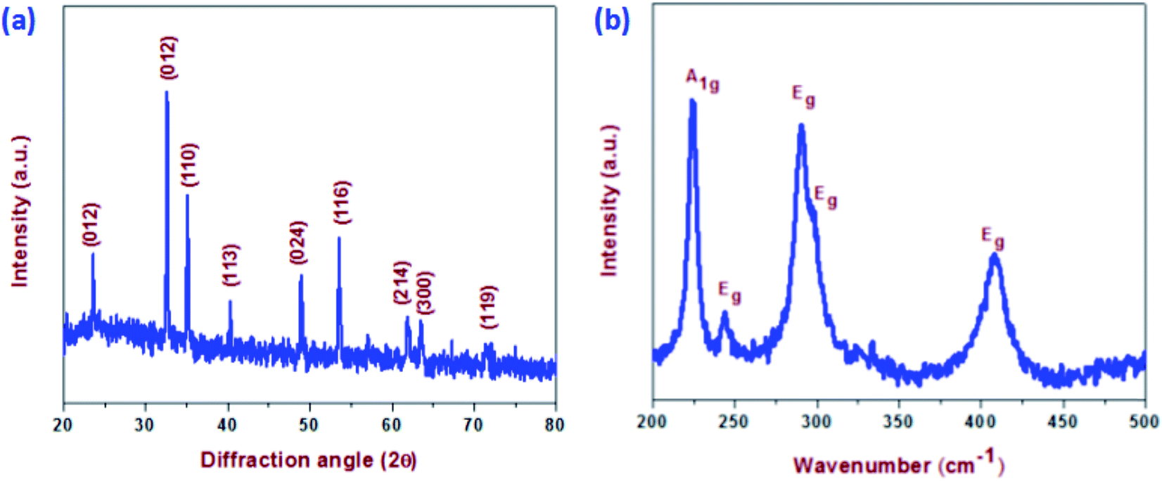
The Fe2O3 nanoparticles used in this study are prepared by sol–gel combustion method using iron nitrate and citric acid (in the ratio 1:1.1) as the starting precursors. Upon mild heat treatment, due to the self combustion of the gel, a black precipitate is formed which results in the formation of Fe2O3 nanoparticles after calcination at a higher temperature. In order to understand the crystalline nature and phase purity of the prepared samples, XRD analysis has been performed. The XRD pattern of the Fe2O3 nanoparticles is shown in Fig. 1(a) which showed the presence of sharp diffraction peaks observed at 2θ = 23.57°, 32.59°, 35.06°, 40.28°, 48.90°, 53.50°, 61.86°, 63.46°, and 72.10° corresponding to the (012), (104), (110), (113), (024), (116), (214), (300) and (119) planes of α-Fe2O3 phase. The observed diffraction pattern and interplanar spacing closely matched with the standard diffraction pattern of α-Fe2O3 (JCPDS no. 079-0007).35 There is no impurity or other iron oxide phases were observed in the XRD pattern, suggesting the formation of high purity Fe2O3 nanoparticles. The average crystallite size of the prepared Fe2O3 nanoparticles was estimated using the Debye–Scherrer equation36 and found to be about 50 nm. The Raman spectrum of the prepared Fe2O3 nanoparticles is shown in Fig. 1(b) which revealed the presence of vibration bands at 220, 239, 286, 294 and 402 cm−1, respectively. The band observed at 220 cm−1 corresponds to the A1g mode and the bands observed at 239, 286, 294 and 402 cm−1 are due to the Eg modes of α-Fe2O3.37 The observed bands are consistent with the earlier report on the Raman spectrum of α-Fe2O3 nanoparticles.38
 |
| | Fig. 1 (a) X-ray diffraction pattern and (b) laser Raman spectrum of Fe2O3 nanoparticles. | |
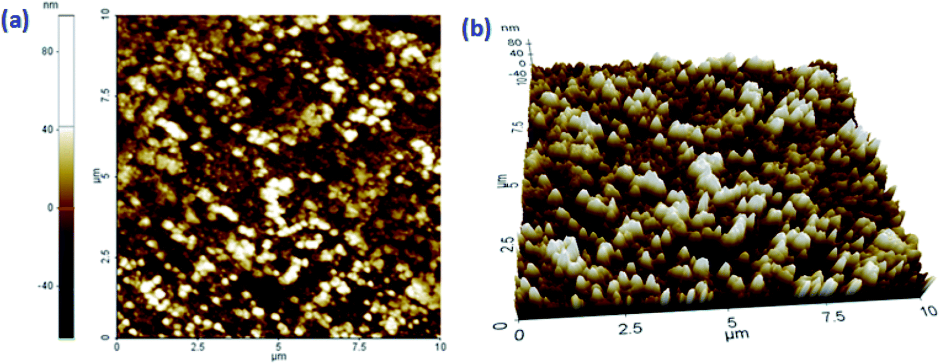
The surface morphology and size of the prepared nanoparticles are examined by atomic force microscopy using non-contact mode. The two- and three-dimensional topographies of the prepared Fe2O3 nanoparticles are shown in Fig. 2. The direct observation of the two dimensional topographic image (Fig. 2(a)) revealed that the nanoparticles are cubic shaped and the 3D topography (Fig. 2(b)) demonstrated that the size of the Fe2O3 nanoparticles is in the range of 30 to 40 nm. The chemical surface states of elements present in Fe2O3 nanoparticles are studied using XPS analysis (as given in Fig. 3). Fig. 3(a) shows the Fe 2p spectrum indicating the presence of two states of Fe in Fe2O3 such as Fe 2p3/2 and Fe 2p1/2 states observed at 718.22 and 731.76 eV, respectively.39 The energy separation between the spin–orbits is about 13.18 eV which indicates the presence of Fe3+ in Fe2O3. Further, a small satellite peak was observed at 725.92 eV which corresponds to the shake-up peak of Fe3+ oxidation state (higher than 7.2 eV binding energy of Fe 2p3/2).40 Fig. 3(b) shows the O 1s spectrum of Fe2O3 suggests the presence of a broad peak with a binding energy of 534.46 eV.35 The XPS analysis clearly reveals that the oxidation state of Fe is about +3 and O is about −2, in the prepared Fe2O3. The observed results are in close agreement with the previous report on the XPS of Fe2O3 nanoparticles.41
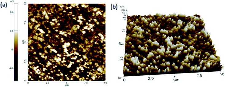 |
| | Fig. 2 Atomic force micrograph of Fe2O3 nanoparticles (a) two dimensional and (b) three dimensional images. | |
 |
| | Fig. 3 X-ray photoelectron spectrum of Fe2O3 nanoparticles (a) Fe 2p and (b) O 1s states. | |
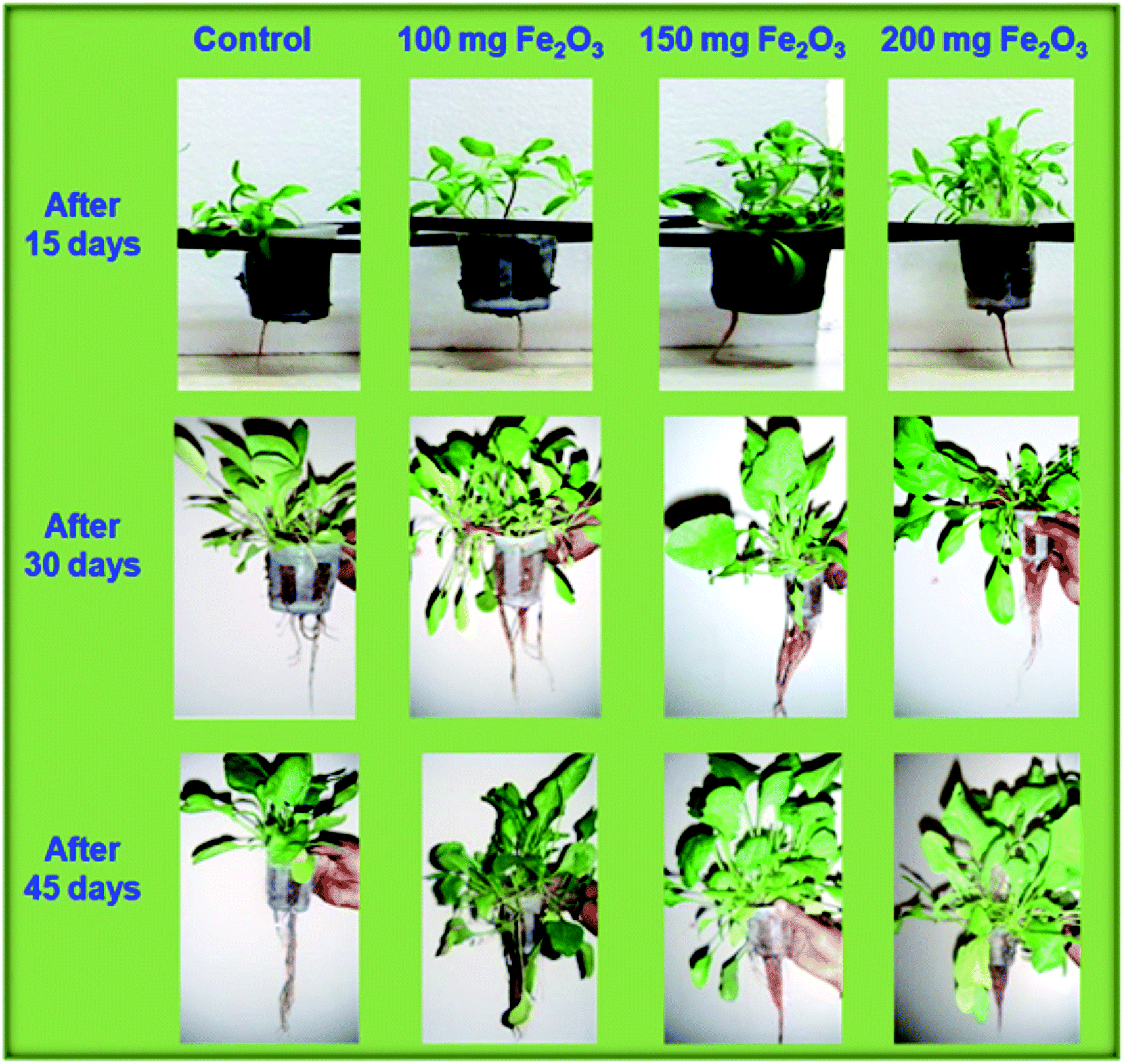
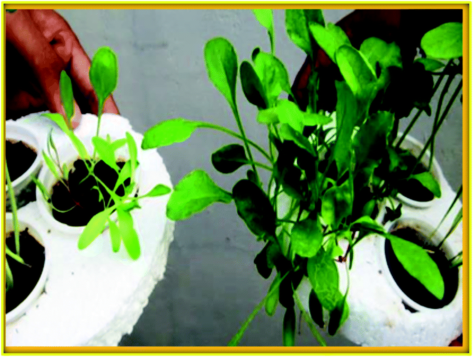
The effect of Fe2O3 nanoparticles on the growth of spinach has been performed in this study. It is well known that hydroponics method is a soil less cultivation method developed for the growth of plants in presence of minerals, nutrients and water.34 In this study, spinach plants were grown hydroponically using Hoagland solution with a mixture of coco peat and sawdust as the anchoring agent (see Fig. S1 in ESI†). The photograph shown in left side of Fig. S1† shows the growth of spinach plant in a Hoagland's solution with thermocol support, whereas the photograph in right side shows the grown plants where the root emerged out at the bottom. The studies on the effect of Fe2O3 nanoparticles on the growth of spinach plant has been carried out via hydroponics method by including the Fe2O3 nanoparticles (at different concentrations) into the Hoagland's solution and the growth kinetics were examined at different time intervals of 15 days upto 45 days. Fig. 4 shows the digital photographs of the spinach plant cultured without Fe2O3 and in presence of Fe2O3 nanoparticles at different concentrations such as 100, 150 and 200 mg. This study suggested that the inclusion of nano Fe2O3 results in enhanced growth rate of spinach in a dose and time dependent manner. Fig. 5 shows the control and nano Fe2O3 treated plants after 45 days, which clearly evidences the use of Fe2O3 for growth promotion of spinach. This study suggests that nano Fe2O3 is safer for the growth of spinach. The length of the roots and stems of the control and treated plants are shown in Fig. 6. It is evident from the graph that the length of the stems (left image in Fig. 6) and roots (right image in Fig. 6) has been increased with respect to the dosage of Fe2O3 and time duration. The length of the stems of spinach plant grown under different concentrations of Fe2O3 such as 100, 150 and 200 mg are about 1.45, 1.91 and 2.27 fold higher than that of the control spinach after a period of 45 days. A similar trend has also been observed for the changes in length of the root as well. The length of the root for 100, 150 and 200 mg of Fe2O3 treated spinach plants shows nearly 1.25, 1.375 and 1.75 fold higher than that of the control spinach. There is no remarkable change in the spinach treated with iron salt (Fe(NO3)3) in the hydroponic medium (about 1.09 and 1.03% of increase in stem and root compared the control spinach plant). This finding is in close agreement with the previous study by Srivastava et al. on the effect of iron salt on the growth promotion of spinach.24 This analysis revealed that both roots and stems showed an increased growth for the nano Fe2O3 treated plants compared to the untreated plant. However, the rate of increase of length in stem is higher than that of the root length which is a beneficial one since leaves are natural solar panels converting sunlight into energy via photosynthesis.
 |
| | Fig. 4 Digital photographs of spinach plant grown without and with Fe2O3 (100, 150, 200 mg) taken after 15, 30 and 45 days. | |
 |
| | Fig. 5 Digital photographs of spinach plant (control-left side and 200 mg Fe2O3 treated right side) after 45 days. | |
 |
| | Fig. 6 Digital photographs of stems and roots of spinach plant after 45 days (left to right indicates the spinach plant – control, 100, 150 and 200 mg Fe2O3 treated plants). Table shows the length of stems and roots after 15, 30 and 45 days. | |
Further, we analyzed the change in biomass of the spinach plant due to the uptake of Fe2O3. After 45th day, the spinach grown in all hydroponic medium with and without Fe2O3 was removed carefully. Except the root portion, the stem and leaves were collected and weighed accurately and named as wet biomass. Then, the biomass was dried in air individually at 30 °C for about 10 days without allowing any dust particles in contact and the dry biomass (without the trace of water molecule) was weighed accurately and the effect of Fe2O3 treatment on the biomass of the spinach is shown in Fig. 7. Compared with the control spinach plant, the wet biomass shows nearly 1.39, 3.24, and 5.04 fold increase for 100, 150, and 200 mg of Fe2O3 treated plants, respectively. The analysis of wet biomass shows nearly 1.36, 3.08, and 4.17 fold increase for increasing concentration of Fe2O3, when compared to the control spinach plant. These studies highlight the enhanced growth rate and productivity of spinach due to the uptake of Fe2O3 nanoparticles. This is in agreement with the previous work which showed increased leaf and pod dry weight of soybean due to uptake of Fe2O3 nanoparticles.42
 |
| | Fig. 7 Biomass ((a) wet and (b) dry) analysis of all spinach grown after 45 days. Results are given as mean ± S.D (n = 3). Values are statistically significant at ****p < 0.0001 Fe2O3 treated plant compared with control group. | |
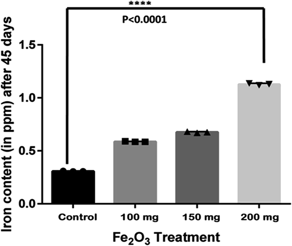
The hysteresis analysis of the dried powder samples obtained for the control and treated spinach plants has been examined by VSM analysis (as shown in Fig. 8) for understanding the existence of iron in the plants. The hysteresis of control samples (Fig. 8(a)) showed the existence of magnetic property with a saturation magnetization of about 0.10221 emu g−1. This suggested the presence of iron mineral in the control plants which is due to the natural bioavailability of iron in spinach plants.43 The hysteresis analysis of spinach plants grown in presence of Fe2O3 (as shown in Fig. 8(b)–(d)) revealed the presence of magnetic property with saturation magnetization of about 0.12155 emu g−1, 0.13291 emu g−1 and 0.14123 emu g−1 for plants grown at 100, 150 and 200 mg of Fe2O3, respectively. A recent study demonstrated the anti-tumor properties of composites containing Au nanorods-natural extracts of spinach leaves.44 Further, assessment of cytotoxicity of these leave extracts will provide useful information on the effect of these spinach for applications in medical sectors. This study substantiates the linear increase saturation magnetization in spinach plants treated with different concentration of Fe2O3 suggesting the iron content up taken by spinach plant might be of different size, or composition which needed to be studied in detail in the near future. The uptake of Fe content by the spinach plant has been examined by the use of ICP analysis as shown in Fig. 9. Naturally, spinach is considered as a reserve of iron.43 The Fe content in the dried mass of spinach (control) is about 0.306 ppm and it is increasing with respect to dose dependent manner in the spinach treated with Fe2O3 nanoparticles. However, there is no significant change in the Fe content was observed in the case of iron salt treated spinach (about 0.317 ppm) compared to that of control. Based on the ICP analysis results, it is inferred that the Fe content found on the spinach (grown in 200 mg Fe2O3) harvested after 45th day shows about 3.6 times higher iron content (1.128 ppm) in compared to that of the control spinach (0.306 ppm). This study substantiates the increase in productivity of spinach attributable to the absorption of iron content occurred via roots and then translocated in various parts of the plant.
 |
| | Fig. 8 Hysteresis analysis of dried powders of spinach grown after 45 days (a) control without Fe2O3, whereas (b), (c), and (d) represents the spinach treated with 100, 150 and 200 mg Fe2O3 nanoparticles. | |
 |
| | Fig. 9 ICP analysis of Fe content in all spinach plant grown after 45 days. Results are given as mean ± S.D (n = 3). Values are statistically significant at ****p < 0.0001 Fe2O3 treated plant compared with control group. | |
The mechanism of Fe uptake by the spinach plant from the Fe2O3 nanoparticles under hydroponic conditions can be explained as follows: in general the uptake of trivalent iron (Fe3+) is found to be pH sensitive and naturally Fe3+ is insoluble which will be slowly converted into Fe2+ under acidic environment. Initially, the hydroponic medium prepared using the distilled water has pH of about 7.8 (slight alkaline). While adding the nutrients, the pH of the medium is getting reduced to about 6.5 (slightly acidic). This is because of the addition of dihydrogen monoammonium phosphate (NH4H2PO4) which is naturally acidic. In this condition, the insoluble Fe3+ might be slowly converted into soluble Fe2+, facilitating the absorption capability of plants. In order understand this issue, the Fe2O3 nanoparticles are suspended in Hoagland's solution alone and their chemical changes were examined via FT-IR spectroscopy. The FT-IR spectrum of bare Fe2O3 nanoparticles (Fig. 10) shows a peak at 514 cm−1 corresponding to the Fe–O stretching vibrations. After incubation of Fe2O3 nanoparticles is in Hoagland's solution over 14 days. The FT-IR spectrum (Fig. 10) revealed the formation of iron phosphates peak at 1040 cm−1. The transformed iron phosphates are taken up by the spinach and translocated into roots, stems and leaves of the spinach. Altogether, this study demonstrated that the uptake of Fe2O3 nanoparticles by the spinach plant via hydroponic method results in positive effects such as increase in root and shoots length, increased biomass as well as an increased Fe content in a dose dependent effect. Further studies on the toxicity and medical applications of this Fe rich spinach needed to be performed in the near future.
 |
| | Fig. 10 Fourier transformed infra-red spectra of pure Fe2O3 and Fe2O3 nanoparticles incubated in Hoagland's solution over a period of 14 days. | |
4. Conclusion
The key findings of this study suggests the positive effects of spinach plant due to uptake of Fe2O3 nanoparticles such as increase in stem and root lengths, biomass production and magnetic properties. An increase in Fe content has been observed in spinach plant in time and dose dependent nature has been observed via ICP analysis. Future works on examining the medical aspects of this iron rich spinach plant such as cytotoxicity can provide new insights on the medical applications of this plant.
Acknowledgements
The authors thank the management and the principal of Mepco Schlenk Engineering College for giving permission and showing constant encouragement to successfully complete the project work.
References
- J. Lee, S. Mahendra and P. J. J. Alvarez, ACS Nano, 2010, 4, 3580–3590 CrossRef CAS PubMed.
- K. Krishnamoorthy, K. Jeyasubramanian, M. Premanathan, G. Subbiah, H. S. Shin and S. J. Kim, Carbon, 2014, 72, 328–337 CrossRef CAS.
- V. H. Nguyen, H.-V. Nguyen and P. Dollfus, Nanotechnology, 2014, 25, 165201 CrossRef PubMed.
- A. Vinodhini, K. Govindaraju, G. Singaravelu, A. M. Sadiq and V. G. Kumar, Colloids Surf., B, 2014, 117, 480–486 CrossRef CAS PubMed.
- K. Kailasam, J. D. Epping, A. Thomas, S. Losse and H. Junge, Energy Environ. Sci., 2011, 4, 4668 CAS.
- K. Krishnamoorthy, M. Premanathan, M. Veerapandian and S. Jae Kim, Nanotechnology, 2014, 25, 315101 CrossRef PubMed.
- T. Ashokkumar, D. Prabhu, R. Geetha, K. Govindaraju, R. Manikandan, C. Arulvasu and G. Singaravelu, Colloids Surf., B, 2014, 123, 549–556 CrossRef CAS PubMed.
- M. S. Mannoor, H. Tao, J. D. Clayton, A. Sengupta, D. L. Kaplan, R. R. Naik, N. Verma, F. G. Omenetto and M. C. McAlpine, Nat. Commun., 2012, 3, 763 CrossRef PubMed.
- J. T. Han, S. Choi, J. I. Jang, S. K. Seol, J. S. Woo, H. J. Jeong, S. Y. Jeong, K.-J. Baeg and G.-W. Lee, Sci. Rep., 2015, 5, 9300 CrossRef PubMed.
- M. P. Genovese, I. V. Lightcap and P. V. Kamat, ACS Nano, 2012, 6, 865–872 CrossRef CAS PubMed.
- F. Natalio, R. André, A. F. Hartog, B. Stoll, K. P. Jochum, R. Wever and W. Tremel, Nat. Nanotechnol., 2012, 7, 530–535 CrossRef CAS PubMed.
- J. D. Judy, J. M. Unrine and P. M. Bertsch, Environ. Sci. Technol., 2011, 45, 776–781 CrossRef CAS PubMed.
- P. Zhang, Y. Ma, Z. Zhang, X. He, J. Zhang, Z. Guo, R. Tai, Y. Zhao and Z. Chai, ACS Nano, 2012, 6, 9943–9950 CrossRef CAS PubMed.
- S. C. C. Arruda, A. L. D. Silva, R. M. Galazzi, R. A. Azevedo and M. A. Z. Arruda, Talanta, 2015, 131, 693–705 CrossRef PubMed.
- B. S. Sekhon, Nanotechnol., Sci. Appl., 2014, 7, 31–53 CrossRef PubMed.
- D. Andreescu, G. Bulbul, R. E. Özel, A. Hayat, N. Sardesai and S. Andreescu, Environ. Sci.: Nano, 2014, 1, 445–458 RSC.
- M. V. Khodakovskaya, K. de Silva, A. S. Biris, E. Dervishi and H. Villagarcia, ACS Nano, 2012, 6, 2128–2135 CrossRef CAS PubMed.
- W.-M. Lee, J. Il Kwak and Y.-J. An, Chemosphere, 2012, 86, 491–499 CrossRef CAS PubMed.
- F. Schwabe, S. Tanner, R. Schulin, A. Rotzetter, W. Stark, A. von Quadt and B. Nowack, Metallomics, 2015, 7, 466–477 RSC.
- M. H. Lahiani, J. Chen, F. Irin, A. A. Puretzky, M. J. Green and M. V. Khodakovskaya, Carbon, 2015, 81, 607–619 CrossRef CAS.
- G. Juhel, E. Batisse, Q. Hugues, D. Daly, F. N. A. M. van Pelt, J. O'Halloran and M. A. K. Jansen, Aquat. Toxicol., 2011, 105, 328–336 CrossRef CAS PubMed.
- Y. Marusenko, J. Shipp, G. A. Hamilton, J. L. L. Morgan, M. Keebaugh, H. Hill, A. Dutta, X. Zhuo, N. Upadhyay, J. Hutchings, P. Herckes, A. D. Anbar, E. Shock and H. E. Hartnett, Environ. Pollut., 2013, 174, 150–156 CrossRef CAS PubMed.
- A. D. Servin, H. Castillo-Michel, J. A. Hernandez-Viezcas, B. C. Diaz, J. R. Peralta-Videa and J. L. Gardea-Torresdey, Environ. Sci. Technol., 2012, 46, 7637–7643 CrossRef CAS PubMed.
- G. Srivastava, C. K. Das, A. Das, S. K. Singh, M. Roy, H. Kim, N. Sethy, A. Kumar, R. K. Sharma, S. K. Singh, D. Philip and M. Das, RSC Adv., 2014, 4, 58495–58504 RSC.
- P. Yang, Y. Ding, Z. Lin, Z. Chen, Y. Li, P. Qiang, M. Ebrahimi, W. Mai, C. P. Wong and Z. L. Wang, Nano Lett., 2014, 14, 731–736 CrossRef CAS PubMed.
- C. Piccirillo, C. Rocha, D. M. Tobaldi, R. C. Pullar, J. A. Labrincha, M. O. Ferreira, P. M. L. Castro and M. M. E. Pintado, J. Mater. Chem. B, 2014, 2, 5999 RSC.
- T. Amna, M. S. Hassan, K.-T. Nam, Y. Y. Bing, N. A. M. Barakat, M.-S. Khil and H. Y. Kim, Int. J. Nanomed., 2012, 7, 1659–1670 CrossRef CAS PubMed.
- L. Li, Y. Chu and Y. Liu, Nanotechnology, 2007, 18, 105603 CrossRef.
- P. Hunter, EMBO Rep., 2008, 9, 15–18 CrossRef CAS PubMed.
- J.-F. Briat, C. Dubos and F. Gaymard, Trends Plant Sci., 2015, 20, 33–40 CrossRef CAS PubMed.
- D. K. Bora, A. Braun and E. C. Constable, Energy Environ. Sci., 2013, 6, 407–425 CAS.
- O. Opuchovic and A. Kareiva, Ceram. Int., 2015, 41, 4504–4513 CrossRef CAS.
- K. Deshpande, A. Mukasyan and A. Varma, Chem. Mater., 2004, 16, 4896–4904 CrossRef CAS.
- J. B. Jones Jr, Hydroponics: A Practical Guide for the Soiless Grower, CRC Press, LLC Boca Raton, Florida, USA, 1997 Search PubMed.
- N. Sabari Arul, D. Mangalaraj, P. Nandha Kumar, E. Kim, P. Devi and J. In Han, Ceram. Int., 2015, 41, 5568–5573 CrossRef CAS.
- H. E. Yong, K. Krishnamoorthy, K. T. Hyun and S. J. Kim, J. Ind. Eng. Chem., 2015, 29, 39–42 CrossRef CAS.
- A. A. Ayachi, H. Mechakra, M. M. Silvan, S. Boudjaadar and S. Achour, Ceram. Int., 2015, 41, 2228–2233 CrossRef CAS.
- A. M. Jubb and H. C. Allen, ACS Appl. Mater. Interfaces, 2010, 2, 2804–2812 CAS.
- A. P. Grosvenor, B. A. Kobe, M. C. Biesinger and N. S. McIntyre, Surf. Interface Anal., 2004, 36, 1564–1574 CrossRef CAS.
- A. G. Nasibulin, S. Rackauskas, H. Jiang, Y. Tian, P. R. Mudimela, S. D. Shandakov, L. I. Nasibulina, S. Jani and E. I. Kauppinen, Nano Res., 2010, 2, 373–379 CrossRef.
- S. Radhakrishnan, K. Krishnamoorthy, C. Sekar, J. Wilson and S. J. Kim, Chem. Eng. J., 2015, 259, 594–602 CrossRef CAS.
- J. L. Gardea-Torresdey, C. M. Rico and J. C. White, Environ. Sci. Technol., 2014, 48, 2526–2540 CrossRef CAS PubMed.
- C. J. Rutzke, R. P. Glahn, M. A. Rutzke, R. M. Welch, R. W. Langhans, L. D. Albright, G. F. Combs and R. M. Wheeler, Habitation, 2004, 10, 7–14 CrossRef PubMed.
- Y. Wang, B. Zhang, L. Zhu, Y. Li, F. Huang, S. Li, Y. Shen and A. Xie, ACS Appl. Mater. Interfaces, 2014, 6, 15000–15006 CAS.
Footnote |
| † Electronic supplementary information (ESI) available. See DOI: 10.1039/c5ra23425e |
|
| This journal is © The Royal Society of Chemistry 2016 |
Click here to see how this site uses Cookies. View our privacy policy here.