
Anti-Malassezia furfur activity of natural surfactant mediated in situ silver nanoparticles for a better antidandruff shampoo formulation†
K. Jagajjanani Rao and
Santanu Paria *
*
Interfaces and Nanomaterials Laboratory, Department of Chemical Engineering, National Institute of Technology, Rourkela-769 008, Orissa, India. E-mail: sparia@nitrkl.ac.in; santanuparia@yahoo.com; Fax: +91 661 246 2999
First published on 14th January 2016
Abstract
Dandruff is a common scalp problem of human beings and a majority of people suffer from this at some point in their life. To prevent dandruff, antidandruff shampoos have become popular in recent years. However, in many cases, a dandruff causing fungus, Malassezia furfur, develops resistance towards commonly used antidandruff drugs. As a result, it is necessary to develop a new class of novel antidandruff shampoos for dual purposes. This study mainly focuses on a silver nanoparticle based green formulation of an antidandruff shampoo. In situ capped silver nanoparticles (Ag NPs) were prepared by green routes using Acacia and Acacia + Aegle marmelos leaf extracts (LEs) with sizes of ∼40 and 13 nm, respectively. The antifungal activity tests using a well diffusion technique show that 13 nm particles have better inhibiting activity (2.43 times) than 40 nm particles, while the TTC assay method shows that the minimum or threshold concentration is the same for both particle sizes used, which is 0.054 mM. The time kill assay shows some synergism in the net antifungal effect by Acacia + LEs and ∼13 nm Ag NPs in aqueous media against M. furfur. However, the promising anti-Malassezia activity of the 40 nm Ag NPs in the Acacia media along with their superior suspension stability against microbial contamination mean they have potential as an active and simple antidandruff shampoo formulation.
1. Introduction
Malassezia fungi are commonly found on human skin, in particular, on sebum-rich areas of the upper body such as the trunk, back, face, and scalp. M. furfur is a lipophilic fungus belonging to a dimorphic fungi group and is well known for its scalp diseases like seborrheic eczema and dandruff on the human skin.1,2 Dandruff is a common scalp disorder, during which flakes of dead skin cells are loosened from the scalp with itching because of a fungal infection. The treatment of dandruff (caused by M. furfur) involves the use of a shampoo containing selenium sulfide, imidazole compounds, etc., and the reported studies show that it is difficult to be completely cured from this disease.2,3 Additionally, M. furfur also causes tinea versicolor (a chronic and superficial skin infection), Pityrosporum folliculitis (an inflammatory skin disorder), seborrheic dermatitis, etc.4 Although antibiotics are the primary agents administered or used to control the infections, they suffer from certain limitations such as side-effects to the patients and development of drug resistance by this human pathogen.5Recent studies point out the ability of nanoparticles (NPs) to tackle multidrug resistance by disease causing microorganisms, and NP based formulations have emerged as novel antimicrobial agents. Despite the fact that different types of NPs such as copper, zinc, titanium, magnesium, gold, alginate, and silver have been reported for their promising antimicrobial properties, nanosilver is widely used as it is most effective against a wide range of bacteria, viruses, and other eukaryotic microorganisms.6,7 The antifungal activity studies of Ag NPs, especially against the potent pathogenic and multidrug resistant strains,8,9 have opened up a challenge to develop a novel formulation which can also be effective against opportunistic human pathogens like M. furfur. To our knowledge, there are limited studies on the anti-Malassezia activity of Ag NPs10,11 and only one study exists with Ag NPs synthesized via a green route against M. furfur.2 The reported green route article presented the use of a fungal cell filtrate mediated synthesis of Ag NPs with the size range of 3–30 nm. Although the authors claimed that their procedure is amenable to large scale antifungal formulation production, the pre-treatment steps of the biomass, recovery of the NPs in more than one step, lack of user friendliness, etc. can affect the formulation feasibility on a larger scale.
In general, user friendly formulations of green shampoos (devoid of harsh chemical agents) have attracted users’ attention rapidly in recent years. In fact, many users have been attracted to plant and herbal remedies for dandruff treatment in recent years. Many of these products are easily available at a low cost and are comparatively safe with no side effects, and as a result, they have been commonly used in good faith.12,13 Under this category, numerous plant extracts and essential oils have been reported to have beneficial effects on hair and are commonly used in shampoos for dandruff treatment.12,13 Some plant extracts such as Aloe vera, Eucalyptus globulus, Phyllanthus emblica, Wrightia tinctoria. Zingiber officinale, Wrightia tinctoria, Cassia alata, Azadirachta indica, etc. have been reported for their good activity against the M. furfur fungus.12–14 In shampoo formulations there is also an increasing trend of natural surfactant (saponin) based shampoos instead of synthetic surfactant based ones. Saponins are also known for their antimicrobial, antioxidant and antidandruff activity, and as a result, they are used alone or in combination with other plant extracts.15 For instance, saponins from Asparagus racemosus, Sapindus mukorossi, Vernonia cinerea, Ricinus communis, and Acacia concinna are used as herbal shampoos because of their surface active properties and additionally they may also have some antidandruff activity.16–20 Despite the mentioned advantages, these agents have certain limitations such as poor stability and low shelf-life in aqueous forms at ambient conditions (they are very susceptible to fungal and bacterial attacks), and they are less surface active (in terms of the wetting behaviour) than the synthetic surfactants. In our previous studies we have shown that natural surfactants are less surface active compared to the commonly used non-ionic surfactants21,22 and also that the presence of nanoparticles can improve the surface activity of natural surfactants.23 From the application perspective, the development of an aqueous-based antidandruff formulation with natural surfactants is a challenging task to date and can replace the use of chemical antifungal agents.
In this study we premise that the addition of Ag NPs can provide an effective solution by improving the shelf-life of the formulation as well as combating the drug resistance of the Malassezia fungi. For the mentioned purpose as well as from the economical view point, the NP synthesis route should be facile with limited purification steps. The green synthesis approach will ensure consumer compliance and marketability. The synthesis of Ag NPs (∼13 and 40 nm) with a narrow size distribution was achieved using the Aegle marmelos leaf extract and/or a plant-based surfactant (Acacia auriculiformis) by statistical optimization of the key parameters reported in our previous study.24 This study highlights the determination of the fungistatic and fungicidal effects of the as-prepared Ag NPs against the selected pathogenic fungus, M. furfur. The combined antifungal effect of A. marmelos, A. auriculiformis (Acacia), and Ag NPs towards M. furfur was evaluated. The addition of Ag NPs arrests the resistance of fungi and the incorporation of natural extracts may limit the total dose required for the antifungal activity. In addition, the stability of the plant extracts with NPs in open air was also monitored to determine the susceptibility towards microbial attack.
2. Materials and methods
2.1 Growth conditions and inoculum quantification
M. furfur was obtained from the Microbial Type Culture Collection and Gene Bank (MTCC), India with the code MTCC 1374. This fungus was grown on either Sabouraud Dextrose Agar (SDA) or Sabouraud Dextrose Broth (SDB) from HiMedia laboratories. The respective media were supplemented with pure Tween 80 (1% v/v) as a lipid source for the growth of the fungus.25 The grown M. furfur was stored in tubes containing SDA at 4 °C and renewed at monthly intervals. The fungus was allowed to grow in Petri dishes containing SDA and incubated at 35 °C for 48 h. Individual colonies were suspended in 10 mL of saline (0.85%) water in glass tubes and the fungal content was determined using turbidity measurements by comparing to the McFarland standard of 0.5 which corresponds to approximately 5 × 10⁶ colony forming units (CFU) mL−1.162.2 Nanoparticle synthesis
The Ag NPs of ∼13 nm size were synthesized using the Aegle marmelos leaf extract (LE) as a green reducing agent (AgNO3/LE ratio: 1 mM/0.3%, pH = 7.0, Acacia solution of 0.428 mM, temperature = 55 °C) as per our reported study.24 The Acacia solution alone also has the ability to form Ag NPs without any additional reducing agent but the size was larger (∼40 nm) in the absence of the LE.2.3 Antimicrobial tests
An in vitro susceptibility test or time-kill assay27 was also performed by growing the fungal cells with constant strength Ag NPs (0.428 mM) in SDB media for a period of 7 days at 35 °C. The strength of the fungal cells for this test was 7 × 10⁵ CFU mL−1. This test provides information regarding the synergism, antagonism or indifference in the microbicidal activity, when more than a single agent is present in the test suspension. Here, as the NPs used were capped (with Acacia or Acacia + LE), their efficiency was also tested against M. furfur. Control experiments with only capping/reducing agents (Acacia, Acacia + LE) in SDB broth were also done in both the ascribed assays devoid of Ag in the reaction media.
2.4 Contact angle measurements
Contact angle measurements give an idea about the hydrophobic/hydrophilic nature of the composite films28 and can also be applied to the microbial films as well. The wetting behaviours of microbial films before and after treatment with Ag NPs, Acacia, and Acacia + LE were tested. An optical contact angle analyzer (OCA-20, Data Physics, Germany) was used to measure the contact angle of water on the surface of the fungal biofilms. A drop of 1 μL of pure water was placed on the surface of the film using a microsyringe and the contact angles on both sides of the drop were measured. Here, the fungal biofilms were prepared from cell suspensions onto a clean glass slide by drop drying (45 °C for 24 h). For this test, fungal cells grown on SDA with an olive oil supplement were scraped and the suspension was prepared according to the McFarland standard as described above.3. Results and discussion
3.1 Well diffusion assay
The scanning electron microscopy images of the synthesized particles used in this study are shown in Fig. 1a and b. Fig. 1c represents the antimicrobial activity of the Ag NPs (13 and 40 nm) qualitatively against M. furfur. The photograph shows that the studied controls (positions 1 and 3) did not show any antifungal activity, however, the diameter of the zone of inhibition (ZOI) increases with a decrease in particle size. The ZOI values for 13 and 40 nm Ag NPs are 8.43 ± 1.3 and 20.54 ± 2.3 mm, respectively, using the same concentration. This test confirms the size dependent toxicity of the Ag NPs towards M. furfur. In addition to the above, the morphological changes before and after exposure of the fungal cells (5 × 10⁵ CFU mL−1) to the NPs are shown in Fig. 2 by SEM analysis. The smooth surface and the bowling pin shape of the cells (Fig. 2a and b) are significantly altered after the treatment with Ag NPs. The cell deformations with surface roughness are observed in parts (c) and (e) of Fig. 2. The structural changes are clearly visible (Fig. 2d) when subjected to 13 nm NPs, and at the same time, surface modifications are also seen when exposed to 40 nm Ag NPs (Fig. 2f). The presence of Ag NPs on cell surfaces was also further confirmed using EDX analysis (Fig. 2g and h).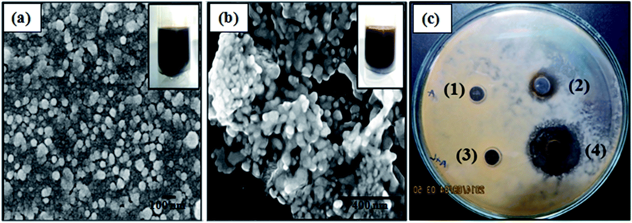 | ||
| Fig. 1 FE-SEM images of (a) ∼40 nm and (b) ∼13 nm AgNPs. Figure insets show the colour of the respective Ag sols. (c) Antifungal activity of Acacia (1), 40 nm Ag NPs (2), Acacia + LE (3), and 13 nm Ag NPs (4). | ||
 | ||
| Fig. 2 SEM images of M. furfur devoid of Ag NPs (a and b) and when exposed to 13 nm (c and d) and 40 nm (e and f) Ag NPs. Figure insets (g and h) show the EDX analysis of the imaged samples (full images are presented in S1, ESI†). The scale bars of (a, c, e), (b, f), and (d) are 50, 10, and 5 μm, respectively. | ||
3.2 TTC assay
The antifungal activity of Ag NPs with different doses against M. furfur was also evaluated using the TTC reagent and the formation of the red formazan colour is shown in the insets of Fig. 3. The higher value of absorbance because of the presence of formazan (at 480 nm) indicates more viable cells in the growth media. The figure also shows that when the dose is less than 10 μL or equivalent to 0.054 mM of the respective NPs (13 or 40 nm) the toxicity is not significant. This result suggests that the stated concentration is the threshold for the cell toxicity of the used NPs. Likewise, significant cytotoxicity was also not observed for systems with only the reaction media (Acacia, Acacia + LE), although mild growth reduction was seen with a high dose (50 μL) of Acacia + LE. The greater reduction in the intensity values and the colour change in the presence of 13 nm particles compared with that in the presence of 40 nm particles (Fig. 3) clearly indicates the size dependent toxicity of NPs. | ||
| Fig. 3 The TTC assay of different samples in the presence of different sizes and concentrations. The secondary X-axis indicates the strengths of only the Ag NPs used. The colour changes due to TTC reduction are given as picture insets. Acacia is abbreviated as Aca. | ||
3.3 In vitro susceptibility test
The synergism or antagonistic effect of two or more agents of a suspension can be determined from a time-kill assay.27 A constant dose of NPs with their respective controls were inoculated with the fungal cells for a period of 7 days and the samples were observed over time for growth by measuring the optical density (OD) at 600 nm wavelength using a spectrophotometer. The changes in the OD values with respect to time, which indicate the growth of M. furfur in that environment, are presented in Fig. 4. The results show that the fungal cells in the reaction media (Acacia and Acacia + LE) devoid of NPs also have some growth reduction unlike the observations found in the above section. This behaviour may be because of the higher concentration of the media used as a control for this test. Further, it is observed that the growth reduction is more in the case of the 13 nm particles (from Acacia + LE) compared to that of the 40 nm particles (from Acacia). The above results indicate that when the concentration of the reaction media is high (Acacia and LE) there is a combined antimicrobial action of the particles as well as the media. Reported studies of some antimicrobial properties of the used plant extracts (Acacia29 and LE30) further support our observation. Reduction in the number of viable cells is given in Fig. S2 of the ESI† as CFU mL−1 after appropriate serial dilutions. | ||
| Fig. 4 Growth of M. furfur in terms of optical density (absorbance at 600 nm wavelength) in various environments (upon seven times dilution). The control in the figure legend refers to only SDB. | ||
3.4 The contact angle investigations
Contact angle measurements were done to get some insight about the wettability of the fungal cell surface in the presence of the nanoparticle suspension. Since the in situ contact angle measurements on fungal cell surfaces is difficult, we performed the contact angle studies after making a fungal film on a glass surface. The results of the initial contact angle of water measurements for the fungal films along with the control are shown in Fig. 5 (the images are shown in Fig. S3, ESI†). The contact angle measurements of the dried fungal films exposed to various conditions (with and without NPs) show the following hydrophilicity order: Acacia > 13 nm Ag NPs ≥ Acacia + LE > 40 nm Ag NPs > fungal film > glass slide. | ||
| Fig. 5 Contact angle values of water on different films dried on the glass slide. | ||
These results indicate that the hydrophilicity of the fungal surface increases, while exposed to lone Acacia or Acacia + LE solutions, which are acting as capping agents as well as reducing agents during the synthesis of the 40 and 13 nm Ag NPs. In comparison, the contact angle of water was slightly more on the 40 nm Ag NP treated fungal film than on that treated with 13 nm particles, while their reaction media (Acacia and Acacia + LE) devoid of fungal film exhibited no significant difference. To address the observed behaviour, film coatings with only capped Ag NPs (13 and 40 mm) on a glass surface devoid of fungal film were also tested for contact angle measurements. To our surprise, the contact angle of water was near zero with immediate spreading for both particle films, similar to that of the pure reaction media (Acacia and Acacia + LE). As NPs are capped in situ during the synthesis process, the effect of the NP size on the contact angle could not be established at this point. Moreover, based on the observations from SEM analysis, the non-uniformity and roughness of the fungal film treated with 13 and 40 nm capped Ag NPs might be responsible for the decrease of the contact angle value of water on its surface (when compared to the native fungal film). The decrease in contact angle or improved wettability on the cell surface enables better contact of the nanoparticles with the surface, which in turn finally enhances the toxicity.
3.5 Stability of the suspensions at ambient conditions
The plant extracts are susceptible to fungal or bacterial attack under ambient conditions. To see the stability of the used suspensions, Acacia (1.2 mM) and Acacia + LE (1.2 mM + 0.9%) in aqueous media were tested for microbial growth under non-aseptic ambient conditions with the inclusion of 13 and 40 nm sized Ag NPs of 0.08 mM strength. The strength of the NPs was selected just above the threshold concentration (from the TTC assay) where they are effective in retarding the growth of M. furfur. Here, the NPs containing plant extracts were periodically opened (1 h day−1) and exposed to the surroundings every day for 15 days. The growth of the microorganisms was monitored in terms of the change in turbidity measured spectroscopically at 600 nm wavelength for various samples as shown in Fig. 6. The figure shows the initial and final O.D values of the different samples tested. The increase in O.D values because of the change in turbidity of the test suspensions is attributed to the aggregation of particles and microbial growth or both together. The appearance and colour change of the NP suspensions and controls are shown in Fig. S4, ESI.† Among the control experiments devoid of NPs, the Acacia solution showed much less increase in turbidity than the LE + Acacia solution (Fig. 6) because of less microbial growth. This indicates the vulnerability of LE towards microbial attack. Moreover, upon comparison of the 13 and 40 nm NP supplemented suspensions, Acacia with 40 nm Ag NPs shows no or negligible change in turbidity, suggesting no aggregation or microbial contamination. Even though 13 nm Ag NPs in the presence of LE + Acacia should show better resistance to microbial attack, the experimental results do not support this. This behaviour is attributed to the aggregation of particles after day 1 (Fig. S4, ESI†) in the presence of polyphenolic molecules such as tannins at the native pH of ∼5.5 (ref. 31) and also because of the presence of the used LE.32 The presence of microbial growth on the upper portion of the solution after 15 days further confirms the aggregation and settling of the NPs in LE + Acacia media (Fig. S3, ESI†). | ||
| Fig. 6 Optical density values in terms of the absorbance of the aqueous samples exposed to ambient non-sterile conditions for a period of 15 days. The Ag NPs prepared in Acacia + LE and Acacia media are referred to as 13 and 40 nm, respectively. | ||
The above observations indicate that the mixture of LE + Acacia media could not have the advantage of long term stability, despite the formation of smaller size (13 nm) Ag NPs. However, a higher dose of NPs may enhance the stability. On the other hand, Acacia assisted synthesis as well as in situ stabilized Ag NPs (40 nm) proved to be resistant to microbial contamination and are a good choice to develop a cost effective and novel anti dandruff surfactant for shampoo formulation. Furthermore, our approach is simple and cheaper than the only reported study on M. furfur with a green route (saponin assisted) synthesis of Ag NPs.2
4. Conclusions
An in situ green protocol was used to synthesize 13 and 40 nm Ag NPs in Acacia + LE and Acacia media, respectively. The obtained in situ capped NPs exhibited good fungitoxic properties against M. furfur. The anti microbial efficacy via well diffusion assays showed the importance of particle size on net toxicity, though the minimum threshold concentration identified was the same (0.054 mM) for both particle sizes used. Liquid media studies showed some synergism between the 13 and 40 nm Ag NPs and their synthesis media (Acacia + LE, Acacia) towards net fungitoxicity. The results from the contact angle experiments revealed an enhancement of fungal film surface hydrophilicity after treatment with Ag NPs, which in turn also helped in enhancing toxicity. The present investigation suggests that although 13 nm Ag NPs in the presence of Acacia + LE showed improved antifungal properties against M. furfur, the superior stability of the 40 nm Ag NP supplemented Acacia media at ambient conditions without microbial contamination has the potential to be used for ready-made antidandruff shampoo formulation.References
- A. K. Gupta, R. Batra, R. Bluhm, T. Boekhout and T. L. Dawson, Skin diseases associated with Malassezia species, J. Am. Acad. Dermatol., 2004, 51(5), 785–798 CrossRef PubMed.
- P. Joshi, S. Bonde, S. Gaikwad, A. Gade, K. Abd-Elsalam and M. Rai, Comparative Studies on Synthesis of Silver Nanoparticles by Fusarium oxysporum and Macrophomina phaseolina and It’s Efficacy Against Bacteria and Malassezia furfur, J. Bionanosci., 2013, 7(4), 378–385 CrossRef CAS.
- A. Sanfilippo and J. C. English, An overview of medicated shampoos used in dandruff treatment, P T, 2006, 31(7), 396 Search PubMed.
- M. Marcon, D. Durrell, D. Powell and W. Buesching, In vitro activity of systemic antifungal agents against Malassezia furfur, Antimicrob. Agents Chemother., 1987, 31(6), 951–953 CrossRef CAS PubMed.
- D. Robson, Malassezia: Mechanisms of possible drug resistance, Australia College of Veterinary Scientists Dermatology Chapter Science Week Proceedings, 6–7 July 2007, pp. 63–67 Search PubMed.
- M. Rai, A. Yadav and A. Gade, Silver nanoparticles as a new generation of antimicrobials, Biotechnol. Adv., 2009, 27(1), 76–83 CrossRef CAS PubMed.
- M. Rai, S. Deshmukh, A. Ingle and A. Gade, Silver nanoparticles: the powerful nanoweapon against multidrug-resistant bacteria, J. Appl. Microbiol., 2012, 112(5), 841–852 CrossRef CAS PubMed.
- Y.-K. Jo, B. H. Kim and G. Jung, Antifungal activity of silver ions and nanoparticles on phytopathogenic fungi, Plant Dis., 2009, 93(10), 1037–1043 CrossRef CAS.
- A. Panáček, M. Kolář, R. Večeřová, R. Prucek, J. Soukupová, V. Kryštof, P. Hamal, R. Zbořil and L. Kvítek, Antifungal activity of silver nanoparticles against Candida spp, Biomaterials, 2009, 30(31), 6333–6340 CrossRef PubMed.
- O. Brandt, M. Mildner, A. E. Egger, M. Groessl, U. Rix, M. Posch, B. K. Keppler, C. Strupp, B. Mueller and G. Stingl, Nanoscalic silver possesses broad-spectrum antimicrobial activities and exhibits fewer toxicological side effects than silver sulfadiazine, Nanomedicine, 2012, 8(4), 478–488 CrossRef CAS PubMed.
- T. Devasena and T. Ravimycin, Ketoconazole coated silver nanoparticles-a point antidandruff agent, Int. J. Plant Sci., 2009, 4(2), 517–520 CAS.
- M. J. Abad, M. Ansuategui and P. Bermejo, Active antifungal substances from natural sources, ARKIVOC, 2007, 7, 116–145 Search PubMed.
- R. Vijayakumar, C. Muthukumar, T. Kumar and R. Saravanamuthu, Characterization of Malassezia furfur and its control by using plant extracts, Indian J. Dermatol. Venereol., 2006, 51(2), 145 CrossRef.
- D. Chandrani, S. Lubaina and M. Soosamma, A review of antifungal effect of plant extract vs. chemical substances against Malassezia spp, Int. J. Pharma Bio Sci., 2012, 3(3), 773–780 Search PubMed.
- E. Barile, G. Bonanomi, V. Antignani, B. Zolfaghari, S. E. Sajjadi, F. Scala and V. Lanzotti, Saponins from Allium minutiflorum with antifungal activity, Phytochemistry, 2007, 68(5), 596–603 CrossRef CAS PubMed.
- C. Onlom, S. Khanthawong, N. Waranuch and K. Ingkaninan, In vitro anti-Malassezia activity and potential use in anti-dandruff formulation of Asparagus racemosus, Int. J. Cosmet. Sci., 2014, 36(1), 74–78 CrossRef CAS PubMed.
- G. R. Waller and K. Yamasaki, Saponins used in food and agriculture. Springer Science & Business Media, 2012, vol. 405 Search PubMed.
- P. Dhanalakshmi, A. J. P. Priya, E. Sagadevan, Y. S. Lakshmi, A. Manimaran, S. Sindhu and P. Arumugam, Evaluation of inhibitory effect of Vernonia cinerea L. leaf extracts on different fungal species, Int. J. Pharm. Pharm. Sci., 2013, 5(2), 414–446 Search PubMed.
- G. Sibi, G. Kaur, G. Devi, K. Dhananjaya, K. Ravikumar and H. Mallesha, Anti-dandruff activity of Ricinus communis L. leaf extract, Int. J. Curr. Pharm. Res., 2012, 4(3), 74–76 Search PubMed.
- S. K. Gediya, R. B. Mistry, U. K. Patel, M. Blessy and H. N. Jain, Herbal Plants: Used as a Cosmetics, J. Nat. Prod. Plant Resour., 2011, 1(1), 24–32 Search PubMed.
- S. Paria, N. R. Biswal and R. G. Chaudhuri, Surface tension, adsorption, and wetting behaviors of natural surfactants on a PTFE surface, AIChE J., 2015, 61(2), 655–663 CrossRef CAS.
- K. J. Rao and S. Paria, Solubilization of Naphthalene in the Presence of Plant – Synthetic Mixed Surfactant Systems, J. Phys. Chem. B, 2008, 113(2), 474–481 CrossRef PubMed.
- K. Chatterjee, Wettability of hair using natural and synthetic surfactants in the presence of silver nanoparticles as additive, M. Tech. thesis, National Institute of Technology, Rourkela, 2012.
- K. J. Rao and S. Paria, Aegle marmelos leaf extract and plant surfactants mediated green synthesis of Au and Ag nanoparticles by optimizing process parameters using Taguchi method, ACS Sustainable Chem. Eng., 2015, 3, 483–491 CrossRef CAS.
- M. Marcon, D. Powell and D. Durrell, Methods for optimal recovery of Malassezia furfur from blood culture, J. Clin. Microbiol., 1986, 24(5), 696–700 CAS.
- C. C. Sollod, A. E. Jenns and M. E. Daub, Cell surface redox potential as a mechanism of defense against photosensitizers in fungi, Appl. Environ. Microbiol., 1992, 58(2), 444–449 CAS.
- C. R. Mahon, D. C. Lehman and G. Manuselis Jr, Textbook of diagnostic microbiology, Elsevier Health Sciences, 2014 Search PubMed.
- J.-W. Rhim, S.-I. Hong, H.-M. Park and P. K. Ng, Preparation and characterization of chitosan-based nanocomposite films with antimicrobial activity, J. Agric. Food Chem., 2006, 54(16), 5814–5822 CrossRef CAS PubMed.
- P. Mandal, S. S. Babu and N. Mandal, Antimicrobial activity of saponins from Acacia auriculiformis, Fitoterapia, 2005, 76(5), 462–465 CrossRef CAS PubMed.
- S. Balakumar, S. Rajan, T. Thirunalasundari and S. Jeeva, Antifungal activity of Aegle marmelos (L.) Correa (Rutaceae) leaf extract on dermatophytes, Asian Pac. J. Trop. Biomed., 2011, 1(4), 309–312 CrossRef CAS PubMed.
- D. Lin, N. Liu, K. Yang, L. Zhu, Y. Xu and B. Xing, The effect of ionic strength and pH on the stability of tannic acid-facilitated carbon nanotube suspensions, Carbon, 2009, 47(12), 2875–2882 CrossRef CAS.
- K. J. Rao and S. Paria, Green synthesis of silver nanoparticles from aqueous Aegle marmelos leaf extract, Mater. Res. Bull., 2013, 48(2), 628–634 CrossRef.
Footnote |
| † Electronic supplementary information (ESI) available. See DOI: 10.1039/c5ra23174d |
| This journal is © The Royal Society of Chemistry 2016 |