
A simple nanoporous silica-based dual mode optical sensor for detection of multiple analytes (Fe3+, Al3+ and CN−) in water mimicking XOR logic gate†
Jafar Afshania,
Alireza Badiei*ab,
Negar Lashgaria and
Ghodsi Mohammadi Ziaranic
aSchool of Chemistry, College of Science, University of Tehran, Tehran, Iran. E-mail: abadiei@khayam.ut.ac.ir; Fax: +98 2161112614; Tel: +98 2161112614
bNanobiomedicine Center of Excellence, Nanoscience and Nanotechnology Research Center, University of Tehran, Tehran, Iran
cDepartment of Chemistry, Alzahra University, Tehran, Iran
First published on 5th January 2016
Abstract
A simple but versatile nanoporous silica-based optical sensor was synthesized and characterized using different techniques such as XRD, BET, TGA, and FT-IR. The sensing ability of the sensor was examined upon addition of a wide variety of cations and anions in water both visually and by fluorescent spectroscopy. The color of the water suspension of the sensor changed from beige to brown in the presence of the Fe3+ ion and its fluorescence intensity enhanced upon addition of Al3+ and CN− following excitation at λ = 390 nm. The rest of the tested ions neither induce remarkable changes in the optical properties of the sensor nor interfere in the detection of the target ions indicating the selectivity of the sensor toward the detected ions. Linear changes of the optical properties of the sensor as a function of the concentrations of the target ions was proved. The detection limits for all three ions were also calculated. Finally, investigation of the logic behavior of the sensor revealed its capability of functioning as a XOR-type fluorescent sensor with Al3+ and CN− ions as inputs.
Introduction
As the most abundant metal-ion in the human body and other mammals, iron plays a vitally important role in biological and environmental systems. Iron is utilized by many proteins for oxygen and electron transportation and as a catalyst in oxidoreductase reactions.1 However, the amount of iron in biological systems must be carefully monitored. On one hand, the deficiency of iron leads to anemia, diabetes, severe damages in the liver and kidneys, and heart diseases.2,3 On the other hand, excess of iron results in cancer and malfunction of certain organs such as the heart, liver and pancreas,4,5 in addition to damage in the central nervous system which is associated with a number of diseases such as Parkinson's, Huntington's, and Alzheimer's.6,7Aluminum is another abundant element in the world (3rd most abundant element) and the most prevalent metal in the earth's crust (approximately 8.3% of its mass).8 Though aluminum is not an essential element for biological systems, its toxicity is of great concern for living beings including humans.9,10 Similar to iron, high amounts of aluminum lead to Parkinson's and Alzheimer's diseases in addition to serious damages to brain and kidney.11–13 Furthermore, aluminum is a potential treat for eco-environmental and biological samples and easily finds its way into the environment due to the widespread utilization of its products in water purification, food additives, occupational dusts, aluminum-based pharmaceuticals, and cooking utensils.14,15 Nevertheless detection of Al3+ ions is quite challenging due to a lack of spectroscopic characteristics and poor coordination ability compared to transition metals.16
CN− is well-known as one of the most toxic species. The toxicity of CN− anion results from its high affinity to bind with iron in cytochrome and interfering with electron transport which in turn leads to hypoxia.17 Widespread utilization of cyanide in various industries such as gold and silver extraction, electroplating, metallurgy, and plastic production18 facilitates the release of CN− anion to the environment. The toxicity of CN− better understood once considering the fact that cyanide-containing substances were used as poison for centuries.
Obviously, detecting and monitoring iron, aluminum and cyanide ions have a direct impact on the environment and particularly human health. Hence, development of new and efficient methods for monitoring these ions is significantly important. A variety of techniques has been developed for detection of Fe3+, Al3+, and CN− ions such as atomic absorption,19–21 voltammetry,22–24 spectrophotometry,25–28 and flow injection27,29,30 which are costly, time consuming, and require sophisticated instruments, complicated sample pretreatment and trained operators. In contrast, optical sensors benefit from low costs, simplicity, real-time monitoring with fast response time, high selectivity and sensitivity.31 A series of colorimetric sensors for Fe3+![[thin space (1/6-em)]](https://www.rsc.org/images/entities/char_2009.gif) 32,33 and fluorescent sensors for Al3+34,35 and CN−27,36 have been developed, but majority of these sensors suffer from poor functionality in water, if any.37–39 Efficiency of a sensor in aqueous media is a highly important factor since biological processes take place in aqueous media. On the other hand, water is the cheapest, the most abundant, and the most available liquid with unique properties for industrial processes which is then released to the environment as waste water. These waste waters usually contain remarkable amounts of dissolved toxic ions which can easily find their ways into biological systems including human body either by direct contact or food chain. Hence, development of optical sensors capable of functioning in water is highly demanded.
32,33 and fluorescent sensors for Al3+34,35 and CN−27,36 have been developed, but majority of these sensors suffer from poor functionality in water, if any.37–39 Efficiency of a sensor in aqueous media is a highly important factor since biological processes take place in aqueous media. On the other hand, water is the cheapest, the most abundant, and the most available liquid with unique properties for industrial processes which is then released to the environment as waste water. These waste waters usually contain remarkable amounts of dissolved toxic ions which can easily find their ways into biological systems including human body either by direct contact or food chain. Hence, development of optical sensors capable of functioning in water is highly demanded.
Ordered nanoporous silica (ONS) materials are of great interest because of their high specific surface area, uniform pores, thick walls, biocompatibility, and high thermal stability.40 These materials have found extensive applications in many fields such as catalysis,41 drug delivery,42 separation43 and optical sensors.44 These materials are optically transparent and non-fluorescence on their own. Besides the straight and large pores of ONS materials facilitate diffusion of target species into the pore channels where they can contact with the attached organic groups. These features make them promising candidates for development of both colorimetric and fluorescent sensors by grafting organic groups onto their pore walls. Though ONS materials have been extensively used for designing optical sensors, only limited efforts have been directed toward preparation of logic gate optical sensors and majority of those who have are of INHIBIT type.45,46 As far as we are aware, a XOR-type logic fluorescent sensor based on nanoporous materials have never been developed.
Herein, preparation and application of a simple but versatile SBA-15-based dual mode optical sensor is reported. This sensor is capable of detecting Fe3+ visually and functioning as a logic gate fluorescent sensor with Al3+ and CN− as inputs in water. To the best of our knowledge, this is the first optical sensor capable of detecting these three ions simultaneously, and the first nanoporous material-based colorimetric sensor for Fe3+ detection and XOR-type logic gate fluorescent sensor.
Experimental
Reagents and materials
Salicylic aldehyde, Co. Pluronic P123 (EO20PO70EO20, MW = ca. 5800), (3-aminopropyl)triethoxysilane, naphthalene (Sigma Aldrich); tetraethylorthosilicate (TEOS), triethylamine, methanol, and toluene (Merck) were used without further purification. Stock solutions of all metal-ions were prepared using their nitrate salts. Those of anions were prepared using their sodium or potassium salts.Apparatus
Low-angle X-ray scattering measurements were performed using an X'Pert Pro MPD diffractometer using Cu Kα radiation (λ = 1.5418 Å). FESEM analysis was performed on a MIRA3 TESCAN operated at 30 kV. N2 adsorption–desorption isotherms were obtained using a BELSORP-miniII instrument at liquid nitrogen temperature (−196 °C). Degassing process for all samples was performed at 100 °C before the measurements. The Brunauer–Emmet–Teller (BET) and Barrett–Joyner–Halenda (BJH) equations were applied on sorption data using BELSORP analysis software to calculate the physical properties of materials such as the specific surface area, pore diameter, pore volume and pore size distribution. Thermogravimetric analyzes (TGA) were carried out by a TGA Q50 V6.3 Build 189 instrument from ambient temperature to 1000 °C with a ramp rate of 10 °C min−1 in the air. FT-IR spectra were obtained in KBr disks on a RAYLEIGH WQF-510A FT-IR spectrometer in 400–4000 cm−1 region. UV-vis spectra were recorded on an Analytik Jena Specord S600 Spectrophotometer. Fluorescence measurements were collected on a Cary Eclipse Fluorescent Spectrophotometer.Synthesis
The sensor was prepared by synthesizing SBA-15 and performing two simple subsequent functionalization steps. The surface of SBA-15 channels was modified using (3-aminopropyl)triethoxysilane groups. The attached propylamine groups onto the walls of SBA-15 channels were then reacted with salicylaldehyde in the next step to yield a Schiff base as the final product. The overall procedure for preparation of the sensor is illustrated in Scheme 1. | ||
| Scheme 1 Synthetic procedure for the preparation of SSA. | ||
Results and discussion
The X-ray powder diffraction (XRD) of SBA-15, SNH, and SSA is provided in Fig. 1. The results demonstrates an intense reflection at around 2θ = 1° diffracted from (100) plane along with two other relatively less intense reflections at around 2θ = 1.6° and 1.8° which are diffracted from (110) and (200) planes, respectively. Appearance of these three reflections in XRD pattern of SBA-15 confirmed the typical two-dimensional hexagonal lattice with P6mm space group.48 Comparing the XRD patterns of SBA-15 with SNH and SSA, the presence of same reflections is observed in all three patterns, though with different intensities. The decrease in the intensities after each step of functionalization can be attributed to introduction of organic moieties into the pore walls of SBA-15, which results in relatively lower long range order. These observations indicate the successful grafting of the organic moieties into the channels of the mesostructure. Furthermore, the FESEM image of SBA-15 is observed in the inset of Fig. 1 which can be used to determine the morphology of the synthesized SBA-15. The image shows particles with sizes of around 400–500 nm. | ||
| Fig. 1 Low angle powder XRD patterns of (a) SBA-15, (b) SNH, and (c) SSA (inset: SEM image of SBA-15). | ||
The porosity variations and surface physical properties of SBA-15 were further studied by N2 adsorption–desorption technique before and after functionalization steps. Fig. 2 provides the N2 adsorption–desorption isotherms for SBA-15, SNH and SSA. SBA-15 showed a type IV standard IUPAC isotherm which corresponds to mesoporous materials. Also the H1 type hysteresis loop at P/P0 of 0.6–0.8 indicates capillary condensation taking place within the uniform pores. These are characteristics of the ordered structure of mesoporous materials.46 Reappearance of these features in the isotherm of SNH and SSA confirms the preservation of the original structure after functionalization steps. Furthermore, the hysteresis loops in the isotherms of SNH and SSA demonstrate that the pore walls are still open and accessible for further applications. However, grafting organic moieties onto the walls of SBA-15 channels has lowered the specific surface area (SBET), average pore diameter (dP), and pore volume (VP) which are observed in the inset table of Fig. 2.
 | ||
| Fig. 2 N2 adsorption–desorption isotherms of (a) SBA-15, (b) SNH, and (c) SSA (inset; structural parameters of SBA-15, SNH, and SSA). | ||
The curves of thermogravimetric analysis (TGA) for SNH and SSA are provided in Fig. S1.† These curves can be used for calculation of the approximate amount of the organic moieties grafted to the pore walls of SBA-15. The initial exceeding of the weight of SSA from 100% is due to the buoyancy effect.49 Both curves demonstrate an initial weigh loss up to around 150 °C which can be assigned to removal of physically absorbed water and other volatiles entrapped within the pores of SBA-15. The subsequent major weigh loss corresponds to elimination of grafted propylamine groups (17%) and the attached salicylaldehyde units (28%). The major weigh loss for SNH and SSA were estimated as ≈2.9 mmol g−1 and 1.7 mmol g−1, respectively.
FT-IR spectroscopy was also used to verify the successful grafting of the organic moieties onto the pore walls of SBA-15. Fig. S2† provides the FT-IR spectra for SBA-15, SNH, and SSA. All three spectra represent two major bands at around 800 to 1100 cm−1, which are attributed to Si–O–Si stretching vibrations. In spectrum b the emerging bands at 2878 and 2937 cm−1 are assigned to aliphatic –CH2 groups which indicate the successful introduction of propylamine groups onto the pore walls of SBA-15. The absence of the characteristic bands of –NH2 groups in spectrum b which were expected to appear around 3300–3600 cm−1 is due to overlapping by the strong stretching band of OH groups of SBA-15. In spectrum c five new bands appeared at 1636, 1498, 1458, 1279 and 759 cm−1 other than the bands already emerged in spectrum b. The band at 1636 cm−1 is assigned to the imine bond formed as a result of the reaction of propylamine groups on the surface of SNH with salicylaldehyde unites. The two bands at 1498 and 1458 cm−1 are attributed to C![[double bond, length as m-dash]](https://www.rsc.org/images/entities/char_e001.gif) C stretching vibrations of the aromatic ring. The shoulder band at 1279 cm−1 can be assigned to phenolic OH deformation vibrations and C–O stretching combination. Also, the characteristic band of o-substituted aromatics emerged at 759 cm−1 in spectrum c. These observations can be interpreted as the successful functionalization of SBA-15 and obtaining the final product as SSA.
C stretching vibrations of the aromatic ring. The shoulder band at 1279 cm−1 can be assigned to phenolic OH deformation vibrations and C–O stretching combination. Also, the characteristic band of o-substituted aromatics emerged at 759 cm−1 in spectrum c. These observations can be interpreted as the successful functionalization of SBA-15 and obtaining the final product as SSA.
Visual sensing
Fig. 3 represents the color changes of SSA (6 mL H2O suspension, 0.2 g L−1) upon addition of a variety of metal-ions (200 μL) including Fe3+, Fe2+, Cu2+, Hg2+, Mn2+, Ni2+, Co2+, Al3+, Cr3+, Mg2+, Na+, K+, Ca2+, Pb2+, Zn2+, and Cd2+. In the presence of Fe3+ ion the beige color of SSA suspension turned brown. Rest of the metal ions hardly influenced the color of SSA suspension. The observed color difference allows the application of SSA as a visual sensor for Fe3+ detection in water. | ||
| Fig. 3 Color change of SSA (6 mL H2O suspension, 0.2 g L−1) upon addition of various metal-ions (200 μL, 1 × 10⁻² M). | ||
UV-vis studies
The UV-vis absorption spectra of SSA (3 mL H2O suspension, 0.2 g L−1) in absence and in the presence of different metal-ions were recorded. As shown in Fig. 4a, the absorption spectrum of SSA suspension showed a broad absorbance extended from visible to UV region with three peaks at 255, 330 and 380 nm. Considerable distortion was observed in the spectrum of SSA upon addition of Fe3+ from that of free SSA in both UV and visible regions which is probably due to metal to ligand charge transfer (MLCT) and explains the observed color change. The absorptions spectrum of SSA in the presence of Fe3+ is clearly distinguishable from that of free SSA and from those treated with other metal-ions. | ||
| Fig. 4 (a) Absorption spectra of SSA (3 mL H2O suspension, 0.2 g L−1) upon addition of various metal-ions (100 μL, 1 × 10⁻² M). (b) Absorbance of SSA (3 mL H2O suspension, 0.2 g L−1) in the presence of Fe3+ (100 μL, 1 × 10⁻² M) and equal amounts of competing metal-ions at 320 nm. | ||
To examine the selectivity of SSA toward Fe3+ ion and to make sure of its capability in Fe3+ detection in multi-analyte environments, UV-vis spectra of SSA with Fe3+ (100 μL, 1 × 10⁻² M) were recorded upon addition of equal amounts of common interfering metal ions. The results in Fig. 4b show negligible interference of competing metal-ions in Fe3+ detection. Hence, SSA can be considered as a selective sensor for Fe3+ detection.
The UV-vis spectrum changes of SSA, was also monitored as a function of Fe3+ concentration which are shown in Fig. 5a. Upon incremental addition of Fe3+ onto 3 mL H2O suspension of SSA, the absorption band at 380 nm disappeared and the overall intensity of absorption increased specially in UV region. The inset of Fig. 5a demonstrates the linear relationship between the absorption and Fe3+ concentration which was obtained by plotting the absorption at 320 nm against the metal-ion concentration. Furthermore, the detection limit of 4.83 × 10⁻⁶ M was calculated for Fe3+ on the basis of DL = 3σ/m where σ is the standard deviation of the blank and m is the slope of the calibration curve at 320 nm.
 | ||
| Fig. 5 (a) Absorption spectra of SSA (3 mL H2O suspension, 0.2 g L−1) upon addition of 30 μL of different concentrations of Fe3+: (1, 2, 3, 4, 5, 6, 7, 8, 9, 10, 20, 30, 40, 50, and 60) × 10−5 M (inset: linear increase of absorption as a function of Fe3+ concentrations at λ = 320 nm). (b) Absorption spectra of SSA, SSA + Fe3+ complex, and the restored SSA (inset: Pictures of (b1) SSA, (b2) SSA + Fe3+ complex, and (b3) the restored SSA powders). | ||
Reversibility of a sensor is an important factor. Thus, this feature was examined for SSA as a colorimetric sensor for Fe3+ detection by addition of EDTA solution. As provided in the inset of Fig. 5b, SSA is a yellow powder (b1) which turns brown after being treated with Fe3+ ion (b2). SSA was suspended in H2O to which Fe(NO3)3 was added. Though the suspension turned brown within a few seconds, it was stirred for additional 5 minutes to ensure the complex formation between SSA and Fe3+ ion. EDTA solution (0.1 M) was then added to the obtained brown suspension. The beige color of the original SSA suspension was once again restored almost immediately after addition of EDTA solution. The powder was filtered off and dried at room temperature (b3). The corresponding absorption spectra of SSA, SSA + Fe3+ complex, and the restored SSA suspensions provided in Fig. 5b match with the visual observations well. These observations reveal the reversibility of SSA as a visual sensor for Fe3+ detection.
The interaction of SSA with Fe3+ was further investigated by FT-IR spectroscopy. Fig. 6 represents the FT-IR spectra for SSA (a), and SSA + Fe3+ (b). The imine band at 1636 cm−1 is observed in both spectra which is considerably broader in SSA + Fe3+ spectrum (b) than that of free SSA (a). This broadening confirms the interaction of Fe3+ with the imine bond of SSA. The band at 1386 cm−1 is assigned to N–O stretching vibrations of NO3− which exists as a counter ion balancing the charge of the complex. Also, disappearance of the band at 1279 cm−1 in b can be attributed to the interaction of Fe3+ with phenolic O–H of SSA. This interaction probably weakens the C–O–H bonds of SSA by withdrawing the electrons of the OH group and subsequently shifts the corresponding band toward lower frequencies where it is covered with the broad band of Si–O–Si vibrations. Also another shift toward lower frequencies is observed from 759 in a to 697 cm−1 in b. This band was assigned to C–H out-of-plane bending vibrations of o-substituted benzene in the characterization section. The shift may be interpreted as the result of complex formation of Fe3+ with SSA and electron withdrawing effect of Fe3+ which decreases the electron density on the aromatic ring and possibly loosens the C–H bonds shifting the corresponding band toward lower frequencies. The suggested complex of SSA and Fe3+ ion is provided in Fig. 7.
 | ||
| Fig. 6 FT-IR spectra of SSA (a) and SSA + Fe3+ complex (b). | ||
 | ||
| Fig. 7 The suggested complex of SSA and Fe3+ ion. | ||
Fluorescent studies
 | ||
| Fig. 8 Suggested chelation of Al3+ with SSA and the fluorescence enhancement mechanism. | ||
To further evaluate the selectivity of SSA toward Al3+ ion, a selectivity experiment was performed in the presence of naturally prevalent metal-ions. The fluorescence intensity variations of SSA (3 mL H2O suspension, 0.2 g L−1) were monitored upon addition of 100 μL of Al3+ ion along with equal amounts of the competing metal-ions. The results in Fig. 9 demonstrate lack of interference by competing ions in Al3+ detection except for Fe3+ and Hg2+ ions. Since Fe3+ can be detected by SSA in case of existing in the environment, Hg2+ is the only metal-ion interfering in Al3+ detection by SSA. Hence, SSA can be considered as a selective sensor for Al3+ detection in water except in the presence of Hg2+.
 | ||
| Fig. 9 Selectivity of SSA (3 mL H2O suspension, 0.2 g L−1) toward Al3+ (100 μL, 1 × 10⁻² M) in the presence of equal amounts of various metal-ions. | ||
Fig. 10 represents the results for the fluorescence emission study of SSA as a function of Al3+ concentration. As the results show, the fluorescence intensity of SSA gradually increased upon incremental addition of Al3+ ion. The linear relationship between Al3+ concentration and the fluorescence intensity is illustrated by plotting the concentration of the ion against the intensity as shown in inset of Fig. 10. The detection limit for Al3+ ion was also calculated as 9.24 × 10⁻⁸ M on the basis of DL = 3σ/m.
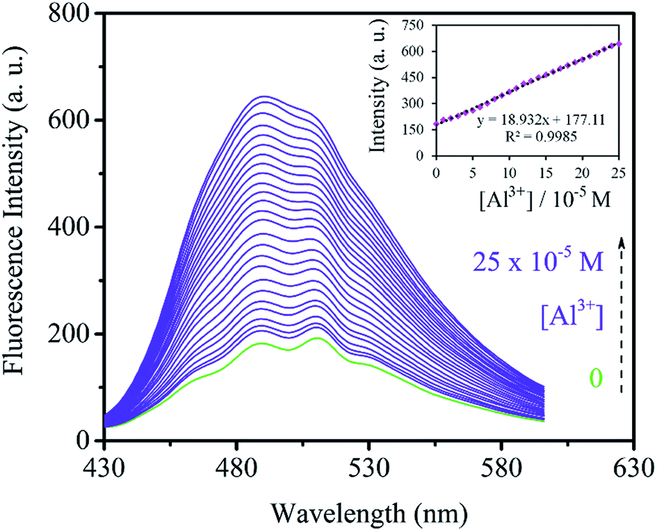 | ||
| Fig. 10 Fluorescence intensity of SSA (3 mL H2O suspension, 0.2 g L−1) upon addition of 30 μL of different concentrations of Al3+: (1, 2, 3, … 25) × 10−5 M (inset: linear increase of fluorescence intensity as a function of Al3+ concentrations). | ||
A selectivity experiment was also performed to evaluate the efficiency of the sensor in CN− detection. The fluorescence intensity of SSA was monitored upon addition of CN− (100 μL, 1 × 10⁻² M) and equivalent amounts of common interfering anions (1 eq.). The results in Fig. 11 demonstrated lack of interference by competing anions in CN− detection, indicating the selectivity of SSA toward CN− anion in water.
 | ||
| Fig. 11 Selectivity of SSA (3 mL H2O suspension, 0.2 g L−1) for CN− (100 μL, 1 × 10⁻² M) in the presence of equal amounts of various metal-ions. | ||
Furthermore, the fluorescence intensity of SSA was studied as a function of CN− concentration. Fig. 12 provides the fluorescence spectra for titration of SSA by CN− anion. The intensity increases gradually upon addition of different amounts of the anion from 0 to 20 × 10−5 M. By plotting the fluorescence intensity against the concentration of CN− anion, a linear plot was obtained which is shown in the inset of Fig. 12. Moreover, the detection limit of 3.61 × 10⁻⁷ M was estimated for CN− anion by the same method used for Al3+.
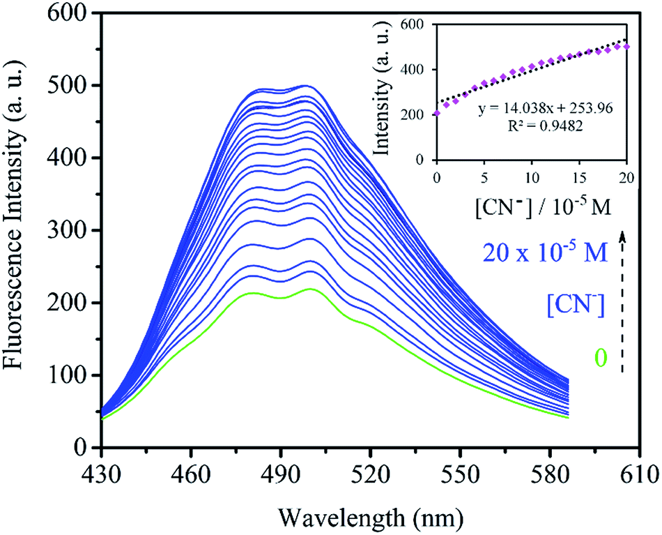 | ||
| Fig. 12 Fluorescence intensity of SSA (3 mL H2O suspension, 0.2 g L−1) upon addition of different concentrations of CN− anion: (1, 2, 3, … 20) × 10−5 M (inset: fluorescent emission as a function of CN− concentrations). | ||
Logic behavior of SSA
Considering the fluorescence responses of SSA discussed above, SSA is capable of acting as a fluorescent logic gate as depicted in Fig. 13. Input 1 and 2 in Fig. 13R represent Al3+ and CN−, respectively. The first row of the truth table shows absence (0 state) of both Al3+ and CN− ions which results in low emission (0 state). The presence (1 state) of Al3+ and absence of CN− leads to high emission (1 state) which is also true for the other way around meaning the presence of CN− ion and absence of Al3+ ion. However, the simultaneous presence of both ions results in no enhancement (0 state). This behavior is assigned to XOR-type logic function. These results indicate that SSA can be used as a XOR-type fluorescent sensor with Al3+ and CN− as inputs. The corresponding fluorescence spectra are provided in Fig. 13L. | ||
| Fig. 13 XOR logic behavior of SSA. L: Fluorescence spectra of (a) SSA, (b) SSA + Al3+, (c) SSA + CN−, and (d) SSA + Al3+ + CN−; R: the truth table and the corresponding logic scheme. | ||
Conclusion
A nanoporous silica-based optical sensor (SSA) was synthesized and successfully characterized. Low angle powder XRD reflections at 2θ around 1, 1.6 and 1.8 degrees appeared in the patterns of SBA-15, SNH, and SSA with steady decrease of intensity after each functionalization step. The N2 absorption–desorption isotherms revealed shrinkage of pore volume, specific surface area, and pore diameter up from SBA-15 down to SSA. By TGA analysis the amount of grafted organic groups onto the pore walls of SBA-15 was estimated as 1.7 mmol g−1. SSA was capable of selective detection of Fe3+ ion visually. The UV-vis studies also demonstrated a totally distinct spectrum for SSA upon addition of Fe3+ from the rest of the metal-ions. The reversibility of SSA in Fe3+ detection was also proved both visually and by absorption spectra. In addition, SSA proved to be a XOR-type logic fluorescent sensor with Al3+ and CN− as inputs. The detection limits of 4.83 × 10⁻⁶, 9.24 × 10⁻⁸, and 3.61 × 10⁻⁷ M were estimated for Fe3+, Al3+ and CN−, respectively which were quite low values. Overall, three ions were selectively detected by two different techniques and all the experiments were performed in water which is a highly valuable factor for a sensor.Acknowledgements
The authors thank the research council of University of Tehran for financial support.References
- J. A. Cowan, Inorganic biochemistry: an introduction, John Wiley & Sons, 1997 Search PubMed.
- N. R. Chereddy, S. Thennarasu and A. B. Mandal, Analyst, 2013, 138, 1334–1337 RSC.
- P. Aisen, M. Wessling-Resnick and E. A. Leibold, Curr. Opin. Chem. Biol., 1999, 3, 200–206 CrossRef CAS PubMed.
- K. Ando and M. Fujita, Clin. Exp. Pharmacol. Physiol., 2012, 39, 111–116 CrossRef CAS PubMed.
- D. Galaris, V. Skiada and A. Barbouti, Cancer Lett., 2008, 266, 21–29 CrossRef CAS PubMed.
- R. Crichton and J. R. Boelaert, Inorganic biochemistry of iron metabolism: from molecular mechanisms to clinical consequences, John Wiley & Sons, 2001 Search PubMed.
- M. J. Behrenfeld, A. J. Bale, Z. S. Kolber, J. Aiken and P. G. Falkowski, Nature, 1996, 383, 508–511 CrossRef CAS.
- S. B. Maity and P. K. Bharadwaj, Inorg. Chem., 2013, 52, 1161–1163 CrossRef CAS PubMed.
- J. R. Sorenson, I. R. Campbell, L. B. Tepper and R. D. Lingg, Environ. Health Perspect., 1974, 8, 3–95 CrossRef CAS PubMed.
- J. M. Donald, M. S. Golub, M. E. Gershwin and C. L. Keen, Neurotoxicol. Teratol., 1989, 11, 345–351 CrossRef CAS PubMed.
- G. D. Fasman, Coord. Chem. Rev., 1996, 149, 125–165 CrossRef CAS.
- R. B. Martin, Acc. Chem. Res., 1994, 27, 204–210 CrossRef CAS.
- M. Sargazi, N. B. Roberts and A. Shenkin, J. Inorg. Biochem., 2001, 87, 37–43 CrossRef CAS PubMed.
- L. Wang, W. Qin, X. Tang, W. Dou, W. Liu, Q. Teng and X. Yao, Org. Biomol. Chem., 2010, 8, 3751–3757 CAS.
- A. Sahana, A. Banerjee, S. Das, S. Lohar, D. Karak, B. Sarkar, S. K. Mukhopadhyay, A. K. Mukherjee and D. Das, Org. Biomol. Chem., 2011, 9, 5523–5529 CAS.
- K. Soroka, R. S. Vithanage, D. A. Phillips, B. Walker and P. K. Dasgupta, Anal. Chem., 1987, 59, 629–636 CrossRef CAS.
- X. Lou, J. Qin and Z. Li, Analyst, 2009, 134, 2071–2075 RSC.
- Z. Xu, X. Chen, H. N. Kim and J. Yoon, Chem. Soc. Rev., 2010, 39, 127–137 RSC.
- A. Ohashi, H. Ito, C. Kanai, H. Imura and K. Ohashi, Talanta, 2005, 65, 525–530 CrossRef CAS PubMed.
- I. Narin, M. Tuzen and M. Soylak, Talanta, 2004, 63, 411–418 CrossRef PubMed.
- A. T. Haj-Hussein, G. D. Christian and J. Ruzicka, Anal. Chem., 1986, 58, 38–42 CrossRef.
- A. Bobrowski, K. Nowak and J. Zarębski, Anal. Bioanal. Chem., 2005, 382, 1691–1697 CrossRef CAS PubMed.
- L. Qiong, W. Lirong, X. Danli and L. Guanghan, Food Chem., 2006, 97, 176–180 CrossRef.
- A. Safavi, N. Maleki and H. Shahbaazi, Anal. Chim. Acta, 2004, 503, 213–221 CrossRef.
- D. Gomes, M. Segundo, J. Lima and A. Rangel, Talanta, 2005, 66, 703–711 CrossRef PubMed.
- T. Guray, Ü. D. Uysal, T. Gedikbey and A. A. Huseyinli, Anal. Chim. Acta, 2005, 545, 107–112 CrossRef CAS.
- J. Ma and P. K. Dasgupta, Anal. Chim. Acta, 2010, 673, 117–125 CrossRef CAS PubMed.
- Z. Zhu and Z. Fang, Anal. Chim. Acta, 1987, 198, 25–36 CrossRef CAS.
- S. Lunvongsa, M. Oshima and S. Motomizu, Talanta, 2006, 68, 969–973 CrossRef CAS PubMed.
- S. H. Sutheimer and S. E. Cabaniss, Anal. Chim. Acta, 1995, 303, 211–221 CrossRef CAS.
- H. N. Kim, W. X. Ren, J. S. Kim and J. Yoon, Chem. Soc. Rev., 2012, 41, 3210–3244 RSC.
- Z.-Q. Liang, C.-X. Wang, J.-X. Yang, H.-W. Gao, Y.-P. Tian, X.-T. Tao and M.-H. Jiang, New J. Chem., 2007, 31, 906–910 RSC.
- S.-P. Wu, Y.-P. Chen and Y.-M. Sung, Analyst, 2011, 136, 1887–1891 RSC.
- S. Kim, J. Y. Noh, K. Y. Kim, J. H. Kim, H. K. Kang, S.-W. Nam, S. H. Kim, S. Park, C. Kim and J. Kim, Inorg. Chem., 2012, 51, 3597–3602 CrossRef CAS PubMed.
- X. Shi, H. Wang, T. Han, X. Feng, B. Tong, J. Shi, J. Zhi and Y. Dong, J. Mater. Chem., 2012, 22, 19296–19302 RSC.
- W.-C. Lin, S.-K. Fang, J.-W. Hu, H.-Y. Tsai and K.-Y. Chen, Anal. Chem., 2014, 86, 4648–4652 CrossRef CAS PubMed.
- C. Bhaumik, S. Das, D. Maity and S. Baitalik, Dalton Trans., 2011, 40, 11795–11808 RSC.
- Y. Jhong, W. H. Hsieh, J.-L. Chir and A.-T. Wu, J. Fluoresc., 2014, 24, 1723–1726 CrossRef CAS PubMed.
- N. Mergu, A. K. Singh and V. K. Gupta, Sensors, 2015, 15, 9097–9111 CrossRef CAS PubMed.
- F. Hoffmann, M. Cornelius, J. Morell and M. Fröba, Angew. Chem., Int. Ed., 2006, 45, 3216–3251 CrossRef CAS PubMed.
- G. Mohammadi Ziarani, N. Lashgari and A. Badiei, J. Mol. Catal. A: Chem., 2015, 397, 166–191 CrossRef CAS.
- Z. Bahrami, A. Badiei, F. Atyabi, H. R. Darabi and B. Mehravi, Mater. Sci. Eng., C, 2015, 49, 66–74 CrossRef CAS PubMed.
- H. Lakhiari, E. Legendre, D. Muller and J. Jozefonvicz, J. Chromatogr. B: Biomed. Sci. Appl., 1995, 664, 163–173 CrossRef CAS PubMed.
- L. Gao, Y. Wang, J. Wang, L. Huang, L. Shi, X. Fan, Z. Zou, T. Yu, M. Zhu and Z. Li, Inorg. Chem., 2006, 45, 6844–6850 CrossRef CAS PubMed.
- Z. Dong, X. Tian, Y. Chen, J. Hou and J. Ma, RSC Adv., 2013, 3, 2227–2233 RSC.
- Z. Dong, Z. Dong, J. Ren, J. Jin, P. Wang, J. Jiang, R. Li and J. Ma, Microporous Mesoporous Mater., 2010, 135, 170–177 CrossRef CAS.
- Z. Bahrami, A. Badiei and F. Atyabi, Chem. Eng. Res. Des., 2014, 92, 1296–1303 CrossRef CAS.
- A. Badiei, H. Goldooz, G. Mohammadi Ziarani and A. Abbasi, J. Colloid Interface Sci., 2011, 357, 63–69 CrossRef CAS PubMed.
- A. E. Newkirk, Anal. Chem., 1960, 32, 1558–1563 CrossRef CAS.
- J. Y. Noh, I. H. Hwang, H. Kim, E. J. Song, K. B. Kim and C. Kim, Bull. Korean Chem. Soc., 2013, 34, 1985–1989 CrossRef CAS.
Footnote |
| † Electronic supplementary information (ESI) available. See DOI: 10.1039/c5ra23136a |
| This journal is © The Royal Society of Chemistry 2016 |