Down shifting and quantum cutting from Eu3+, Yb3+ co-doped Ca12Al14O33 phosphor: a dual mode emitting material

R. V. Yadav, R. S. Yadav, A. Bahadur and S. B. Rai*
Department of Physics, Institute of Science, Banaras Hindu University, Varanasi-221005, India. E-mail: sbrai49@yahoo.co.in; Fax: +91 542 236 9889; Tel: +91 542 230 7308

Received 3rd November 2015 , Accepted 8th January 2016

First published on 12th January 2016


Abstract

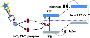
We report the quantum cutting (QC) in a Eu3+, Yb3+ co-doped Ca12Al14O33 phosphor synthesized through combustion method. The X-ray diffraction (XRD), transmission electron microscopy (TEM) and scanning electron microscopy (SEM) measurements reveal the crystalline nature of the phosphor sample. The photoluminescence (PL) spectra of the different samples have been studied using 266 and 394 nm radiation, which give an intense red emission at 612 nm due to 5D07F2 transition. The addition of Yb3+ in the Eu3+ doped sample reduces the emission intensity of Eu3+ bands in the visible region continuously whereas in the NIR region, the intensity of the Yb3+ band first increases up to 2 mol% and then decreases. This is due to cooperative downconversion energy transfer from Eu3+ to Yb3+ ions and concentration quenching. The decay curve analyses of different samples reveal an efficient energy transfer from Eu3+ to Yb3+ ions. The quantum efficiency (QE) has been calculated for different concentrations of Yb3+ ions and is estimated to be 197%. The possible mechanisms involved in different transitions and energy transfer processes can be understood using a schematic energy level diagram. The phosphor sample is a potential candidate for enhancing the efficiency of c-Si solar cells.


1. Introduction

Rare earth doped phosphor materials have been a subject of focus in recent years due to their broad range of potential applications such as blue light-emitting diodes (LEDs), field emission display devices (FEDs), biosensors, magnetic resonance imaging (MRI), medical diagnostics, solar cells, etc.1–5 Among these, the solar cell is one of the interesting applications of rare earth ions used for spectral conversion of ultraviolet (UV)/visible (vis) light into near infrared (NIR) light. In a solar cell, electron–hole (e–h) pairs are produced on irradiation with NIR photons having energy higher than the band gap of the solar cell. In the case of UV/vis light the excess energy of the UV/vis photons transferred to the e–h pair is dissipated in the form of heat, which degrades the efficiency of the materials.6,7 However, the NIR photons obtained after spectral conversion through quantum cutting (QC) contain energy, which is sufficient to generate an e–h pair only. Lot of efforts have been made to improve the solar spectral conversion efficiency but the quantum efficiency is still poor (∼37%). The quantum efficiency determines the optical effectiveness of the material and needs further attention.8

In recent years, researches have been focussed on improving efficiency of solar cell by photon conversion process and use the environment friendly energy source to meet demand, which can reduce the utilization of resources of fossil fuels.9 It is well known that the theoretical photon conversion efficiency of crystalline Si solar cell is 30% having bandgap of 1.12 eV.10,11

The research work based on rare earth doped phosphors in recent years is mainly motivated in two broad areas viz. bio-imaging and energy harvesting. In bio-imaging, the upconverted lanthanide ions are used as bio-marker to improve the image quality from different point of view i.e. penetration depth, lower scattering, negligible auto-fluorescence, high signal to noise ratio, etc.12–15 The second aspect i.e. the energy harvesting in which the photoluminescence (PL), upconversion (UC) and downconversion (DC)/quantum cutting (QC) emission characteristics of lanthanide ions are used. Lanthanides have wide range of absorption of solar light from UV-visible to NIR in better ways. The rare earth doped phosphors emit radiation in NIR region through DC/QC process. The generation of NIR emission is highly useful for solar cell research and are being actively explored worldwide.16,17 Downshifting (DS) is normal photoluminescence process in which conversion of a high energy absorbed photon (266 or 394 nm) into a low energy photon may be in visible region. Therefore, the conversion efficiency is always less than 100% and the excess energy is lost non-radiatively or by emission of photons with small energy. However, QC is a process in which a high energy absorbed photon is converted into two (or more) low energy photons, which increases the quantum efficiency more than 100%.7,18 Thus, there is a large demand of QC materials due to their novel application in the field of solar energy.

The QC phenomenon was first observed in singly Pr3+ doped fluorides host.19,20 This approach is further shifted towards two ion systems in which one ion transfers its excitation energy to other through cooperative downconversion process. There are various combinations of rare earth ions such as Tb3+–Yb3+, Tm3+–Yb3+, Eu2+–Yb3+, Nd3+–Yb3+, etc., co-doped in different fluoride and oxide host materials, which show QC phenomenon to enhance the efficiency of solar cell.8,10,21–27 The QC mechanism in Eu3+–Yb3+ combination is reported very little.28,29 There is no other report seen for the QC mechanisms from Eu3+–Yb3+ pair in Ca12Al14O33 phosphor, which motivated us to design the experiment and study the QC in this combination. We have used Eu3+ and Yb3+ rare earth ions combination in which Eu3+ acts as donor whereas Yb3+ as an acceptor. The lifetime studies confirm that Eu3+ ion transfers its energy from 5D2 level to two Yb3+ ions in 2F7/2 level through cooperative energy transfer process to promote them to 2F5/2 level. Ca12Al14O33 is a host with low phonon frequency (∼800 cm−1) which supports the radiative transitions. This enhances the energy transfer efficiency. Thus, this material can be a potential candidate to improve the efficiency of solar cell.

In this paper, we have synthesized Eu3+, Yb3+ co-doped single cubic Ca12Al14O33 phosphor. The structural characterizations of the synthesized samples have been carried out using X-ray diffraction (XRD), transmission electron microscopy (TEM) and scanning electron microscopy (SEM) measurements. The photoluminescence spectra of the synthesized sample have been recorded using 266 nm from Nd-YAG laser and 394 nm source from Xe-lamp. The lifetime of 5D0 level of Eu3+ in different samples have been measured to verify the energy transfer between Eu3+ and Yb3+ ions. The possible mechanisms involved in the QC have been discussed using energy level diagram.

2. Experimental

2.1 Preparation of Eu3+/Yb3+ co-doped calcium aluminate phosphors

The Eu3+/Yb3+ co-doped calcium aluminate phosphors have been synthesized through solution combustion method. The CaCO3 (99.0%), Al(NO3)3·9H2O (98%), Eu2O3 (99.99%) and Yb2O3 (99.9%) were taken as starting materials in stoichiometric ratio. Urea has been used as organic fuel for combustion. A detailed synthesis procedure has been described earlier.5,25 The concentration of Eu3+ was varied from 0.5 mol% to 7 mol% and optimized at 1 mol%. The concentration of Yb3+ was varied from 1 mol% to 15 mol% with the optimized concentration of Eu3+ ion. These ions have been replaced by Al3+ ions in the host.

2.2 Characterization

The identification of phase and crystallinity in the phosphor samples were confirmed using X-ray diffraction pattern (XRD) using Philips Model PW1830, Cu Kα (λ = 1.54056 Å) radiation at 40 kV and 40 mA. XRD pattern of the synthesized sample was recorded in between 15°–80° range at an interval of 3° per minutes. The transmission electron microscope (TEM) and scanning electron microscope (SEM) micrographs of the sample have been monitored to see the morphological behaviour of the sample. The UV-vis-NIR spectra of the samples have been recorded using diffuse reflectance method with Perkin Elmer UV-vis-NIR spectrometer (Lambda 750). The photoluminescence excitation (PLE) and photoluminescence (PL) measurements have been carried out using a Fluorolog-3 spectrofluorometer equipped with 450 W xenon flash lamp and Nd:YAG laser. The lifetime measurements have been carried out using a pulsed xenon lamp (25 W). The downconversion and quantum cutting spectra were recorded using 266/394 nm excitations.

3. Results and discussion

3.1 Structural characterizations

The XRD pattern of 1 mol% Eu3+, 2 mol% Yb3+ co-doped Ca12All4O33 has been recorded to identify the phase, crystallinity and average crystallite size and is shown in Fig. 1(a). The diffraction patterns match well with pure cubic Ca12All4O33 with lattice constants a = b = c = 11.98 Å and space group I[4 with combining macron]3d (220). The XRD peaks have been assigned using JCPDS file no. 09-0413 (see Fig. 1(b)). This confirms the formation of pure Ca12All4O33 phosphor without any effect of doping concentration. The average crystallite size (D) for the three most intense peaks have been calculated by Debye Scherrer equation:
image file: c5ra23117e-t1.tif
where, λ is the wavelength of incident X-ray [Cu Kα (1.54056 Å)], β is the FWHM (full width at half maxima) and θ is the diffraction angle. The FWHM of the peaks were taken by their Lorentzian peak fitting. The average crystallite size was found to be 64 ± 1 nm.

image file: c5ra23117e-f1.tif
Fig. 1 X-ray diffraction (XRD) pattern (a), its JCPDS file no. 09-0413 (b), transmission electron microscopy (TEM) (c) and scanning electron microscopy (d) images of the annealed 1 mol% Eu3+, 2 mol% Yb3+ co-doped calcium aluminate phosphor sample.

Furthermore, we have recorded the TEM micrograph of the phosphor sample, which shows the nano-particles agglomerated with each other (see Fig. 1(c)). The TEM micrograph further reveals that the particles of Ca12All4O33 co-doped phosphor are spherical in shape with average particle sizes ≈100 ± 5 nm. The spherical shape of the particles is also confirmed by SEM micrograph (see Fig. 1(d)).

3.2 Optical characterizations

3.2.1 UV-visible-NIR diffuse reflection spectroscopy. The UV-vis-NIR diffuse reflectance spectrum of the Eu3+/Yb3+ co-doped Ca12Al14O33 phosphor is shown in Fig. 2. The bands of Eu3+ are very weak at lower concentration and they could be observed with distinct features only at 7 mol% of Eu3+. The absorption band observed in 200–350 nm range is due to the overlap of charge transfer band (CTB) of the host and Eu3+–O2− ions. The spectrum contains bands at 363, 382, 397 and 467 nm of Eu3+ ions due to 7F05L8, 7F05L7, 7F05L6 and 7F05D2 transitions, respectively and they are shown as inset in figure. The spectrum also contains a peak centred at 978 nm in NIR region, which is attributed due to 2F7/22F5/2 transition of Yb3+ ion. This is in good agreement with the excitation spectra of Eu3+/Yb3+ co-doped Ca12Al14O33 phosphor.
image file: c5ra23117e-f2.tif
Fig. 2 The UV-vis-NIR diffuse reflectance spectrum of the 7 mol% Eu3+, 2 mol% Yb3+ co-doped Ca12Al14O33 phosphor.
3.2.2 Photoluminescence excitation spectra of Eu3+/Yb3+ co-doped Ca12Al14O33 phosphor. The photoluminescence excitation (PLE) spectra of the different samples have been recorded in the range of 220–550 nm at λemi = 612 nm and are shown in Fig. 3. The spectra show a broad band in between 230–310 nm due to overlapped charge transfer band (CTB) of Eu3+–O2− and Yb3+–O2−, which is centred at 260 nm.26 The spectra also contain intense peaks at 322, 363, 382, 394, 464 and 535 nm, which are assigned to arise due to the 7F05H6, 7F05L8, 7F05L7, 7F05L6, 7F05D2 and 7F05D1 transitions of Eu3+ ion, respectively. The strongest band is observed at 394 nm due to 7F05L6 transition.
image file: c5ra23117e-f3.tif
Fig. 3 Photoluminescence excitation (PLE) spectra of 1 mol% Eu3+, xYb3+ co-doped Ca12Al14O33 phosphor at different concentration (i.e. x = 0, 1, 2, 4, 6, 10 and 15 mol%) of Yb3+ at λemi = 612 nm.
3.2.3 Photoluminescence and quantum cutting emission in Eu3+/Yb3+ co-doped Ca12Al14O33 phosphor. The photoluminescence spectra of 1 mol% Eu3+, 2 mol% Yb3+ doped and co-doped Ca12Al14O33 phosphors have been recorded in 550–1030 nm region on excitation with 266 nm laser radiation separately and are shown in Fig. 4. It excites the charge transfer band of Eu3+ ion and gives various transitions. The Eu3+ doped and Eu3+, Yb3+ co-doped samples contain several emission peaks at 591, 612, 652 and 700 nm, which are attributed due to 5D07F1, 5D07F2, 5D07F3 and 5D07F4 transitions of Eu3+, respectively, in which 5D07F2 transition appears with maximum intensity. The Eu3+ ions in charge transfer state (CTS) relax through discrete excited states of Eu3+ ion non-radiatively to populate 5Dn states and ultimately the lowest 5D0 states to give various transitions. On addition of Yb3+ in Eu3+ the emission intensity of the Eu3+ decreases. The Eu3+, Yb3+ co-doped sample also contains a broad NIR band at 976 nm due to 2F5/22F7/2 transition of Yb3+. The CTB of Yb3+ is very deep and transfers its excitation energy to 2F5/2 of Yb3+ ion.26 Therefore, the singly Yb3+ doped sample on excitation with 266 nm radiation gives also a broad NIR band centred at 982 nm due to 2F5/22F7/2 transition.
image file: c5ra23117e-f4.tif
Fig. 4 Photoluminescence (PL) spectra of 1 mol% Eu3+ doped; 1 mol% Eu3+, 2 mol% Yb3+ co-doped and 2 mol% Yb3+ doped Ca12Al14O33 phosphor on excitation with 266 nm.

As mentioned above when different concentrations of Yb3+ are doped with the Eu3+ doped sample the emission intensity of Eu3+ bands decreases continuously with Yb3+ concentrations. This is due to energy transfer from Eu3+ to Yb3+ ions. As we add Yb3+, the emission intensity of the Eu3+ peaks initially decreases rapidly upto 2 mol%; and then slowly depending on the energy transfer rate. The PL spectra of phosphor for different concentration of Yb3+ ion with a fixed concentration of Eu3+ recorded in 550–1030 nm range are shown in Fig. 5. On the other hand, the emission intensity of Yb3+ initially increases upto 2 mol% and then decrease for its higher concentration.


image file: c5ra23117e-f5.tif
Fig. 5 Photoluminescence (PL) spectra of 1 mol% Eu3+, xYb3+ co-doped Ca12Al14O33 phosphor at different concentration (i.e. x = 0, 1, 2, 4 and 6 mol%) of Yb3+ on excitation with 266 nm radiation.

The 266 nm radiation excites the CTB of Eu3+ as well as Yb3+ ions to give fluorescence. We therefore selected 394 nm radiation, which excites Eu3+ ion only. We have recorded the photoluminescence spectra of Eu3+, Yb3+ doped and co-doped Ca12Al14O33 phosphors at different concentration of Yb3+ ion with a fixed concentration of Eu3+(at 1 mol%) in 550–1030 nm region on excitation with 394 nm and they are shown in Fig. 6.


image file: c5ra23117e-f6.tif
Fig. 6 Photoluminescence (PL) spectra of 1 mol% Eu3+ doped; 1 mol% Eu3+, 2 mol% Yb3+ co-doped and 2 mol% Yb3+ doped Ca12Al14O33 phosphor on excitation with 394 nm.

It is observed that the emission spectra show similar emission peaks in the visible region with larger emission intensity compared to previous case because Eu3+ ion has larger absorption cross section for 394 nm photon. There is no peak seen in NIR region on excitation with 394 nm from the pure Yb3+ doped sample because the charge transfer band of Yb3+ ion lies far apart from 394 nm excitation. Therefore, the 394 nm wavelength does not excite Yb3+ ion. The Yb3+ ion is only excited in Yb3+:Eu3+ sample by cooperative downconversion energy transfer from Eu3+ to Yb3+ and results a broad band at 976 nm. The emission intensity of Eu3+ bands decreases on increasing the concentration of Yb3+ ions exactly in the same way as in earlier case and the effect of concentration of Yb3+ ions are shown in Fig. 7. The spectrum has no peak in the NIR region in the absence of Yb3+ ion. As soon as Yb3+ is added in sample, a broad NIR peak appears in 930–1030 nm range centred at 976 nm due to 2F5/22F7/2 transition of Yb3+ ion. The emission intensity of Eu3+ bands also decreases with Yb3+ concentration. The decrease in emission intensity is due to energy transfer from Eu3+ to Yb3+ ions through QC process. We have also optimized the emission intensity in NIR region with concentration of Yb3+ ions and it is maximum at 2 mol%. If the concentration of Yb3+ ions is increased further, the emission intensity of Yb3+ peak decreases due to concentration quenching.


image file: c5ra23117e-f7.tif
Fig. 7 Photoluminescence (PL) spectra of 1 mol% Eu3+, xYb3+ co-doped Ca12Al14O33 phosphor at different concentration (i.e. x = 0, 1, 2, 4 and 6 mol%) of Yb3+ on excitation with 394 nm radiation.

As mentioned earlier the intensity of the bands due to Eu3+ decreases regularly whereas in the NIR region due to Yb3+ initially increases and then decreases on increasing the concentration of Yb3+ ions. The variation in the emission intensity strongly depends on the separation between Eu3+ and Yb3+ ions, which determines the energy transfer efficiency. Thus for a particular concentration of Yb3+, the separation between the Eu3+ and Yb3+ ions are in the range of critical distance. This enhances the energy transfer rate and thereby the emission intensity of Yb3+ ions. The emission intensity in NIR region is optimum at 2 mol% concentration of Yb3+ ions. When the concentration of Yb3+ ions is increased further, the emission intensity decreases due to concentration quenching by Yb3+ ion. Actually in presence of Yb3+ ions at higher concentration, one Eu3+ ion in 5D2 level interacts with two Yb3+ ions and transfer its energy to them to promote from 2F7/2 to 2F5/2 level. The Yb3+ ions when relax radiatively to 2F7/2, emit photons of 976 nm. The probability of interaction of one Eu3+ donor with two Yb3+ acceptor ions increases with increasing concentration and leads to QC phenomenon. This enhances the energy transfer and quantum cutting efficiency at higher Yb3+ concentration. However, the concentration quenching by Yb3+ ions start playing role after 2 mol% concentration of Yb3+ and therefore the emission intensity starts decreasing in NIR region.

The mechanism involved in QC can be understood on the basis of schematic energy level diagram shown in Fig. 8. When the sample is excited using 266 nm wavelengths, the ions are promoted to charge transfer states of Eu3+ as well as Yb3+. The Eu3+ ions relax to 5L6 level non-radiatively, which relax further to populate 5D2 excited level. Since the lifetime of this level is relatively large and behaves as a metastable state. The ions in the 5D2 level can relax in two different ways. One, it can relax non-radiatively to populate 5D0 level via 5D1 level. The ions in 5D0 level emit photons of different wavelengths and give transitions to various low lying levels. The other way may be that the ions in 5D2 level transfer their energy to two Yb3+ ions simultaneously through cooperative energy transfer process and populates 2F5/2 level of Yb3+ (because the energy of 5D2 level of Eu3+ matches well with the energy of two Yb3+ ions in 2F5/2 level). The Yb3+ ions when relax to ground state, emit two NIR photons (quantum cutting). A similar result has also been reported by Liu et al.7


image file: c5ra23117e-f8.tif
Fig. 8 Schematic energy level diagram of Eu3+/Yb3+ co-doped calcium aluminate phosphor, illustrating the probable cooperative energy transfer mechanism of NIR QC from 5D2 level of Eu3+ to Yb3+ ions in ground state. The solid arrows indicate the excitation and the emission processes and the dotted arrows represent the non-radiative transition and energy-transfer processes.

On the other hand the 266 nm radiation populates the charge transfer state of Yb3+ ion also. The charge transfer state of Yb3+ is very deep and it crosses the 2F5/2 level of Yb3+ ion. Thus, when the Yb3+ ions are excited to its charge transfer state it populates 2F5/2 level, which gives a band at 982 nm due to 2F5/22F7/2 transition. Thus, the 266 nm excitation gives fluorescence due to Yb3+ as well as Eu3+.

On the other hand when we excite the sample with 394 nm radiation, it selectively excites the 5L6 level of Eu3+ (not CTS of Yb3+). Thus, in this case we get bands due to Eu3+ ions only. However, if the concentration of Yb3+ is increased, the emission due to Yb3+ is also seen at 982 nm through cooperative energy transfer process. Since the population in 5D0 level arise due to relaxation from 5D2 level. The population in 5D0 is affected due to the presence of another channel of energy transfer. It will not only affect the intensity of spectral lines arising from 5D0 but also the lifetime of this level.

In order to understand the effect of energy transfer we measured lifetime of the 5D0 state at 612 nm in presence and absence of Yb3+ and calculated the corresponding energy transfer and quantum cutting efficiency for different concentrations of Yb3+ ion exciting the sample with 266 as well as 394 nm radiations.

3.2.4 Lifetime measurements of Eu3+/Yb3+ co-doped Ca12Al14O33 phosphor. We have monitored the fluorescence decay curves of 5D07F2 transition for Eu3+ doped and Eu3+, Yb3+ co-doped Ca12Al14O33 phosphor at different concentration of Yb3+ on excitation with 266 and 394 nm and they are shown in Fig. 9. The decay curves were found to fit well with single exponential equation:
image file: c5ra23117e-t2.tif
where, I0 and I are the intensities at time 0 and t s, respectively and τ is the lifetime. The lifetimes thus obtained are given in Table 1.

image file: c5ra23117e-f9.tif
Fig. 9 Decay curves for 5D07F2 transition of Eu3+ ion in Eu3+/Yb3+ co-doped Ca12Al14O33 phosphors for different concentration of Yb3+ on excitations with (a) 266 nm and (b) 394 nm.
Table 1 Lifetime, ET efficiency and quantum efficiency of Eu3+, Yb3+ co-doped Ca12Al14O33 phosphors for different concentration of Yb3+ on excitations with 266 and 394 nm
Yb3+ concentration (mol%) (λemi = 612 nm)
(λexc = 266 nm) (λexc = 394 nm)
Lifetime (μs) ηET (in %) ηQE (in %) Lifetime (μs) ηET (in %) ηQE (in %)
0 1558 0 100 1442 0 100
1 844 45 145 832 42 142
2 610 60 160 527 63 163
6 576 63 163 515 64 164
10 411 73 173 177 87 187
15 85 94 194 42 97 197


It is clear from the table that the lifetime of 5D0 level of Eu3+ decreases continuously on increasing the concentration of Yb3+ ions in the phosphor.28,29 The QC emission is mainly due to cooperative energy transfer (CET) from 5D2 level of Eu3+ to 2F7/2 level of Yb3+. As the concentration of Yb3+ ion increases the emission intensity of Eu3+ bands decreases and the corresponding lifetime also decreases simultaneously. The decrease in lifetime also depends on population/depopulation rates of 5D2 level. Since the relaxation of ions and energy transfer take place from 5D2 level of Eu3+, it is assumed that the rate of relaxation of ions from 5D2 level of Eu3+ to 5D0 decrease with the increase of Yb3+ ion and thereby the lifetime of 5D0 level also decrease vice versa, a decrease in emission intensity of bands and lifetime of 5D0 level supports the CET from 5D2 level of Eu3+ to 2F7/2 level of Yb3+. It is well known that CET process is not efficient at very low or very high concentration due to the intrinsic properties of Yb3+ ion. It can be more efficient in between the two. When the concentration of Yb3+ is increased from 0 to 2 mol% the corresponding lifetime is decreased from 1558 to 610 μs and from 1442 to 527 μs in the two cases. Therefore, the decay curves show non-exponential behaviour due to involvement of additional decay pathways, which suggests the involvement of CET process (see Table 1). Therefore, the excitation emission and lifetime studies reveal the conversion of one 266/394 photon into two NIR photons.26 The energy transfer efficiency from Eu3+ to Yb3+ (ηET) can be calculated by the following equation:

image file: c5ra23117e-t3.tif
where, τx and τ0 are the fluorescence lifetimes of Eu3+/Yb3+ co-doped and singly Eu3+ doped phosphors, respectively.

The value of ηET calculated for different concentration of Yb3+ ion using above relation is summarized in Table 1.

The table shows that energy transfer (ET) efficiency increases with increasing Yb3+ concentration, and its value is found to be 97%, which is maximum for 15 mol% of Yb3+. The internal quantum efficiency (ηQE) for the phosphor sample can be calculated by:

 
ηQE = ηEu(1 − ηET) + 2ηET (1)

The first term in the relation belongs to the visible photons emitted by Eu3+ ions and the second term refers to the corresponding NIR photons emitted by Yb3+. Here, ηEu represents the quantum efficiency of emission of Eu3+ ions. Assuming that the non-radiative transitions to be zero, ηEu = 1, the theoretically estimated ηQE for 15 mol% concentration of Yb3+ is found to be 197%.

This suggests that the absorption of 100 photons (i.e. 394 nm radiation) produces a total number of 197 photons (NIR and visible both), out of which 194 photons are emitted in 900–1050 nm (i.e. NIR region) and the remaining 3 photons are emitted in visible region for Ca12Al14O33:1% Eu3+, 15% Yb3+ phosphor. The calculated value for the internal QE increases with increasing concentration of the Yb3+ as shown in Table 1.

The UV/visible to NIR QC in phosphor materials are of great interest for c-Si solar cell applications. It is well known that the c-Si solar cells have poor efficiency in UV and visible regions. The maximum spectral conversion efficiency of c-Si solar cell is in 900–1030 nm region.6 The NIR QC in the synthesized phosphor can be achieved on excitation with UV/visible photons. The utilization of these NIR photons leads to generate e–h pairs in silicon, which enhances the current in c-Si solar cell, etc. A working model illustrating the quantum cutting process and mechanism to enhance solar cell efficiency is summarized in Fig. 10. If an electron is excited from valence band to conduction band by 266/394 nm, almost half of the excitation energy is lost by thermal vibration/relaxation. However, if the energy of incidence photon is divided to emit two NIR photons by QC process there will not be any energy loss. The two NIR photons (λ = 976 nm, = 1.26 eV) emitted by the QC from 5D2 level of Eu3+ through cooperative downconversion process populates the 2F5/2 level of Yb3+. As a result two electrons together exceeds the band gap (Eg = 1.12 eV) of single-crystalline Si, and the electric current thus becomes double theoretically. In this way, a highly efficient silicon-based solar cell could be realized through the NIR QC process.


image file: c5ra23117e-f10.tif
Fig. 10 The working model represents the application of QC phosphor in c-Si solar cell. The NIR photons are emitted through cooperative energy transfer, which matches with the band gap (1.12 eV) of Si. They are absorbed to produce e–h pair, which enhances the flow of solar current.

4. Conclusions

The Eu3+/Yb3+ co-doped calcium aluminate phosphors have been synthesized through combustion method. The structural studies reveal crystalline nature of the synthesized sample. The phosphor sample gives efficient visible and NIR emissions via downconversion (DC)/quantum cutting (QC) and downshifting (DS) processes on excitation with 266 and 394 nm wavelengths. The presence Yb3+ in the Eu3+ doped samples decreases the emission intensity of Eu3+ in visible region continuously. However in the NIR region, the emission intensity increases initially upto 2 mol% concentration of Yb3+ ions and then starts decreasing due to concentration quenching. An efficient cooperative downcoversion energy transfer has been observed from Eu3+ to Yb3+ ions, which leads to quantum cutting phenomenon. The energy transfer efficiency has been calculated for different concentration of Yb3+ ions and is found to be maximum at 15 mol% concentration of Yb3+ ions as 97% whereas the corresponding QC efficiency is achieved maximum as 197%. Thus, the Eu3+/Yb3+ co-doped calcium aluminate phosphor is a promising candidate for energy conversion in c-Si solar cell applications.

Acknowledgements

The authors are thankful to Department of Science and Technology, New Delhi, India (Grant no. SR/S2/LOP-023/2012) for providing financial assistance.

References

  1. K. Korthout, P. F. Smet and D. Poelman, Appl. Phys. Lett., 2011, 98, 261919–261921 CrossRef.
  2. S. E. Brinkley, N. Pfaff, K. A. Denault, Z. Zhang, H. T. B. Hintzen, R. Seshadri, S. Nakamura and S. P. DenBaars, Appl. Phys. Lett., 2011, 99, 241106–241108 CrossRef.
  3. X. Wang, O. S. Wolfbeis and R. J. Meier, Chem. Soc. Rev., 2013, 42, 7834–7869 RSC.
  4. E. Hemmer, N. Venkatachalam, H. Hyodo, A. Hattori, Y. Ebina, H. Kishimoto and K. Soga, Nanoscale, 2013, 5, 11339–11361 RSC.
  5. R. V. Yadav, S. K. Singh and S. B. Rai, RSC Adv., 2015, 5, 26321–26327 RSC.
  6. T. Trupke, M. A. Green and P. Wurfel, J. Appl. Phys., 2002, 92, 1668–1674 CrossRef CAS.
  7. X. Huang, S. Han, W. Huang and X. Liu, Chem. Soc. Rev., 2013, 42, 173–201 RSC.
  8. J. Sun, W. Zhou, Y. Sun and J. Zeng, Opt. Commun., 2013, 296, 84–86 CrossRef CAS.
  9. M. Zhang, Y. Lin, T. J. Mullen, W. F. Lin, L. D. Sun, C. H. Yan, T. E. Patten, D. Wang and G. Y. Liu, J. Phys. Chem. Lett., 2012, 3, 3188–3192 CrossRef CAS PubMed.
  10. J. Sun, Y. Sun, C. Cao, Z. Xia and H. Du, Appl. Phys. B, 2013, 111, 367–371 CrossRef.
  11. D. Serrano, A. Braud, J. L. Doualan, W. Bolanos, R. Moncorge and P. Camy, Phys. Rev. B, 2013, 88, 205144–205154 CrossRef.
  12. H. Guo and S. Sun, Nanoscale, 2012, 4, 6692–6706 RSC.
  13. X. Li, R. Wang, F. Zhang, L. Zhou, D. Shen, C. Yao and D. Zhao, Sci. Rep., 2013, 3, 3536–3542 Search PubMed.
  14. Y. Sun, J. Peng, W. Feng and F. Li, Theranostics, 2013, 3, 346–353 CrossRef PubMed.
  15. S. K. Singh, RSC Adv., 2014, 4, 58674–58698 RSC.
  16. Y. Z. Wang, D. C. Yu, H. H. Lin, S. Ye, M. Y. Peng and Q. Y. Zhang, J. Appl. Phys., 2013, 114, 203510–203514 CrossRef.
  17. S. Lian, C. Rong, D. Yin and S. Liu, J. Phys. Chem. C, 2009, 113, 6298–6302 CAS.
  18. L. Li, X. Zhou, X. Wei, C. G. Ma and M. G. Brik, Mater. Chem. Phys., 2014, 147, 860–866 CrossRef CAS.
  19. W. W. Piper, J. A. DeLuca and F. S. Ham, J. Lumin., 1974, 8, 344–348 CrossRef CAS.
  20. J. L. Sommerdijk, A. Bril and A. W. de Jager, J. Lumin., 1974, 8, 341–343 CrossRef CAS.
  21. T. C. Liu, G. Zhang, X. Qiao, J. Wang, H. J. Seo, D. P. Tsai and R. S. Liu, Inorg. Chem., 2013, 52, 7352–7357 CrossRef CAS PubMed.
  22. X. Liu, S. Ye, Y. Qiao, G. Dong, B. Zhu, D. Chen, G. Lakshminarayana and J. Qiu, Appl. Phys. B, 2009, 96, 51–55 CrossRef CAS.
  23. S. Ye, B. Zhu, J. Chen, J. Luo and J. R. Qiu, Appl. Phys. Lett., 2008, 92, 141112–141114 CrossRef.
  24. L. Xie, Y. Wang and H. Zhang, Appl. Phys. Lett., 2009, 94, 061905–061907 CrossRef.
  25. R. V. Yadav, S. K. Singh, R. K. Verma and S. B. Rai, Chem. Phys. Lett., 2014, 599, 122–126 CrossRef CAS.
  26. J. Zhou, Y. Zhuang, S. Ye, Y. Teng, G. Lin, B. Zhu, J. Xie and J. Qiu, Appl. Phys. Lett., 2009, 95, 141101–141103 CrossRef.
  27. Z. Liu, N. Dai, L. Yang and J. Li, Appl. Phys. A, 2015, 119, 553–557 CrossRef CAS.
  28. J. Liao, D. Zhou, S. Liu, H. R. Wen, X. Qiu and J. Chen, Phys. B, 2014, 436, 59–63 CrossRef CAS.
  29. M. K. Lau and J. H. Hao, Energy Procedia, 2012, 15, 129–134 CrossRef CAS.

This journal is © The Royal Society of Chemistry 2016