Branched polyethyleneimine modified with hyaluronic acid via a PEG spacer for targeted anticancer drug delivery

Chen Chena, Benqing Zhoua, Xiaoyue Zhua, Mingwu Shena and Xiangyang Shi*ab
aCollege of Chemistry, Chemical Engineering and Biotechnology, Donghua University, Shanghai 201620, People's Republic of China. E-mail: xshi@dhu.edu.cn
bState Key Laboratory for Modification of Chemical Fibers and Polymer Materials, Donghua University, Shanghai 201620, People's Republic of China

Received 2nd November 2015 , Accepted 12th January 2016

First published on 15th January 2016


Abstract

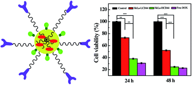
It is generally required to develop a nanocarrier system that is able to improve the water solubility of an anticancer drug and enable targeted delivery of the drug to cancer cells via a receptor-mediated endocytosis pathway. In this work, polyethyleneimine (PEI) was sequentially modified with dual functional polyethylene glycol (NH2–PEG–COOH), hyaluronic acid (HA), and fluorescein isothiocyanate (FI). The prepared PEI–FI–(PEG–HA) conjugate was then used as a nanoplatform to encapsulate the anticancer drug doxorubicin (DOX). We show that the formed PEI–FI–(PEG–HA) conjugate is able to encapsulate approximately 19 DOX molecules within each multifunctional PEI, and the formed PEI–FI–(PEG–HA)/DOX complexes can release DOX in a pH-dependent manner with a higher DOX release rate under an acidic pH condition than under a physiological pH condition. In addition, the PEI–FI–(PEG–HA)/DOX complexes are able to specifically target cancer cells overexpressing CD44 receptors as confirmed via flow cytometric analysis and confocal microscopic observation, and thus deliver DOX to the target cancer cells to inhibit their growth. The developed HA-targeted PEI may hold great promise to be used as an efficient nanoplatform for the targeted delivery of different anticancer drugs.


Introduction

Chemotherapy has been considered as one of the most important approaches used for cancer treatment. However, most of the current anticancer drugs are not able to achieve satisfactory clinical efficacy because of their poor water solubility, limited bioavailability, side effects to harm normal tissues, and poor release kinetics.1–3 It is of great importance to develop an effective nanoplatform/carrier for anticancer drug delivery to overcome the above limitations.4–11

Branched polyethyleneimine (PEI) possessing a large number of amine groups has excellent water solubility and is able to be used as a golden standard for gene delivery.12–14 In addition, PEI can also be used as a template or stabilizer to synthesize different nanoparticle (NP) systems for biomedical applications.13,15–17 For instance, PEI-stabilized iron oxide NPs can be synthesized under a hydrothermal condition18 and the formed NPs can be further biofunctionalized via PEI amine-mediated conjugation chemistry for magnetic resonance (MR) imaging of tumors.15,19 PEI with surface amines modified with polyethylene glycol (PEG) can be used as a template to synthesize Au NPs for computed tomography imaging of tumors and blood pool imaging.16 In the latter case, it seems that PEGylation of PEI surface amines is an effective approach to enlarge the periphery of PEI, to reduce its cytotoxicity, and to render the particles with prolonged blood circulation time, similar to dendrimers.20 Hence, the interior of PEGylated PEI is able to effectively encapsulate Au NPs. Based on these prior successes, it is reasonable to hypothesize that PEGylated PEI may be used as a nanocarrier to encapsulate anticancer drugs to significantly improve the water solubility and bioavailability of the drugs. Simultaneously, the release kinetics of the anticancer drugs may also be effectively improved by encapsulate the drug molecules within PEGylated PEI as a carrier system, similar to other drug delivery systems.21–23

For targeted anticancer drug delivery, it is essential to modify targeting ligands onto the NP systems.2,3 Hyaluronic acid (HA) is a broad-spectrum targeting agent that can target CD44 receptor-overexpressing cancer cells.24–31 Besides, HA also has good biocompatibility, hence the modification of HA onto the particle surface can further improve the biocompatibility of the carrier system. Our previous work has shown that HA can be readily modified onto the surface of iron oxide NPs via PEI amine-mediated 1-ethyl-3-(3-dimethylaminopropyl) carbodiimide hydrochloride (EDC) chemistry for targeted MR imaging of CD44 receptor-expressing tumors.19,32 In addition, the structure of PEGylated PEI is quite similar to that of dendrimers in terms of their hydrophobic branched interiors. In this context, the drug encapsulation and release mechanisms using PEGylated PEI as a delivery system could be similar to those using dendrimers as a delivery system.33–35 Therefore, we also hypothesize that PEGylated PEI can be modified with HA for targeted drug delivery to inhibit CD44 receptor-overexpressing cancer cells.

In this present study, PEI was modified with HA via a PEG spacer. It is known that PEGylation modification of NPs is able to render the NPs with improved biocompatibility and antifouling property.16,20,33,36–39 The formed PEI–PEG was then linked with HA via EDC chemistry, followed by modification of fluorescein isothiocyanate (FI) in order to track cellular binding of the conjugate. The formed PEI–FI–(PEG–HA) conjugate was used to encapsulate an anticancer drug doxorubicin (DOX) (Scheme 1). The prepared PEI–FI–(PEG–HA) conjugate and the PEI–FI–(PEG–HA)/DOX complexes were characterized via different techniques. The DOX release from the PEI–FI–(PEG–HA)/DOX complexes was investigated under different pH conditions. Finally, the antitumor efficacy and the performance of HA-mediated targeted delivery of the PEI–FI–(PEG–HA)/DOX complexes were investigated via cell viability assay, flow cytometric analysis, and confocal laser scanning microscopic (CLSM) observation, respectively. To the best of our knowledge, this is the first report to develop HA-targeted PEI as a nanocarrier for anticancer drug delivery applications.


image file: c5ra23022e-s1.tif
Scheme 1 Schematic illustration of the formation of the PEI–FI–(PEG–HA)/DOX complexes.

Experimental

Materials

Branched PEI (Mw = 25[thin space (1/6-em)]000) was purchased from Aldrich (St. Louis, MO). Dual functional PEG with one end of carboxyl group and the other end of amine group (NH2–PEG–COOH, Mw = 2000) was from Shanghai Yanyi Biotechnology Corporation (Shanghai, China). EDC and N-hydroxysuccinimide (NHS) were from J&K Chemical, Ltd. (Shanghai, China). Doxorubicin hydrochloride (DOX·HCl) was from Beijing Huafeng Pharmaceutical Co., Ltd. (Beijing, China). HeLa cells (a human cervical cancer cell line) was supplied from Institute of Biochemistry and Cell Biology (the Chinese Academy of Sciences, Shanghai, China). Dulbecco's Modified Eagle Medium (DMEM), fetal bovine serum (FBS), penicillin, and streptomycin were from Hangzhou Jinuo Biomedical Technology (Hangzhou, China). All chemicals and materials were used as received. Water used in all experiments was purified using a Milli Q Plus 185 water purification system (Millipore, Bedford, MA) with a resistivity higher than 18.2 MΩ cm. Regenerated cellulose dialysis membranes (molecular weight cut-off, MWCO = 14[thin space (1/6-em)]000) were acquired from Fisher (Pittsburgh, PA).

Synthesis of PEI–FI–(PEG–HA) conjugate

Under magnetic stirring, NH2–PEG–COOH (40.0 mg) dissolved in 5 mL water was activated by EDC (19.2 mg) and NHS (11.5 mg) for 3 h. Then the activated NH2–PEG–COOH was dropped into a PEI solution (25.0 mg, in 10 mL DMSO) at room temperature, and stirred for 3 days to complete the reaction. The reaction mixture was extensively dialyzed against phosphate buffered saline (PBS, 3 times, 2 L) and water (6 times, 2 L) for 3 days using a dialysis membrane (MWCO = 14[thin space (1/6-em)]000) to remove the excess reactants, followed by a freeze-drying process to obtain the PEI–PEG conjugate.

HA was then modified onto the surface of PEI–PEG via EDC chemistry according to protocols described in our previous work.19,32 In brief, the carboxyl groups of HA (95.8 mg, in 10 mL water) were activated by EDC (15.3 mg, in 5 mL DMSO) for 3 h under vigorous stirring. Then the activated HA was dropwise added to a PEI–PEG solution (25.0 mg, in 10 mL water) under stirring for 3 days, followed by addition of FI (10 mol equiv. of PEI, 2.0 mg, in 2 mL DMSO) under stirring for 24 h. Then the mixture was purified and lyophilized using the same procedure described above to obtain the final product of PEI–FI–(PEG–HA) conjugate. The intermediate products of PEI–PEG and PEI–(PEG–HA) were collected and purified to quantify the modification degree of PEG and HA on each PEI.

Encapsulation of DOX within PEI–FI–(PEG–HA) conjugate

DOX was encapsulated within the PEI–FI–(PEG–HA) conjugate according to our previous work.33,34 In brief, DOX·HCl (1.0 mg mL−1, 5 mL) with 20 mol equiv. of PEI–FI–(PEG–HA) conjugate was dissolved in 300 μL methanol, and neutralized by addition of 10 μL triethylamine. The DOX solution was then dropped into an aqueous solution of PEI–FI–(PEG–HA) (10.0 mg mL−1, 10 mL) under vigorous stirring for 12 h to evaporate the methanol solvent. Then, the mixture was centrifuged (5 min, 8000 rpm) to remove the non-encapsulated free DOX, which is a precipitate. Lastly, the supernatant was lyophilized to obtain the PEI–FI–(PEG–HA)/DOX complex.

Characterization techniques

1H NMR spectra were recorded using Bruker AV400 nuclear magnetic resonance spectrometer (Karlsruhe, Germany). Samples were dissolved into D2O before NMR measurements. UV-vis spectra were acquired by a Lambda 25 UV-vis spectrophotometer (PerkinElmer, Waltham, MA). Samples were dissolved in water, methanol, or PBS before measurements. Transmission electron microscopy (TEM) was performed using a JEOL 2010F analytical electron microscope (JEOL, Tokyo, Japan) operating at 200 kV. TEM samples were prepared by dropping a dilute particle suspension (5 μL) onto a carbon-coated copper grid and air-dried before measurements. Zeta potential and dynamic light scattering (DLS) measurements were carried out using a Malvern Zetasizer Nano ZS model ZEN 3600 (Worcestershire, UK) equipped with a standard 633 nm laser. Fourier transform infrared (FTIR) spectroscopy was performed using a Nicolet Nexus 670 FTIR spectrophotometer (Nicolet Thermo, Waltham, MA).

In vitro drug release study

The in vitro DOX release from the PEI–FI–(PEG–HA)/DOX complexes was investigated under two different pH conditions. In brief, PEI–FI–(PEG–HA)/DOX complex (5.0 mg) was dissolved in 1 mL of PBS (pH = 7.4) or acetate buffer (pH = 5.4), placed in a dialysis bag with an MWCO of 14[thin space (1/6-em)]000, and immersed in 9 mL corresponding buffer medium. All the samples were placed in a vapor bathing constant temperature vibrator at 37 °C. We took out 1 mL buffer medium from the outer phase of each sample vial at each predetermined time point and equal volume of the corresponding buffer was replenished. The concentration of the released DOX was tested by UV-vis spectroscopy. Release of free DOX·HCl was also tested under similar experimental conditions.

Cell culture

HeLa cells were cultured in DMEM supplemented with 10% FBS, 100 U mL−1 penicillin, and 100 μg mL−1 streptomycin at 37 °C and 5% CO2. The HeLa cells cultured in HA-free medium expressed high-level CD44 receptors (denoted as HeLa-HCD44), while the HeLa cells cultured in HA-containing medium (5.0 μM) expressed low-level CD44 receptors (denoted as HeLa-LCD44). Unless otherwise stated, the term HeLa cells always represents the HeLa-HCD44 cells.

Cytotoxicity assay and morphological observation

To detect the therapeutic efficacy of the PEI–FI–(PEG–HA)/DOX complexes, MTT assay of HeLa cell viability was performed. HeLa cells were first seeded into a 96-well culture plate with a density of 1 × 10⁴ cells per well, and cultured with 200 μL fresh medium for 24 h. Then the medium was replaced with 200 μL fresh medium containing free DOX in 20 μL normal saline (NS) at a DOX concentration of 4.0, 8.0, 10.0, 15.0, or 20.0 μg mL−1, respectively, or PEI–FI–(PEG–HA)/DOX complex with the same final DOX concentrations. Cells treated with the PEI–FI–(PEG–HA) conjugate were also tested after treated with equivalent PEI concentration to the corresponding PEI–FI–(PEG–HA)/DOX complex for comparison. In addition, cells treated with NS were used as the negative control. After incubated for 24 h, the medium was removed, and each well was washed with PBS for 3 times, and added with 200 μL fresh medium containing 20 μL MTT solution (5.0 mg mL−1 in PBS). After the cells were incubated for another 4 h, the medium was then discarded carefully, and DMSO (200 μL) was added into each well. The absorbance at 570 nm in each well was measured by a Thermo Scientific Multiskan MK 3 ELISA reader (Thermo Scientific, Waltham, MA). Mean and standard deviation of triplicate wells were reported for each sample.

After treated with free DOX, PEI–FI–(PEG–HA), or PEI–FI–(PEG–HA)/DOX complex for 24 h, the morphology of cells was observed by a Leica DM IL LED inverted phase contrast microscope (Wetzlar, Germany) with a magnification of 200× for each sample.

Flow cytometric analysis

To detect the uptake of DOX within HeLa cells, the DOX fluorescence within the cells was analyzed by a FACS Calibur flow cytometer (Becton Dickinson, Mountain View, CA). Briefly, both HeLa-HCD44 and HeLa-LCD44 cells were separately seeded into a 24-well plate at a density of 1 × 10⁵ cells per well. After overnight incubation, the medium was replaced with refresh medium containing PEI–FI–(PEG–HA)/DOX complexes at different DOX concentrations (8.0, 10.0, or 15.0 μg mL−1, respectively). After incubated for 4 h, the medium was discarded, and each well was washed with PBS for 3 times. Then the cells were trypsinized, centrifuged, and resuspended in 1 mL of PBS. Each sample was measured for 3 times, and 1 × 10⁴ cells were counted. HeLa-HCD44 cells treated with NS were used as the blank control.

Confocal microscopic observation

The targeted uptake of PEI–FI–(PEG–HA)/DOX complexes by HeLa cells was observed with CLSM (Carl Zeiss LSM 700, Jena, Germany). Coverslips with a diameter of 14 mm were pretreated with 5% HCl for 4 h, 30% HNO3 for 12 h, washed with water for 10 times, sterilized, and fixed in a 12-well culture plate. After that, both HeLa-HCD44 and HeLa-LCD44 cells were separately plated into the 12-well culture plate at a density of 2 × 10⁵ cells per well, and cultured with fresh medium for 24 h to allow the cells to adhere onto the coverslips. The medium was replaced with fresh medium containing the PEI–FI–(PEG–HA)/DOX complexes at a final DOX concentration of 12.5 μg mL−1 for 4 h. Then the cells were fixed with glutaraldehyde (2.5%) for 15 min at 4 °C, and counterstained with 4′,6-diamidino-2-phenylindole (DAPI, 1 μg mL−1, in 300 μL PBS) for 20 min at 37 °C using a standard procedure. Finally, samples were imaged using a 63× oil-immersion objective lens. HeLa-HCD44 cells treated with NS were used as control.

Targeted antitumor efficacy of the PEI–FI–(PEG–HA)/DOX complexes

To evaluate the targeted cancer cell inhibition efficacy of the PEI–FI–(PEG–HA)/DOX complexes, both HeLa-HCD44 and HeLa-LCD44 cells were separately plated into a 96-well culture plate at a density of 8 × 10³ cells per well for 24 h at 37 °C and 5% CO2. Then the medium was replaced with fresh medium containing the PEI–FI–(PEG–HA)/DOX complexes at a DOX concentration of 15 μg mL−1. After incubated for 4 h, the cells were washed with PBS for 3 times and cultured with fresh medium for another 24 or 48 h at 37 °C and 5% CO2. Finally, the cell viability was measured by MTT assay according to the protocols described above.

Statistical analysis

One-way ANOVA statistical method was performed to evaluate the significance of the experimental data. A value of 0.05 was selected as the significance level, and the data were indicated with (*) for p < 0.05, (**) for p < 0.01, and (***) for p < 0.001, respectively.

Results and discussion

Synthesis and characterization of the PEI–FI–(PEG–HA) conjugate

In this work, PEI was modified with HA via a PEG spacer (Scheme 1). The formed PEI–FI–(PEG–HA) conjugate was used as a nanocarrier to encapsulate DOX. The PEI–FI–(PEG–HA) conjugate and the PEI–FI–(PEG–HA)/DOX complex were characterized via different techniques.

First, the intermediate products of PEI–PEG and PEI–(PEG–HA) were characterized using 1H NMR spectroscopy (Fig. S1a, ESI). The peaks at 2.0–3.4 ppm were associated with the –CH2– proton signals of PEI, and that at 3.6 ppm was assigned to the –CH2– proton signal of PEG. By NMR peak integration, we were able to deduce the number of PEG moieties modified onto each PEI to be 15. Subsequent HA modification onto the PEI–PEG led to the appearance of –CH3 proton signals at 1.6–2.2 ppm (Fig. S1b, ESI). By comparing the NMR peak integration of the –CH3 proton peaks of HA and the –CH2– proton signals of PEI, the number of HA moieties attached onto each PEI was calculated to be 27, which is close to the initial molar feeding ratio (HA[thin space (1/6-em)]:[thin space (1/6-em)]PEI = 30[thin space (1/6-em)]:[thin space (1/6-em)]1). It should be noted that HA could react with both PEI surface amines and PEG amines. However, due to the spacer effect of PEG, HA may have a priority to first react with PEG amines, and then react with PEI surface amines.

UV-vis spectroscopy was used to confirm the FI modification onto the PEI surface. The formed PEI–FI–(PEG–HA) displays a peak at 500 nm (Fig. S2, ESI), suggesting that FI has been successfully conjugated onto the PEI surface, in agreement with the literature.35 FTIR was also performed to analyze these samples (Fig. S3, ESI). Compared with the spectrum of PEI, the PEI–PEG spectrum shows an obvious peak at 1079 cm−1, which could be assigned to the asymmetrical stretching vibration of PEG ether group. This confirms the successful PEGylation modification of PEI. In addition, by comparison of the spectrum of PEI–PEG, a peak at 1614 cm−1 emerges for both PEI–PEG–HA and PEI–FI–PEG–HA, which is attributed to stretching of carbonyl group of HA.

The PEI surface modification was also characterized by zeta potential and DLS measurements (Table 1). Clearly, the surface potential of PEI–PEG is reduced from 23.5 ± 1.53 mV to −19.4 ± 1.50 mV after HA modification. This further confirms the success of the HA modification. After further modification of FI, the surface potential of the PEI–FI–PEG–HA conjugate changed to −17.2 ± 1.08 mV.

Table 1 Zeta potential and hydrodynamic size of the samples
Samples Zeta potential (mV) Hydrodynamic size (nm)
PEI–PEG 23.5 ± 1.53 97.0 ± 0.13
PEI–(PEG–HA) −19.4 ± 1.50 195.7 ± 0.32
PEI–FI–(PEG–HA) −17.2 ± 1.08 218.6 ± 0.49
PEI–FI–(PEG–HA)/DOX −16.8 ± 1.20 232.4 ± 0.60


The hydrodynamic sizes of PEI–PEG, PEI–(PEG–HA), and PEI–FI–(PEG–HA) were characterized by DLS (Table 1). It is clear that the hydrodynamic size of PEI–PEG (97.0 ± 0.13 nm) increases after further HA modification to form PEI–(PEG–HA) (195.7 ± 0.32 nm). Further FI modification does not seem to lead to a significant change in the particle size (218.6 ± 0.49 nm). TEM was further used to observe the morphology of the PEI–FI–(PEG–HA). Clearly, PEI–(PEG–HA) possesses a relatively narrow size distribution with a mean diameter of 1.8 nm (Fig. 2a).


image file: c5ra23022e-f1.tif
Fig. 1 UV-vis spectrum of PEI–FI–(PEG–HA)/DOX complexes.

image file: c5ra23022e-f2.tif
Fig. 2 TEM images and size distribution histograms of PEI–FI–(PEG–HA) conjugates (a) and PEI–FI–(PEG–HA)/DOX complexes (b), respectively.

The PEI–FI–(PEG–HA) displayed a good colloidal stability, and did not precipitate after dispersed in water, PBS, and cell culture medium for 7 days (Fig. S4, ESI). Likewise, the surface potential and hydrodynamic size of the PEI–FI–(PEG–HA) dispersed in water did not show apparent change after being stored for 1, 2, 4, and 7 days (Table S1, ESI), further confirming their good colloidal stability.

DOX encapsulation and release

Using a protocol similar to our previous work,22,35 DOX was encapsulated within the PEI–FI–(PEG–HA) conjugate. The formed PEI–FI–(PEG–HA)/DOX complexes were characterized with UV-vis spectroscopy (Fig. 1). After DOX loading, the PEI–FI–(PEG–HA)/DOX complexes display an absorption shoulder at 480 nm. However, due to the overlapping with the FI absorption, it is hard to distinguish the characteristic absorption peak of DOX. Based on the standard curve of DOX, the number of DOX encapsulated within each PEI–FI–(PEG–HA) conjugate was calculated to be 19. The DOX loading percentage and encapsulation efficiency were calculated to be 4.7 ± 0.43% and 95.0 ± 0.17%, respectively. When compared to PEI–FI–(PEG–HA) conjugate, the encapsulation of DOX does seem to significantly change the surface potential (−16.8 ± 1.20 mV) and hydrodynamic size (232.4 ± 0.60 nm) of the carrier (Table 1). Likewise, TEM image shows that the size of the PEI–FI–(PEG–HA)/DOX complexes (2.1 nm) is quite similar to the PEI–FI–(PEG–HA) conjugate before DOX loading (Fig. 2b).

The release kinetics of DOX from the PEI–FI–(PEG–HA)/DOX complexes was evaluated under two different pH conditions (Fig. 3). It can be seen that DOX is able to be released from the PEI–FI–(PEG–HA)/DOX complexes in a sustained manner with 52.1% and 60.6% DOX released under pH 7.4 and pH 5.0, respectively at 48 h. The DOX release rate from the PEI–FI–(PEG–HA)/DOX complexes is faster under an acidic pH condition (pH 5.0) than under a physiological pH condition (pH 7.4). The pH-responsive release behavior is presumably due to the fact that DOX is protonated under an acidic pH condition, and quite hydrophilic. In contrast, DOX is deprotonated under a physiological pH condition, and quite hydrophobic. The pH-responsive behavior of DOX from the PEI–FI–(PEG–HA)/DOX complexes is similar to that from other delivery systems reported in the literature.22,33 Due to the unique internal structure of PEI, which is different from that of other systems, the difference of the release rate under acidic and physiological pHs is quite low. In contrast, free DOX is released fast, and the maximum accumulative release of free DOX is attained at 5 h, in agreement with our previous report.33


image file: c5ra23022e-f3.tif
Fig. 3 Cumulative release of DOX from the PEI–FI–(PEG–HA)/DOX complexes in PBS (pH 7.4) and acetate buffer (pH 5.0) at 37 °C as a function of time.

Therapeutic efficacy of the PEI–FI–(PEG–HA)/DOX complexes

The therapeutic efficacy of the PEI–FI–(PEG–HA)/DOX complexes was evaluated by MTT assay of HeLa cells treated with the complexes (Fig. 4). It can be seen that compared to cells treated with NS, the cells treated with the PEI–FI–(PEG–HA) conjugate at different equivalent DOX concentrations display a quite high cell viability (>80%), suggesting that the PEI–FI–(PEG–HA) conjugate displays low cytotoxicity. In contrast, both the PEI–FI–(PEG–HA)/DOX complexes and free DOX are able to cause a significant loss of cell viability with the DOX concentration. At the DOX concentration of 20 μg mL−1, the viability of cells treated with the PEI–FI–(PEG–HA)/DOX complexes and free DOX is 40.2 ± 0.23% and 20.4 ± 0.18%, respectively. The half-maximal inhibitory concentration (IC50) value of the PEI–FI–(PEG–HA)/DOX complexes (16.5 μg mL−1) is higher than that of free DOX (7.5 μg mL−1). This could be due to the fact that DOX has to be released from the PEI–FI–(PEG–HA)/DOX complexes before exerting therapeutic function, and at a given time point the concentration of DOX released from the complexes is lower than that of free DOX.
image file: c5ra23022e-f4.tif
Fig. 4 MTT assay of HeLa cells treated with PEI–FI–(PEG–HA), PEI–FI–(PEG–HA)/DOX complexes, and free DOX at different DOX or equivalent DOX concentrations for 24 h.

To further confirm the therapeutic efficacy of the PEI–FI–(PEG–HA)/DOX complexes, the morphology of HeLa cells was also observed (Fig. 5). It is clear that a significant portion of the cells becomes rounded and detached after the cells were treated with the PEI–FI–(PEG–HA)/DOX complexes for 24 h, indicating the cell death, similar to cells treated with free DOX. In contrast, no rounded and detached cells are able to be visualized after the cells treated with the PEI–FI–(PEG–HA) conjugate without DOX loading. This suggests that the therapeutic efficacy of the complexes is solely associated to the loaded DOX, and the results corroborate the MTT assay data.


image file: c5ra23022e-f5.tif
Fig. 5 Phase contrast photomicrographs of HeLa cells treated with free DOX (a–c), PEI–FI–(PEG–HA) (d–f), and PEI–FI–(PEG–HA)/DOX complexes (g–i) at the DOX or equivalent DOX concentrations of 10.0 μg mL−1 (a, d, and g), 15.0 μg mL−1 (b, e, and h), 20.0 μg mL−1 (c, f, and i) for 24 h at 37 °C, respectively. Cells treated with NS were used as control (j).

Targeting specificity of the PEI–FI–(PEG–HA)/DOX complexes

HA was selected as a targeting ligand for specific delivery of DOX to CD44 receptor-overexpressing cancer cells. By virtue of the fluorescence of DOX, the binding specificity of the complexes was tested via flow cytometry (Fig. S5, ESI). An apparent enhancement of mean fluorescence in both HeLa-HCD44 and HeLa-LCD44 cells can be observed after the cells were treated with the complexes (Fig. 6). However, at the same DOX concentration, HeLa-HCD44 cells treated with the complexes display much stronger mean fluorescence than HeLa-LCD44 cells (p < 0.001). The enhanced uptake of the complexes in HeLa-HCD44 cells could be due to the HA-mediated targeting to CD44 receptor-expressing cancer cells, in agreement with the literature.19,32
image file: c5ra23022e-f6.tif
Fig. 6 Mean fluorescence of HeLa-HCD44 and HeLa-LCD44 cells treated with the PEI–FI–(PEG–HA)/DOX complexes at the DOX concentrations of 8.0, 10.0, and 15.0 μg mL−1, respectively for 4 h at 37 °C.

The HA-mediated targeting specificity of the complexes was further confirmed by CLSM imaging (Fig. 7). It can be clearly seen that the HeLa-HCD44 cells treated with the complexes display strong green (due to FI conjugation) and red (due to the loaded DOX) fluorescence signals. In sharp contrast, HeLa-LCD44 cells treated with the complexes display quite weak green and red fluorescence signals at the same DOX concentration, and the control cells even do not display any green and red fluorescence signals. These results further suggest that the PEI–FI–(PEG–HA) conjugate is able to deliver the loaded DOX specifically to CD44-overexpressing cancer cells, corroborating the flow cytometry assay data.


image file: c5ra23022e-f7.tif
Fig. 7 CLSM images of HeLa-HCD44 (c) or HeLa-LCD44 (b) cells treated with the PEI–FI–(PEG–HA)/DOX complexes at the DOX concentration of 12.5 μg mL−1 for 4 h at 37 °C. HeLa-HCD44 cells treated with NS were used as control (a).

Targeted antitumor efficacy of the PEI–FI–(PEG–HA)/DOX complexes

We next explored the HA-mediated targeting therapy of CD44 receptor-expressing cancer cells treated with the PEI–FI–(PEG–HA)/DOX complexes (Fig. 8). It can be seen that the viability of HeLa-HCD44 cells treated with the complexes for 24 h or 48 h was much lower than that of HeLa-LCD44 cells at the same time point, especially at the 48 h time point. Therefore, the PEI–FI–(PEG–HA)/DOX complexes afford a targeted inhibition of the growth of cancer cells via a receptor-mediated binding pathway.
image file: c5ra23022e-f8.tif
Fig. 8 MTT assay of HeLa-HCD44 and HeLa-LCD44 cells treated with the PEI–FI–(PEG–HA)/DOX or free DOX at the DOX concentration of 15 μg mL−1 for 4 h, followed by replacing the medium with a DOX-free fresh medium and incubating the cells for another 24 or 48 h, respectively.

Conclusion

In summary, we developed a facile approach to synthesizing a new cost-effective anticancer drug delivery system based on HA-modified PEI platform. In our study, HA was modified on the surface of PEI via a PEG spacer. The formed PEI–FI–(PEG–HA) conjugate is able to effectively encapsulate DOX with high loading percentage and efficiency. Likewise, the formed carrier/drug complexes are able to release DOX in a sustained manner with a higher release rate under an acidic pH condition than under a physiological pH condition. Furthermore, via an HA-mediated targeting pathway, the PEI–FI–(PEG–HA)/DOX complexes can specifically target CD44 receptor-expressing cancer cells and exert specific therapeutic efficacy to the target cells. The developed PEI–FI–(PEG–HA) conjugate may hold great promise to be used for targeted cancer therapy applications.

Acknowledgements

This research is financially supported by the National Natural Science Foundation of China (21273032 and 21405012) and the Program for Professor of Special Appointment (Eastern Scholar) at Shanghai Institutions of Higher Learning. X. Zhu thanks the financial support from Shanghai Pujiang Program (14PJ1400400). M. Shen thanks the support from the Fund of the Science and Technology Commission of Shanghai Municipality (15520711400).

Notes and references

  1. M. T. Morgan, Y. Nakanishi, D. J. Kroll, A. P. Griset, M. A. Carnahan, M. Wathier, N. H. Oberlies, G. Manikumar, M. C. Wani and M. W. Grinstaff, Cancer Res., 2006, 66, 11913–11921 CrossRef CAS PubMed.
  2. M. M. Gottesman, Annu. Rev. Med., 2002, 53, 615–627 CrossRef CAS PubMed.
  3. S. H. van Rijt and P. J. Sadler, Drug Discovery Today, 2009, 14, 1089–1097 CrossRef CAS PubMed.
  4. T. M. Allen and P. R. Cullis, Science, 2004, 303, 1818–1822 CrossRef CAS PubMed.
  5. Y. Malam, M. Loizidou and A. M. Seifalian, Trends Pharmacol. Sci., 2009, 30, 592–599 CrossRef CAS PubMed.
  6. J. Shi, A. R. Votruba, O. C. Farokhzad and R. Langer, Nano Lett., 2010, 10, 3223–3230 CrossRef CAS PubMed.
  7. T. P. Thomas, I. J. Majoros, A. Kotlyar, J. F. Kukowska-Latallo, A. Bielinska, A. Myc and J. R. Baker, J. Med. Chem., 2005, 48, 3729–3735 CrossRef CAS PubMed.
  8. D. Peer, J. M. Karp, S. Hong, O. C. Farokhzad, R. Margalit and R. Langer, Nat. Nanotechnol., 2007, 2, 751–760 CrossRef CAS PubMed.
  9. S. Prakash, M. Malhotra, W. Shao, C. Tomaro-Duchesneau and S. Abbasi, Adv. Drug Delivery Rev., 2011, 63, 1340–1351 CrossRef CAS PubMed.
  10. X. Shi, S. H. Wang, S. D. Swanson, S. Ge, Z. Cao, M. E. Van Antwerp, K. J. Landmark and J. R. Baker Jr, Adv. Mater., 2008, 20, 1671–1678 CrossRef CAS.
  11. J. F. Kukowska-Latallo, K. A. Candido, Z. Cao, S. S. Nigavekar, I. J. Majoros, T. P. Thomas, L. P. Balogh, M. K. Khan and J. R. Baker, Cancer Res., 2005, 65, 5317–5324 CrossRef CAS PubMed.
  12. U. Lungwitz, M. Breunig, T. Blunk and A. Göpferich, Eur. J. Pharm. Biopharm., 2005, 60, 247–266 CrossRef CAS PubMed.
  13. L. Zhang, T. Wang, L. Li, C. Wang, Z. Su and J. Li, Chem. Commun., 2012, 48, 8706–8708 RSC.
  14. M. Elfinger, C. Pfeifer, S. Uezguen, M. M. Golas, B. Sander, C. Maucksch, H. Stark, M. K. Aneja and C. Rudolph, Biomacromolecules, 2009, 10, 2912–2920 CrossRef CAS PubMed.
  15. J. Li, L. Zheng, H. Cai, W. Sun, M. Shen, G. Zhang and X. Shi, Biomaterials, 2013, 34, 8382–8392 CrossRef CAS PubMed.
  16. B. Zhou, L. Zheng, C. Peng, D. Li, J. Li, S. Wen, M. Shen, G. Zhang and X. Shi, ACS Appl. Mater. Interfaces, 2014, 6, 17190–17199 CAS.
  17. A. Masotti, A. Pitta, G. Ortaggi, M. Corti, C. Innocenti, A. Lascialfari, M. Marinone, P. Marzola, A. Daducci and A. Sbarbati, Magn. Reson. Mater. Phys., Biol. Med., 2009, 22, 77–87 CrossRef CAS PubMed.
  18. H. Cai, X. An, J. Cui, J. Li, S. Wen, K. Li, M. Shen, L. Zheng, G. Zhang and X. Shi, ACS Appl. Mater. Interfaces, 2013, 5, 1722–1731 CAS.
  19. J. Li, Y. He, W. Sun, Y. Luo, H. Cai, Y. Pan, M. Shen, J. Xia and X. Shi, Biomaterials, 2014, 35, 3666–3677 CrossRef CAS PubMed.
  20. C. Peng, L. Zheng, Q. Chen, M. Shen, R. Guo, H. Wang, X. Cao, G. Zhang and X. Shi, Biomaterials, 2012, 33, 1107–1119 CrossRef CAS PubMed.
  21. H. Maeda, G. Bharate and J. Daruwalla, Eur. J. Pharm. Biopharm., 2009, 71, 409–419 CrossRef CAS PubMed.
  22. Y. Wu, R. Guo, S. Wen, M. Shen, M. Zhu, J. Wang and X. Shi, J. Mater. Chem. B, 2014, 2, 7410–7418 RSC.
  23. M. Zhang, R. Guo, M. n. Kéri, I. n. Bányai, Y. Zheng, M. Cao, X. Cao and X. Shi, J. Phys. Chem. B, 2014, 118, 1696–1706 CrossRef CAS PubMed.
  24. S. Arpicco, C. Lerda, E. Dalla Pozza, C. Costanzo, N. Tsapis, B. Stella, M. Donadelli, I. Dando, E. Fattal and L. Cattel, Eur. J. Pharm. Biopharm., 2013, 85, 373–380 CrossRef CAS PubMed.
  25. M. Kamat, K. El-Boubbou, D. C. Zhu, T. Lansdell, X. Lu, W. Li and X. Huang, Bioconjugate Chem., 2010, 21, 2128–2135 CrossRef CAS PubMed.
  26. X. Wei, T. H. Senanayake, G. Warren and S. V. Vinogradov, Bioconjugate Chem., 2013, 24, 658–668 CrossRef CAS PubMed.
  27. M. Yu, S. Jambhrunkar, P. Thorn, J. Chen, W. Gu and C. Yu, Nanoscale, 2013, 5, 178–183 RSC.
  28. K. H. Bae, J. J. Yoon and T. G. Park, Biotechnol. Prog., 2006, 22, 297–302 CrossRef CAS PubMed.
  29. K. Y. Choi, K. H. Min, J. H. Na, K. Choi, K. Kim, J. H. Park, I. C. Kwon and S. Y. Jeong, J. Mater. Chem., 2009, 19, 4102–4107 RSC.
  30. G. Jiang, K. Park, J. Kim, K. S. Kim, E. J. Oh, H. Kang, S. E. Han, Y. K. Oh, T. G. Park and S. Kwang Hahn, Biopolymers, 2008, 89, 635–642 CrossRef CAS PubMed.
  31. T. Lee, E.-K. Lim, J. Lee, B. Kang, J. Choi, H. S. Park, J.-S. Suh, Y.-M. Huh and S. Haam, Nanoscale Res. Lett., 2013, 8, 1–9 CrossRef PubMed.
  32. J. Li, Y. Hu, J. Yang, P. Wei, W. Sun, M. Shen, G. Zhang and X. Shi, Biomaterials, 2015, 38, 10–21 CrossRef CAS PubMed.
  33. F. Fu, Y. Wu, J. Zhu, S. Wen, M. Shen and X. Shi, ACS Appl. Mater. Interfaces, 2014, 6, 16416–16425 CAS.
  34. Y. Wang, X. Cao, R. Guo, M. Shen, M. Zhang, M. Zhu and X. Shi, Polym. Chem., 2011, 2, 1754–1760 RSC.
  35. Y. Wang, R. Guo, X. Cao, M. Shen and X. Shi, Biomaterials, 2011, 32, 3322–3329 CrossRef CAS PubMed.
  36. H. Liu, H. Wang, Y. Xu, M. Shen, J. Zhao, G. Zhang and X. Shi, Nanoscale, 2014, 6, 4521–4526 RSC.
  37. H. Liao, H. Liu, Y. Li, M. Zhang, H. Tomás, M. Shen and X. Shi, J. Appl. Polym. Sci., 2014, 131, 40358 Search PubMed.
  38. E. K. Larsen, T. Nielsen, T. Wittenborn, H. Birkedal, T. Vorup-Jensen, M. H. Jakobsen, L. Østergaard, M. R. Horsman, F. Besenbacher and K. A. Howard, ACS Nano, 2009, 3, 1947–1951 CrossRef CAS PubMed.
  39. J. Xie, C. Xu, N. Kohler, Y. Hou and S. Sun, Adv. Mater., 2007, 19, 3163–3166 CrossRef CAS.

Footnote

Electronic supplementary information (ESI) available: Additional experimental results. See DOI: 10.1039/c5ra23022e

This journal is © The Royal Society of Chemistry 2016