DOI:
10.1039/C5RA22352K
(Communication)
RSC Adv., 2016,
6, 1902-1906
Improved cycling performance of sulfur nanoparticles with a mussel inspired polydopamine coating†
Received
25th October 2015
, Accepted 15th December 2015
First published on 21st December 2015
Abstract
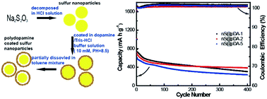
In this paper, a mussel inspired polydopamine (pDA) coating was successfully used to improve the electrochemical performance of sulfur nanoparticles. Sulfur nanoparticles with a pDA coating of a suitable thickness (i.e. 9.5 nm) could retain a capacity of 385.1 mA h g−1, 54% of the initial capacity at 0.5C (1C = 1670 mA g−1) after 400 charge/discharge cycles. Their cycling performance was greatly improved compared with that of uncoated sulfur nanoparticles due to the stabilization of sulfur nanoparticles by a pDA coating. Due to simplicity of the coating process and strong adhesion of the pDA coating, the process could be easily applied to the surface modification of other electrode materials.
Introduction
Lithium ion batteries (LIBs) have been attracting increasing attentions due to their wide range of applications in electric vehicles and portable electronic devices. However, traditional cathode materials such as LiCoO2 and LiFePO4 cannot meet the urgent demand of high-capacity and high-power LIBs.1–3 In this regard, sulfur has emerged as a promising cathode material due to its high theoretical specific capacity of ca. 1673 mA h g−1 and energy of ca. 2600 W h kg−1.4–6 Meanwhile, sulfur itself has some advantages including high abundance, low toxicity and low cost.7,8 However, Li–S batteries suffered from rapid capacity fading and low coulombic efficiency, impeding its practical applications. Above problems were partially caused by the dissolution of intermediate polysulfide into electrolyte during the charge/discharge process.6,9 To mitigate polysulfide dissolution, extensive efforts have been devoted to encapsulate/coat the sulfur particles with different materials. They included carbon materials (e.g. mesoporous carbon, graphene/graphene oxide, hollow carbon fiber), polymers (e.g. PEDOT, PANI, PVP) and metal oxide/sulfide (e.g. TiO2, TiS2).10–23 Such encapsulation/coating could greatly improve the stability of Li–S batteries. Here, we reported a simple process to encapsulate sulfur nanoparticles with polydopamine (pDA) coating. The coating inspired by mussels contains both amine and catechol groups, which exhibit strong adhesion ability on various surfaces.24 It has good stability in a broad range of pH values. At pH = 8.5 and under atmosphere with oxygen, pDA coating can be self-polymerized from dopamine, a commercially available biological neurotransmitter.25,26 Thus, the whole polymerization process of pDA was simple and environmentally benign. It also exhibited a variety of applications.24–28 In fact, pDA was coated on polyethylene separator to modify its ionic conductivity, electrolyte uptake and wettability.29–31 The modified separator could greatly enhance whole LIB's power performance and long-cycle stability.31 However, few studies were conducted to utilize pDA coating directly to modify the electrochemical performance of the electrode materials. Considering the strong adhesion and high stability of pDA coating, herein we studied electrochemical performance of pDA coated sulfur nanoparticles as cathode materials. The sulfur particles with suitable pDA coating (i.e. 9.5 nm) could retain a capacity of 385.1 mA h g−1, 54% of initial capacity after 400 cycles at 0.5C. Influence of pDA thickness on the electrochemical performance of the sulfur nanoparticles were also investigated. The improved electrochemical performance was ascribed to pDA coating, which could mitigate dissolution of the lithium polysulfides in electrolyte, and modify the surface property of sulfur nanoparticles.
Experimental
Synthesis of pDA coated sulfur nanoparticles
The aqueous solution of Na2S2O3 (500 ml, 0.04 M) was mixed with PVP (0.02 wt%). Concentrated HCl (3.35 ml, 37 wt%) was then added to the mixture under magnetic stirring. After reaction for 2 h at room temperature, the formed sulfur nanoparticles were then collected and washed by centrifugation, and dried at 60 °C in an oven for 24 h. To coat sulfur nanoparticles with pDA, different amounts of dopamine were dissolved in (10 mM, pH = 8.5) buffer solution to form the solutions with concentrations of 0.1, 0.2, 0.5 mg ml−1, respectively. Sulfur nanoparticles were then dispersed into the solution and stirred for 24 h. The pDA coated sulfur nanoparticles were washed, collected and redispersed into the mixed solution of deionized water, isopropanol and toluene under stirring for 4 h, followed by centrifugation and washed using deionized water. After drying, the powders of nS@pDA were obtained.
Characterization and electrochemical measurements
X-ray diffraction (XRD) patterns were collected with BRUKER D8 Advance. Scanning electron microscopy (SEM) images, transmission electron microscopy (TEM) images and element mapping were recorded with HITACHI S-4800 and JEOL 2100F, respectively. The functional groups on surface of materials were characterized by Fourier transform infrared (FTIR) spectroscopy (Nicolet 6700). Thermogravimetric (TG) analyses were conducted on a Discovery TGA from 50 °C to 500 °C with a heating rate of 5 °C min−1. The sulfur contents in the solution were determined with a inductively coupled plasma mass spectrometer (Vista MPX ICP). To fabricate the working electrodes, 80 wt% nS@pDA composite, 10 wt% carbon black (Super P) and 10 wt% PVDF were mixed in NMP to form a slurry. This slurry was then coated onto a carbon-coated Al foil using doctor blade. The electrode film was dried in an oven at 60 °C overnight and then punched into round disks with a diameter of 12 mm. The 2016-type coin cells were assembled with polypropylene membrane as separator and lithium foil as the counter electrode in a glove box filled with Ar. The electrolyte contained 1 M lithium bis(tri-fluoromethanesulfonyl)imide (LiTFSI) in a mixture of 1,3-dioxolane (DOL) and dimethoxymethane (DME) (1
:
1 v/v) with 0.2 M LiNO3. Galvanostatic discharge/charge tests were performed by a battery tester (Land 2100A) from 1.5–2.6 V versus Li+/Li at room temperature. The specific capacity was calculated on the basis of the total weight of the nS@pDA composite material. The sulfur mass loading was 0.5–0.6 mg cm−2. Electrochemical impedance (EIS) measurement was carried out with Autolab 302N electrochemical workstation (5 mV, 100 kHz to 0.01 Hz).
Results and discussion
The whole process coating the sulfur nanoparticles with pDA was illustrated in Fig. 1. The sulfur nanoparticles were synthesized through disproportion reaction of Na2S2O3 in acid solution with a small amount of PVP.13 The PVP addition could alleviate the aggregation of hydrophobic sulfur nanoparticles in aqueous solution. Then the sulfur nanoparticles from the obtained milky aqueous suspension were coated in Tris–HCl buffered aqueous solution with a dopamine concentration of 0.1 mg ml−1 for 24 h. The pDA coated sulfur nanoparticles were then separated, dried and immersed in toluene to partially dissolve sulfur, forming the void between the coated pDA layer and sulfur nanoparticles.17 The obtained pDA coated sulfur nanoparticle product was labelled as nS@pDA-1. Fig. 2a and b showed typical SEM images of nS@pDA-1. The size of nS@pDA-1 varied from several to ten hundred nanometers. Though the spherical templating agent, PVP,13 was added during the synthesis of sulfur nanoparticles, the expected fully spherical shape was not observed. The appearance of irregular shapes might be due to formation and coalesce of aggregates during drying or the lack of sufficient PVP. Compared with the uncoated sulfur nanoparticle, the coated one did not exhibit obvious morphology change, indicating possible existence of conformal coating on sulfur nanoparticles. XRD spectra (Fig. S1†) of the sulfur nanoparticles confirmed their polycrystalline nature. After the coating, no peaks from impurity were observed. FTIR spectra (Fig. S2†) of the coated sulfur nanoparticles showed additional vibrations at 3250, 2950 and 1650 cm−1 compared with uncoated sulfur. It indicated that pDA was successfully coated on the surface of sulfur nanoparticles. The thickness of pDA coating was determined to be ca. 3.9 nm from TEM images (Fig. 2c and d). The STEM image and mapping of related elements of nS–pDA-1 were shown in Fig. S3.† Among them, carbon, nitrogen and oxygen signals were from pDA coating. The mapping areas of these elements fully covered the mapping area of sulfur, indicating the pDA continuously covered the sulfur nanoparticles. To study the influence of initial dopamine concentration on the coated pDA thickness, sulfur nanoparticles were also coated in aqueous solutions with dopamine concentrations of 0.2 and 0.5 mg ml−1. The obtained products were labelled as nS@pDA-2 and nS@pDA-5, respectively. The pDA thicknesses of nS@pDA-2 and nS@pDA-5 were ca. 9.5 and 13.0 nm determined from TEM images (Fig. S4†), respectively. The higher concentration of dopamine led to thicker pDA coatings. It might be due to the higher collision possibility of monomers in the solution with higher concentrations of dopamine. The above results indicated that the thickness of coated pDA could be easily adjusted by the initial concentration of dopamine solution. TG analyses (Fig. S5†) were conducted to determine the composition and thermal stability of pDA coated sulfur nanoparticles. For nS–pDA-2, the weight percentage of pDA coating was determined to 1.5% since 82 wt% was remained when the pure pDA was heated up to 296 °C.32 However, the pDA coating did not improve the thermal stability of sulfur nanoparticles. For bare sulfur nanoparticles, they lost weight completely at 345 °C. While, for pDA coated sulfur nanoparticles, they lost weight at lower temperature and completely lost sulfur at 296 °C. The weight loss of sulfur at lower temperature was also observed in samples of PPy coated sulfur particles or TiO2 coated sulfur nanoparticles.17,33
 |
| Fig. 1 Schematic of mussel inspired pDA coating process on the sulfur nanoparticles. | |
 |
| Fig. 2 SEM images of sulfur nanoparticles (nS@pDA-1) coated with pDA in 0.1 mg ml−1 dopamine solution at low magnification (a) and high magnification (b). (c) TEM image of the nS@pDA-1 and (d) high magnification TEM image of the red rectangle area in (c). | |
To examine their electrochemical performance and evaluate the influence of pDA thickness on their performance, nS@pDA composites were assembled into coin cells with lithium foil as the counter electrode. Fig. 3a showed cycle voltammetry of the nS@pDA for the first three cycles at a scan rate of 0.2 mV s−1 in a potential range of 1.5 to 3.0 V. In the first cathodic scan, two reductions peaks at 2.26 and 1.93 V were observed, corresponding to the main multistep reduction of sulfur. The first peak was attributed to transformation of the elemental sulfur to soluble long-chain lithium polysulfides (i.e., Li2Sx, 4 ≤ x < 8). The second peak was ascribed to the further reduction of long-chain polysulfides to the short-chain insoluble lithium polysulfides (i.e. Li2S2 and LiS2). In the subsequent anodic scan, the oxidation peak at 2.53 V was related to the oxidation of Li2S2 and Li2S into lithium polysulfides and elemental sulfur.18,20,34,35 In the second and third scans, the peak positions were almost unchanged, confirming good reversibility of the reaction. The discharge voltage profiles at the current density of 0.1C were shown in Fig. 3b. In these voltage profiles, two plateaus around 2.27 and 1.95 V were observed. The upper plateau was associated with the formation of high-order polysulfides and the lower plateau corresponded to conversion of the high-order polysufides to the Li2S2 and Li2S. The voltage profiles were consistent with the cycle voltammetry curves. It indicated that pDA coating did not change the electrochemical-reaction nature of sulfur. The initial discharge specific capacities of the coin cells were 1140, 1016 and 1000 mA h g−1 for nS@pDA-1, nS@pDA-2, nS@pDA-5 composites at a rate of 0.1C. The slight difference might be due to the less ratio of active material for sulfur nanoparticles coated with thicker PDA coating. Rate performance of pDA coated sulfur was shown in Fig. 3c. Under low current density (i.e. 0.5C), the capacities for nS@pDA-1, nS@pDA-2 and nS@pDA-5 were 553.7, 560.4 and 556.8 mA h g−1, respectively. The difference in capacities among three samples was not obvious. However, under higher current rates (e.g. 2C), their specific capacities were 408, 411, 376 mA h g−1 for ns@PDA-1, nS@pDA-2, nS@pDA-5, respectively. The difference in capacities became larger. It indicated the thin pDA coating did not dramatically influence their performance at the low current rate. However, when the current rate became high, the time for electron transport and ionic diffusion through the thick pDA coating layer should be considered. As a result, the thick pDA coating would deteriorate the rate performance of sulfur nanoparticles.25 The cycling performance of sulfur was shown in Fig. 3d. Although the degradation of capacities of all samples was observed, their capacities could be stabilized. However the capacity of bare sulfur nanoparticles dropped dramatically from 496.4 mA h g−1 to 123.0 mA h g−1 in just 50 cycles at 0.5C (Fig. S6†). It indicated that pDA coating could improve the stability of sulfur cathode. nS@pDA-2 showed the best stability among all samples. After 400 cycles at 0.5C, it still retained a capacity of 385.1 mA h g−1, 54% of the initial discharge capacity. As cycling proceeded, pDA coating layer with the suitable thickness not only protected the polysulfides from dissolving in electrolyte but also balanced the Li+ ionic and electronic conductivity.25 When the pDA coating was too thin, it would not become a valid barrier for soluble lithium sulfide, which resulted in the larger capacity loss in nS@pDA-1. The improved stability was also ascribed to the internal void which mitigated the stress caused by volume change during cycling.15,17 The coulombic efficiency of nS@pDA could approach 100%, which also proved that the shuttle effect of the polysufides was confined by the suitable pDA coating layer on the sulfur surface.36 To determine the sulfur content dissolved in electrolyte after discharging, the two coin after discharging were disassembled in glove box, then 1,3-dioxolane (DOL) was used to collect the polysulfides dissolved in the electrolyte. ICP analyses on the collected solutions showed that 83.7% and 39.7% sulfur was dissolved in electrolytes for the electrode with bare sulfur nanoparticles after 40 cycles and nS–pDA-2 for 400 cycles. The less sulfur loss in nS–pDA-2 proved the protection of dopamine coating.
 |
| Fig. 3 (a) Cycle voltammetry of nS@pDA-2 at a scan speed of 0.2 mV s−1 in the potential range of 1.5–3 V. Discharge/charge profiles for the initial cycle (b), rate performance (c) and cycling performance (d) of nS@pDA. | |
To better understand the electrochemical performance of the nS@pDA composites, EIS analysis was conducted. Before cycling, Nyquist plots (Fig. 4a) of electrodes were composed of a semicircle. After three cycles, additional semicircle appeared in the plot (Fig. 4b) due to the formation of the solid electrolyte interphase (SEI). The middle frequency-semicircle could be related to the charge-transfer resistance and its relative capacitance while the semicircle at high frequency might be related to the interphase resistance.37–39 The resistances (radius) in the two frequency regions of nS@pDA-1 and nS@pDA-2 were much smaller than the resistances (radius) of nS@pDA-5 according to the parameters in the fitting model (Fig. 4 and Table S1†). It indicated lower charge-transfer resistance and interphase resistance in sulfur samples with thinner pDA coatings. The pDA layer has both catechol and amine groups which can absorb amount of liquid electrolyte. It could modify the wetting property of sulfur nanoparticles, improve the ionic conductivity at the interface, facilitating diffusion of Li+.29–31,36 However, when pDA coating got thicker, the distance for lithium-ion diffusion got longer and the time for diffusion was increased. Meanwhile, due to the insulating nature of the pDA film, the thick pDA coating also blocked the electron's conducting, which also increased the resistance. The above result was consistent with their cycling and rate performance.
 |
| Fig. 4 Nyquist plots and fitted spectra of the electrodes based on nS@pDA (a) before and (b) after cycling for three times (inset, equivalent circuit). | |
Conclusions
In summary, mussel inspired pDA coating was successfully used to improve the electrochemical performance of sulfur nanoparticles. The sulfur nanoparticles with suitable pDA coatings (i.e. 9.5 nm) could retain a capacity of 385.1 mA h g−1, 54% of the initial discharge capacity at 0.5C. The performance was far better than that of naked sulfur nanoparticles. Such improvement might be ascribed to that stable pDA coating could alleviate the loss of soluble polysulfide during the charge/discharge process. The surface modification of sulfur nanoparticles by pDA coating also could contribute to such improvement. Due to simplicity of the coating process and strong adhesion of pDA coating, the process could be easily applied to the surface modification of other electrode materials.
Acknowledgements
This work was supported by the Scientific Research Foundation for Returned Scholars, the Ministry of Education of China, Key Basic Research Projects of Science and Technology Commission of Shanghai (no. 11JC1412900), the National Science Foundation of China program (no. 21271140, 51472182) and Shanghai Natural Science Foundation of China (no. 13ZR1428200).
Notes and references
- I. D. Scott, Y. S. Jung, A. S. Cavanagh, Y. Yan, A. C. Dillon, S. M. George and S. H. Lee, Nano Lett., 2011, 11, 414–418 CrossRef CAS PubMed.
- Z.-X. Chi, W. Zhang, F.-Q. Cheng, J.-T. Chen, A.-M. Cao and L.-J. Wan, RSC Adv., 2014, 4, 7795 RSC.
- C. Gong, F. Deng, C.-P. Tsui, Z. Xue, Y. S. Ye, C.-Y. Tang, X. Zhou and X. Xie, J. Mater. Chem. A, 2014, 2, 19315–19323 CAS.
- G. Zheng, Q. Zhang, J. J. Cha, Y. Yang, W. Li, Z. W. Seh and Y. Cui, Nano Lett., 2013, 13, 1265–1270 CrossRef CAS PubMed.
- A. Manthiram, Y. Fu, S. H. Chung, C. Zu and Y. S. Su, Chem. Rev., 2014, 114, 11751–11787 CrossRef CAS PubMed.
- R. Elazari, G. Salitra, A. Garsuch, A. Panchenko and D. Aurbach, Adv. Mater., 2011, 23, 5641–5644 CrossRef CAS PubMed.
- Y. M. Lee, N.-S. Choi, J. H. Park and J.-K. Park, J. Power Sources, 2003, 119–121, 964–972 CrossRef CAS.
- L. Chen and L. L. Shaw, J. Power Sources, 2014, 267, 770–783 CrossRef CAS.
- S. Lim, R. L. Thankamony, T. Yim, H. Chu, Y. J. Kim, J. Mun and T. H. Kim, ACS Appl. Mater. Interfaces, 2015, 7, 1401–1405 CAS.
- X. Li, Y. Cao, W. Qi, L. V. Saraf, J. Xiao, Z. Nie, J. Mietek, J.-G. Zhang, B. Schwenzer and J. Liu, J. Mater. Chem., 2011, 21, 16603 RSC.
- H. Wang, Y. Yang, Y. Liang, J. T. Robinson, Y. Li, A. Jackson, Y. Cui and H. Dai, Nano Lett., 2011, 11, 2644–2647 CrossRef CAS PubMed.
- G. Zheng, Y. Yang, J. J. Cha, S. S. Hong and Y. Cui, Nano Lett., 2011, 11, 4462–4467 CrossRef CAS PubMed.
- W. Li, G. Zheng, Y. Yang, Z. W. Seh, N. Liu and Y. Cui, Proc. Natl. Acad. Sci. U. S. A., 2013, 110, 7148–7153 CrossRef CAS PubMed.
- G.-C. Li, G.-R. Li, S.-H. Ye and X.-P. Gao, Adv. Energy Mater., 2012, 2, 1238–1245 CrossRef CAS.
- W. Zhou, Y. Yu, H. Chen, F. J. DiSalvo and H. D. Abruna, J. Am. Chem. Soc., 2013, 135, 16736–16743 CrossRef CAS PubMed.
- Z. W. Seh, J. H. Yu, W. Li, P. C. Hsu, H. Wang, Y. Sun, H. Yao, Q. Zhang and Y. Cui, Nat. Commun., 2014, 5, 5017 CrossRef CAS PubMed.
- Z. Wei Seh, W. Li, J. J. Cha, G. Zheng, Y. Yang, M. T. McDowell, P. C. Hsu and Y. Cui, Nat. Commun., 2013, 4, 1331 CrossRef PubMed.
- L. Xiao, Y. Cao, J. Xiao, B. Schwenzer, M. H. Engelhard, L. V. Saraf, Z. Nie, G. J. Exarhos and J. Liu, Adv. Mater., 2012, 24, 1176–1181 CrossRef CAS PubMed.
- Y. S. Su and A. Manthiram, Nat. Commun., 2012, 3, 1166 CrossRef PubMed.
- X. Yang, L. Zhang, F. Zhang, Y. Huang and Y. Chen, ACS Nano, 2014, 8, 5208–5215 CrossRef CAS PubMed.
- L. Ji, M. Rao, H. Zheng, L. Zhang, Y. Li, W. Duan, J. Guo, E. J. Cairns and Y. Zhang, J. Am. Chem. Soc., 2011, 133, 18522–18525 CrossRef CAS PubMed.
- G. Zhou, S. Pei, L. Li, D. W. Wang, S. Wang, K. Huang, L. C. Yin, F. Li and H. M. Cheng, Adv. Mater., 2014, 26, 625–631 CrossRef CAS PubMed.
- N. Jayaprakash, J. Shen, S. S. Moganty, A. Corona and L. A. Archer, Angew. Chem., 2011, 50, 5904–5908 CrossRef CAS PubMed.
- H. Lee, S. M. Dellatore, W. M. Miller and P. B. Messersmith, Science, 2007, 318, 426–430 CrossRef CAS PubMed.
- C. Lei, F. Han, D. Li, W. C. Li, Q. Sun, X. Q. Zhang and A. H. Lu, Nanoscale, 2013, 5, 1168–1175 RSC.
- R. Liu, S. M. Mahurin, C. Li, R. R. Unocic, J. C. Idrobo, H. Gao, S. J. Pennycook and S. Dai, Angew. Chem., 2011, 50, 6799–6802 CrossRef CAS PubMed.
- J. Ou, J. Wang, D. Zhang, P. Zhang, S. Liu, P. Yan, B. Liu and S. Yang, Colloids Surf., B, 2010, 76, 123–127 CrossRef CAS PubMed.
- N. Liu, H. Wu, M. T. McDowell, Y. Yao, C. Wang and Y. Cui, Nano Lett., 2012, 12, 3315–3321 CrossRef CAS PubMed.
- M. H. Ryou, Y. M. Lee, J. K. Park and J. W. Choi, Adv. Mater., 2011, 23, 3066–3070 CrossRef CAS PubMed.
- J. Jiang, L. Zhu, L. Zhu, B. Zhu and Y. Xu, Langmuir, 2011, 27, 14180–14187 CrossRef CAS PubMed.
- M.-H. Ryou, D. J. Lee, J.-N. Lee, Y. M. Lee, J.-K. Park and J. W. Choi, Adv. Energy Mater., 2012, 2, 645–650 CrossRef CAS.
- H. Hu, B. Yu, Q. Ye, Y. Gu and F. Zhou, Carbon, 2010, 48, 2347–2351 CrossRef CAS.
- Y. Zhu and A. Manthiram, J. Phys. Chem. C, 2012, 116, 8910–8915 Search PubMed.
- F. Wu, S. Wu, R. Chen, J. Chen and S. Chen, Electrochem. Solid-State Lett., 2010, 13, A29 CrossRef CAS.
- L. Chen, Y. Liu, M. Ashuri, C. Liu and L. L. Shaw, J. Mater. Chem. A, 2014, 2, 18026–18032 CAS.
- W. Zhou, X. Xiao, M. Cai and L. Yang, Nano Lett., 2014, 14, 5250–5256 CrossRef CAS PubMed.
- C. Barchasz, J.-C. Leprêtre, F. Alloin and S. Patoux, J. Power Sources, 2012, 199, 322–330 CrossRef CAS.
- L. Yuan, X. Qiu, L. Chen and W. Zhu, J. Power Sources, 2009, 189, 127–132 CrossRef CAS.
- L. Yuan, H. Yuan, X. Qiu, L. Chen and W. Zhu, J. Power Sources, 2009, 189, 1141–1146 CrossRef CAS.
Footnote |
† Electronic supplementary information (ESI) available: FTIR, TEM and cycling performance for the sample. See DOI: 10.1039/c5ra22352k |
|
This journal is © The Royal Society of Chemistry 2016 |
Click here to see how this site uses Cookies. View our privacy policy here.