Yongping Liuab,
Peng Chenb,
Yuan Chenb,
Huidan Lu*ab,
Jixiang Wangb,
Zhishu Yangb,
Zhenhuan Lu*ab,
Ming Liab and
Liang Fangc
aGuangxi Key Laboratory of Electrochemical and Magneto-chemical Functional Materials, Guilin University of Technology, Guilin, P.R. China. E-mail: lhuidangl@163.com; zhenhuanlu@glut.edu.cn
bCollege of Chemistry and Bioengineering, Guilin University of Technology, Guilin, P.R. China
cState Key Laboratory Breeding Base of Nonferrous Metals and Specific Materials Processing, Guilin University of Technology, Guilin 541004, P.R. China
First published on 6th January 2016
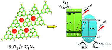
In this paper, free standing graphitic carbon nitride (g-C3N4) nanosheets have been synthesized by mixed solvents (water/IPA = 2/1) liquid phase exfoliation, and a series of SnS2/g-C3N4 heterojunctions with different contents of SnS2 have been prepared via a simple ion-exchange process. Exfoliated g-C3N4 presents a two-dimension sheet-like structure with the thickness of 2.8 nm and small SnS2 nanoparticles with diameter of 5–10 nm are well anchored on the surface of g-C3N4 nanosheets, which was proved by transmission electron microscopy (TEM) and atomic force microscope (AFM). The 4.0-SnS2/g-C3N4 sample shows the highest photocurrent density of 13.66 μA cm−2 at 0.8 V, which is about 1.5 time of the g-C3N4 nanosheets and 2 times of the bulk g-C3N4, respectively. Photocatalytic measurement also demonstrated that constructing heterojunction of SnS2/g-C3N4 can improve the photocatalytic efficiency as compared to pure g-C3N4 and g-C3N4 nanosheets. The highly effective photoelectrochemical and photocatalytic activities of SnS2/g-C3N4 heterojunctions are attributed to the efficient separation of photogenerated hole–electron pairs. This work may provide a novel concept for the rational design of high performance g-C3N4-based photocatalysts.
However, the photocatalytic activity of pure g-C3N4 is low owing to its poor light-absorbance and fast recombination of photo-generated electron–hole pairs.6 Recent investigations show that single or several atom thickness g-C3N4 nanosheets have improved photocatalytic property, strong electrochemical luminescence activity, good biocompatibility and high fluorescent quantum yield compared with their bulk counterparts.7–9 Usually, g-C3N4 nanosheets are prepared by liquid phase separation method in water or organic solvent by ultrasonic exfoliating process.7,10,11 Zhao et al. prepared single atomic layer of g-C3N4 nanosheet and adopt it for degradation rhodamine B,12 as a result, they found that the photocatalytical activity of single layer graphitic g-C3N4 was about 3 and 10 times higher than that of few layer g-C3N4 and bulk g-C3N4.
Constructing heteroconjection is another way to improve the photocatalytic performance of C3N4. Among various carbon nitride crystal forms, g-C3N4 possesses the smallest band gap, so it is easy to form a heterojunction,13,14 for instance ZnO/g-C3N4,15 Bi2WO6/g-C3N4,16 g-C3N4/TiO2 (ref. 17) and g-C3N4/SrTiO3.18 Katsumata et al. prepared the g-C3N4/Ag3PO4 heterojunction with different weight ratios of g-C3N4 by a facile in situ precipitation method, which showed highly effective photocatalytic activity under visible light irradiation. Hou et al. constructed 2D porous graphitic C3N4 nanosheets/nitrogen-doped graphene/layered MoS2 ternary nanojunction.19 Chen et al. used ion exchange process to prepare the g-C3N4/NiS composite material and found that the sample containing 1.5% NiS exhibited the highly photocatalytic activity;20 Young-Si Jun et al. adopt a new approach to assemble low-dimensional g-C3N4 into a three-dimensional structure, which significantly improved photocatalytic properties.21
SnS2 is a nontoxic and low-priced semiconductor material with a visible-light band gap of 2.2–2.35 eV. So, we choose SnS2 to form heteroconjections with few layer g-C3N4 nanosheets for increasing photon-generated carrier transfer. In this paper, we have prepared free standing g-C3N4 nanosheets by liquid phase exfoliation, and successfully synthesized SnS2/g-C3N4 nanosheet heterojunction via a simple ion-exchange process. Moreover, we explored the photoelectrochemical response and photocatalytic performance of SnS2/g-C3N4 heterojunction, which exhibited the higher photocurrent and photocatalytic efficiency compared with pure g-C3N4 nanosheets. Additionally, a smaller charge transfer resistance was observed for the heterojunction under visible light irradiation.
The g-C3N4 nanosheets were obtained by liquid exfoliation of as-prepared bulk g-C3N4 in various solvents via sonication. Typically, 20 mg of bulk g-C3N4 was added into 20 mL flask with 15 mL different solvents such as isopropyl alcohol (IPA), water, ethanol, formamide, N-methyl-pyrrolidone (NMP), or the mixture solvents (water and IPA). Then, the flask was ultrasounded for 10 h. The initial formed suspension was centrifuged at about 3000 rpm to remove aggregates, and then, the supernatant was collected, the exfoliated g-C3N4 nanosheets were obtained, centrifuged and dried in vacuum for further characterization.
The SnS2/g-C3N4 heterojunctions were prepared through an ion-exchange method at room temperature, as shown in Scheme 1. Firstly, 30, 60, 90 or 120 μL of 0.01 M SnCl4 solution was dropped in the 10 mL g-C3N4 nanosheets dispersion (a concentration of 0.27 mg mL−1). After stirring 20 min, 60, 120, 180 or 240 μL of 0.01 M Na2S solution was added into the above mixed dispersion. The mixed solution was stirred for another 5 minutes, then the solution was centrifuged at 14000 rpm for 10 min, and the precipitates were collected and washed with ethanol and deionized water 3 times, respectively. The products were dried for 12 h in a vacuum oven at 80 °C. The mass ratio of SnS2 to g-C3N4 nanosheets are 2%, 4%, 6%, and 8% in the final samples, thus they are denoted as 2.0-SnS2/g-C3N4, 4.0-SnS2/g-C3N4, 6.0-SnS2/g-C3N4 and 8.0-SnS2/g-C3N4, respectively.
Bulk SnS2 were synthesized by precipitation method. Briefly, 20 mL of 0.01 M SnCl4 solution was dropped in 50 mL of 0.01 M Na2S solution and the mixed solution was stirred for 30 minutes, then the precipitate was collected by centrifuged.
The photocatalytic activity of powder samples was evaluated by the degradation of methyl orange (MO) The optical system for the degradation reaction included a 350 W Xe lamp, a cutoff filter of 420 nm. In each experiment, 20 mg photocatalyst was added to 100 mL dye aqueous solution (10 mg L−1). Before irradiation, the suspensions were magnetically stirred in the dark for 30 min to ensure the establishment of an adsorption/desorption equilibrium between the photocatalyst and the dye. During the reaction process under visible light illumination, 3 mL of suspension was sampled at given time intervals and centrifuged to remove the photocatalyst particles. The concentration of MO was determined by monitoring the height of the maximum of the absorbance in ultraviolet-visible spectra.
To explore the photo-induced radical species in the photocatalytic degradation of MO, the reactive species trapping experiments were performed. The benzoquinone and IPA were utilized as the scavenger of the superoxide radical (·O2−) and hydroxyl (·OH), respectively. For the h+ hole, disodium ethylenediaminetetraacetate (EDTA–2Na) was used as quencher. The trapping experiment taken was similar to the above photocatalytic activity test except that the different scavengers were added into the MO solution.
The crystal structure of the obtained samples was investigated by X-ray diffraction (XRD). As seen clearly from Fig. 2, the pure g-C3N4 nanosheets and SnS2/g-C3N4 heterojunction have two distinct diffraction peaks at 27.5° and 13.2°, which can be indexed for graphite-like materials as the (002) and (100) diffraction plane. The (002) peak at 27.5° and the (100) peak at 13.2° corresponding to distance of 0.324 and 0.670 nm reflects the characteristic inter-planar stacking peak of conjugated aromatic systems and in-plane structural packing motif, respectively.25 In addition, the characteristic diffraction peaks of SnS2 appear at 51.8°. Seen from the illustration of Fig. 2, the diffraction peak at 51.8° gradually increase with the increasing amount of SnS2 for SnS2/g-C3N4 heterojunctions.
The atomic structure of bulk g-C3N4 and g-C3N4 nanosheets were characterized by FT-IR spectroscopy. As shown in Fig. 3, the absorption band of 3000–3600 cm−1 is due to NH stretching vibration mode, and absorption band of 1200–1700 cm−1 is ascribed to the stretching vibration mode of CN hetero-ring.26 Note that, strong absorption peak at 809 cm−1 is the bending vibration peak of the s-triazine ring.27 In the case of SnS2 nanosheets, a broadband peaked at 545 cm−1 is originated to the vibration of Sn–S bands.28 However, this feature band is difficult to distinguish on the curve of SnS2/g-C3N4 heterojunction nanosheets due to the low content of SnS2 nanoparticle.
The chemical compositions of the as-prepared photocatalysts are characterized by XPS spectra, as shown in Fig. 4. The appearance of elements Sn, S, C, N in survey XPS spectrum of 4.0-SnS2/g-C3N4 hybrid confirms the presence of both components C3N4 and SnS2, as shown in Fig. 4a. The high-resolution spectrum of C 1s (Fig. 4b) can be deconvoluted into three peaks at 284.6 eV, 285.8 eV and 288.1 eV, corresponding to sp2-bonded aromatic structure (N–CN), graphitic carbon (C–C), and residual C–O, respectively.10,29 As to the N 1s spectrum of g-C3N4 nanosheets (Fig. 4c), N 1s peak at 398.5 eV corresponds to sp2 hybridized aromatic N bonded to carbon atoms (C
N–C). The peak at 399.56 eV is assigned to the tertiary N bonded to carbon atoms in the form of N–(C)3 or H–N–(C)2. The weaker peak with a high binding energy at 400.9 eV is attributed to quaternary N bonded to three carbon atoms in the aromatic cycles and the peak centered at 404.3 eV correspondes to the π excitations.30,31 The evolution of Sn 3d with two strong peaks is observed at 486.5 and 494.9 eV (Fig. 4d), which were attributed to Sn 3d5/2 and Sn 3d3/2, respectively.32 However, the peak relating to the C
N–C bonds of g-C3N4 nanosheets is shifted slightly to the higher binding energy side after loading SnS2 and the S 2p peaks of SnS2, reflecting to S–Sn bond,32 are also moved to the low binding energy side (Fig. 4e), when coupling with g-C3N4 nanosheets. It is indicated that there might be some interactions between the “nitrogen pots” of g-C3N4 with the Sn species of SnS2. The observed binding energies of Sn 3d and S 2p spectra are in good accordance with Sn4+ and S2− of SnS2.
![]() | ||
Fig. 4 XPS survey spectrum (a), high resolution of C 1s (b), N 1s (c), Sn 3d (d), S 2p (e) spectra of as-prepared photocatalysts. |
TEM image of the g-C3N4 nanosheets (Fig. 5c) clearly presents a two-dimension sheet-like structure with relatively smooth surface. For revealing microstructures and the thickness, the diluted dispersion of the products obtained after ultrasonication treatment of bulk g-C3N4 was further investigated by AFM analyses. The AFM image (Fig. 5a) shows the exfoliated products are free-standing nanosheets with a large lateral size of 300–400 nm. Fig. 5b presents the thickness of the products is about 2.8 nm, suggesting 8–9 atomic monolayers.33,34 In addition, the exfoliated sample can be formed to stable colloidal dispersion over a period of two weeks and exhibits a typical Tyndall effect. The TEM image of SnS2/g-C3N4 hybrid is displayed in Fig. 5d, which shows that some small SnS2 nanoparticles with diameter of 5–10 nm are well anchored on the surface of g-C3N4 nanosheets. These results indicated that SnS2 has combined with ultrathin g-C3N4 nanosheets to form effective SnS2/g-C3N4 heterojunction.
![]() | ||
Fig. 5 AFM image (a) and the corresponding height analyses of g-C3N4 nanosheets (b), the TEM image of the g-C3N4 nanosheets (c) and the SnS2/g-C3N4 hybrid (d). |
The UV-vis diffuse reflection spectra of the SnS2/g-C3N4 hybrids with different mass percentage of SnS2, together with those of pure g-C3N4 nanosheets and pure SnS2, were investigated and shown in Fig. 6a. The pure g-C3N4 nanosheets show an absorption edge at 450 nm, corresponding to band gap of ∼2.65 eV presented in Fig. 6b, and it is well be in consistent with the value of previously reported literature.35,36 The pure SnS2 displays light absorption over nearly entire visible light range. After introducing SnS2, the SnS2/g-C3N4 samples show the similar absorption edge and a broader absorption in the visible region as comparison to pure g-C3N4 nanosheets. Moreover, the absorption intensity of SnS2/g-C3N4 samples in the visible region is gradually strengthened with increasing SnS2 contents.
![]() | ||
Fig. 6 (a) UV-vis diffuse reflection spectra of pure g-C3N4 nanosheets, pure SnS2 and SnS2/g-C3N4 heterojunctions; (b) band gap energies of SnS2 and g-C3N4 nanosheets. |
The band gap energy of the semiconductors can be calculated by the following equation:37–39
αhv = A(hv − Eg)n/2 | (1) |
The band edge positions of CB and VB for semiconductor can be determined by the following equations:44–46
EVB = X − Ee + 0.5Eg | (2) |
ECB = EVB − Eg | (3) |
To investigate photoelectrochemical performance of SnS2/g-C3N4 heterojunction, typical current–voltage measurements of various samples were performed under light switch (Fig. 7). The 4.0-SnS2/g-C3N4 heterojunction shows the highest photocurrent density of 13.66 μA cm−2 at 0.8 V, which is about 1.5 and 2 times of g-C3N4 nanosheets (9.02 μA cm−2) and bulk g-C3N4 (6.82 μA cm−2), respectively.
![]() | ||
Fig. 7 Current–potential plots for bulk g-C3N4, pure g-C3N4 nanosheets and SnS2/g-C3N4 hybrids under light switch. |
To gain deeper insights into the charge transport behavior in the system, we conducted electrochemical impedance spectroscopy (EIS) measurements (Fig. 8). In the Nyquist diagram, the radius of each arc is associated with the charge-transfer process at the corresponding electrode/electrolyte interface, and a smaller radius corresponds to a lower charge-transfer resistance. In Fig. 8a, the impedance of the g-C3N4 nanosheets is significantly lower than the bulk material, and when the light source turned on, the impedance is further reduced, which also confirms that the g-C3N4 nanosheets has better photoelectric properties. Obviously, the 4.0-SnS2/g-C3N4 heterojunction exhibits minimal charge transfer resistance under simulated sunlight irradiation, indicating that effective shuttling of charges between the electrode and the electrolyte. Faster interfacial charges transfer occurred at composite interface, thereby generating the increasing photocurrent. So, SnS2 may serve as electron capture centres, speeding up charges transfer.
Fig. 9a shows a comparison of the visible light photocatalytic activity of bulk g-C3N4, SnS2 nanoparticles, g-C3N4 nanosheets and SnS2/g-C3N4 composites. Prior to irradiation, the MO solution containing the catalyst was kept in the dark for 30 min to reach an adsorption/desorption equilibrium. As we can see, with absence of any photocatalyst the decomposition of MO hardly occurred when subjected to visible light irradiation. It is also suggested that the SnS2 nanoparticles content has a significant effect on the photocatalytic activity of g-C3N4 nanosheets. In the case of bulk C3N4, approximately 82.6% of MO was decomposed after visible light irradiation for 40 min. When it comes to the pure g-C3N4 nanosheets, the degradation rate of MO is 90.7% and higher than that of SnS2 nanoparticles which only display only 87.4% after irradiation for 40 min. However, the photoactivities of g-C3N4 nanosheets are obviously enhanced with the presence of SnS2 nanoparticles on its surface. The photoactivities of SnS2/g-C3N4 heterojunctions are higher than that of g-C3N4 nanosheets and SnS2 nanoparticles. Especially, 4.0-SnS2/g-C3N4, can decompose 95.3% of MO after only 25 min for visible light irradiation. The average decomposition rate of the MO dye over bulk g-C3N4, SnS2 nanoparticles, g-C3N4 nanosheets and 4.0-SnS2/g-C3N4 heterojunction is estimated to be about 0.0135 mg min−1, 0.0166 mg min−1, 0.0200 mg min−1 and 0.0324 mg min−1. It reveals that constructing heterojunction of SnS2/g-C3N4 could improve the photocatalytic efficiency of g-C3N4 nanosheets.
![]() | ||
Fig. 9 (a) Photocatalytic degradation of MO with various powder samples, (b) the cycling runs of 4.0-SnS2/g-C3N4 heterojunction for the photodegradation of MO under visible light. |
The reusability and stability of SnS2/g-C3N4 heterojunction was evaluated by recycling experiments. By utilizing the same photocatalyst and a fresh MO solution, five consecutive cyclic photodegradation experiments were carried out for the 4.0-SnS2/g-C3N4 heterojunction. As shown in Fig. 9b, MO was decomposed and decreased in every cycling run. The percentage of degradation of MO after the first cycle was 94.9% by 25 min visible light irradiation; while it was 89.9% at the fifth cycle with the same time irradiation. Only a little decrease in catalytic activity was observed after the fifth-cycle decomposition process. So SnS2/g-C3N4 nanosheets heterojunction could be regarded as a stable and durable photocatalyst.
It is well known that the ·O2−, ·OH and h+ are the major reactive species for the photocatalytic decomposition. To uncover the possible photocatalytic mechanism of 4.0-SnS2/g-C3N4 nanosheets heterojunction, the radical species trapping experiments were carried out to explore the main reactive species in the process of photocatalytic degradation. Herein, three chemicals including benzoquinone (BQ), disodium ethylenediaminetetraacetate (EDTA–2Na) and isopropyl alcohol (IPA) were utilized as the scavenger of ·O2−, h+ holes and ·OH, respectively. As listed in Fig. 10a, the degradation efficiency of MO is 95% with absence of scavenger. When the IPA was added into the reaction solution, the degradation efficiency of MO is down to 73.5%. And adding the EDTA–2Na into the reaction solution, the degradation efficiency of MO is decreased to 43%. The degradation efficiency of MO is even whittled down into 28% with the presence of BQ. The phenomenon suggested that the photocatalytic activity of 4.0-SnS2/g-C3N4 is extremely suppressed by BQ and EDTA–2Na, while slightly inhibited by IPA. It also can be concluded that ·O2−, ·OH and h+ are the major reactive species for the 4.0-SnS2/g-C3N4 nanosheets photocatalyst. The influencing degree is belonged to ·O2− > h+ > ·OH.
![]() | ||
Fig. 10 (a) Effects of various scavengers on the photocatalytic efficiency of 4.0-SnS2/g-C3N4 heterojunction, (b) schematic model of the photocatalytic mechanism for SnS2/g-C3N4 heterojunction. |
On the basis of radical species trapping experiments and the aforementioned band gap energy analysis, the possible photocatalytic mechanism is speculated. As illustrated in Fig. 10b, the g-C3N4 nanosheets and SnS2 semiconductor possess overlapping band potentials, indicating that the band potentials of g-C3N4 nanosheets and SnS2 nanoparticles can be well matched to form an effective heterojunction. Under visible light irradiation, both components are readily excited to generate photo-induced electrons–holes pairs. And the separation process of photo-induced electrons–holes can be vividly described in Fig. 10b. When the SnS2/g-C3N4 nanosheets heterojunctions absorb the visible light and then are excited, the quick and strong combination is occurred between the photo-induced electrons in the conduction band (CB) of SnS2 and photo-induced holes in the valence band (VB) of g-C3N4 nanosheets. Meanwhile, the photo-induced electrons in the CB of g-C3N4 nanosheets, which have more negative potential than the standard redox potential of E°(O2/·O2−) = −0.33 eV (vs. NHE), can react with the O2 to produce ·O2−. And then the generated holes in the VB of SnS2 can directly decompose the MO molecules. Moreover, the generated holes in the VB of SnS2, which have more positive potential than the standard redox potential of E°(·OH/OH−) = 1.99 eV (vs. NHE),48–52 can oxidize OH− to yield ·OH radicals. Therefore, a typical Z-scheme separation mechanism of photo-induced electrons–holes pairs is proposed and favourable for the generation of ·O2− and ·OH reactive species. As a result, efficient separation of photo-generated hole–electron pairs is achieved by the distribution of electrons and holes on different semiconductors, thus enhance the corresponding photocatalytic performance.
Footnote |
† Electronic supplementary information (ESI) available. See DOI: 10.1039/c5ra21506d |
This journal is © The Royal Society of Chemistry 2016 |