DOI:
10.1039/C5RA21262F
(Paper)
RSC Adv., 2016,
6, 17446-17455
In vitro antileishmanial activity of aza-scorpiand macrocycles. Inhibition of the antioxidant enzyme iron superoxide dismutase†
Received
13th October 2015
, Accepted 28th January 2016
First published on 4th February 2016
Abstract
The in vitro leishmanicidal activity of a series of nine aza-scorpiand-like macrocycles, recently synthesized, was tested on Leishmania infantum, Leishmania braziliensis and Leishmania donovani parasites, using promastigotes and intracellular amastigotes forms. The cytotoxicity of the tested compounds on J774.2 macrophage cells was also measured. Four of the tested compounds (1, 2, 8 and 9) showed selectivity indexes higher than those of the reference drug Glucantime for the three Leishmania species. Moreover, the data on infection rates and on amastigotes showed that compounds 1, 2, 8 and 9 are the most active against the three Leishmania species. The changes in the excretion product profile of parasites treated with the four compounds (1, 2, 8 and 9) were also consistent with substantial cytoplasmic alterations. On the other hand, the most active compounds were potent inhibitors of Fe-SOD in the three parasite species considered, whereas their impact on human CuZn-SOD was low. The high activity, low toxicity, stability, low cost of the starting materials and straightforward synthesis make these compounds appropriate molecules for the development of affordable anti-leishmanicidal agents.
Introduction
Leishmaniasis is a parasitic disease caused by protozoan parasites of the genus Leishmania and transmitted by sandflies commonly found in tropical and subtropical regions of the world. It is estimated that 350 million people are at risk of infection worldwide and the annual incidence of the disease has been calculated to range between 1.5 and 2 million with about 70
000 deaths every year.1 It is widely spread in almost 100 countries of Latin America, the Indian subcontinent, east Africa and the Mediterranean Basin.2 However, the principal etiological agents, vectors, animal reservoirs and clinical syndromes may be different in each continent. However, it has to be stressed that these data are probably very conservative because of under reporting and misdiagnosis. There are different clinical manifestations of this disease. The most widespread, but also the least dangerous is cutaneous leishmaniasis (CL), which triggers localized self-healing skin lesions. In mucocutaneous leishmaniasis (ML), also known as espundia, the infection spreads in the mucous membranes of the mouth and nose, becoming fatal in some cases. However, the most serious among them is undoubtedly visceral leishmaniasis (VL), also known as kala azar, which is often lethal if untreated, since the parasites infect the liver and spleen and cause anaemia by severely affecting the immune system.3 Those clinical syndromes are caused by different Leishmania subspecies. The most representative are: (i) L. major, which originates CL and is found in North Africa, India, China or the Middle East; (ii) L. braziliensis, one of the main species causing ML, most common in South America (Bolivia, Brazil and Peru), (iii) L. donovani complex, responsible for the fearsome VL, which is mainly constituted by zoonotic L. infantum, found in Europe, North Africa and Latin America and (iv) anthroponotic L. donovani, present in East Africa and the Indian subcontinent.4,5
Because leishmaniasis is a severe health problem, mainly (but not only) in developing countries, it is necessary to develop drugs that adequately treat and prevent the disease. The current list of available drugs is limited in number and clearly unsatisfactory due to toxicity, generation of resistance and/or the very high costs involved. Pentavalent antimonials, mainly meglumine antimoniate (Glucantime) and sodium stibogluconate, have been the usual drugs of choice in the treatment of VL and CL for more than 60 years. However, acquired resistance has been reported, especially in India, with failure rates of 65% and side-effects including serious cardiotoxicity in about 10% of patients.1,6 Second-line drugs, like pentamidine and amphotericin B have not experienced widespread use due to toxicity and cost. Pentamidine is associated with hypotension, hypoglycemia and diabetes, and also causes nephrotoxicity. Miltefosine is used in the treatment of VL infections and is less harmful than pentamidine, but the ease with which resistant mutants are obtained is worrying.7 The most recent line of drugs are fluconazole, paromomycin and sitamaquine, but they also exhibit toxicity problems (i.e. hepatotoxicity with paromomycin or fluconazole, methemoglobinemia with sitamaquine) and it remains to be seen how these agents stand up in face of the pandemic of resistance.8,9 Due to the growing resistance to chemotherapy, which is a drawback shown by most of the compounds mentioned above, drug combinations are now being explored as a path to delay or prevent the emergence of resistance, increase efficacy and shorten the course of treatment.9
In the search of new effective anti-leishmanial drugs able to broaden the narrow spectrum of compounds currently available, potential targets that perform functions essential for parasite survival are being investigated. Among them, antioxidant enzymes preventing oxidative stress that would otherwise lead to death of the parasite are in the spotlight. Within that group, trypanothione reductase is currently receiving special consideration.10,11 However, another basic defense mechanism against oxidation is provided by the iron superoxide dismutase enzyme (Fe-SOD), which is located in the cytosol, the mitochondria and glycosomes of the parasite and plays an important role in the dismutation of harmful superoxide radicals into oxygen and hydrogen peroxide.12 One further advantage of Fe-SOD as a drug target is that it is not present in humans, who use copper/zinc and manganese superoxide dismutase enzymes (Cu, Zn and Mn-SOD) for the same purposes. Therefore, molecules able to inhibit selectively Fe-SOD could be promising candidates as leishmanicidal agents.13 Since their prosthetic groups are essential in all processes regulated by enzymes, alterations in the active centre induced by dissociation of the iron atom or by modifications in the coordination geometry could effectively deactivate its antioxidant action and, therefore, influence both the growth and survival of the parasite cells.
Regarding this point, in the last few years, our research group has designed a new family of polyamine compounds consisting of a macrocyclic pyridinophane core substituted with side chains containing additional donor atoms (Fig. 1).14 These macrocycles are termed scorpiand ligands because the side chain can fold towards the macrocyclic core following the binding of target guest species such as protons, anions or metal ions.14,15
 |
| Fig. 1 Chemical structure of the tested aza-scorpiand-like macrocycles compound complexes. | |
Recently, we have studied the in vitro action of several of aza-scorpiand-like macrocyclic compounds against Trypanosoma cruzi and two species of Leishmania (L. infantum and L. braziliensis). The results showed that these macrocycles are potentially promising agents for the treatment of trypanosomatid infections.16,17 In the present work, we tested the in vitro leishmanicidal (against L. infantum, L. braziliensis and L. donovani) activity of another new 9 macrocycles of this family and their toxicity against macrophages. We compared these new compounds with Glucantime as a reference drug. The effect of the compounds on the ultrastructure of Leishmania spp. was also studied by transmission electronic microscopy (TEM) experiments to confirm the type of damage caused to the parasite. 1H-NMR analysis of the nature and percentage of excreted metabolites was performed to obtain information about the inhibitory effect of our compounds on the glycolytic pathway since this represents the prime energy source of the parasite. Previously, we have demonstrated that these derivatives are good inhibitors of Fe-SOD, not only in T. cruzi,17 but also in Leishmania spp.16 An evaluation of their effectiveness as putative inhibitors of Fe-SOD in relation to human CuZn-SOD is presented.
Results and discussion
Synthesis of the compounds
The synthesis of 1–9 was accomplished as described previously14,18,19 following a modification of the Richman–Atkins procedure.20 First, pertosylated polyamine tris(2-aminoethyl)amine was reacted with 2,6-bis(bromomethyl)pyridine in a 1
:
1 molar ratio using potassium carbonate (K2CO3) as the base in refluxing acetonitrile (CH3CN). Detosylation was carried out with HBr/HAc/PhOH. Compounds 1, 2, 3, 4 and 9 were obtained by reacting this intermediate, in its free amine form, with the corresponding carboxaldehydes in dry ethanol followed by in situ reaction with sodium borohydride (NaBH4). The compounds were finally precipitated as hydrochloride salts (Scheme S1, ESI†).
The reaction of the pertosylated intermediate with N-(3-bromopropyl)phthalimide in a 1
:
1 molar ratio using K2CO3 as the base in refluxing CH3CN followed by deprotection and detosylation, carried out with hydrazine monohydrate and HBr/HAc/PhOH, respectively, yielded a second intermediate (Scheme S1, ESI†). Compounds 5, 6, 7 and 8 were obtained by reacting this intermediate, in its free amine form, with the corresponding carboxaldehydes in dry ethanol followed by in situ reaction with NaBH4. The compounds were finally precipitated as the hydrochloride salts.
Interaction with iron
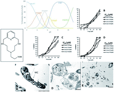
As discussed in the introduction, one of the potential targets of new anti-leishmanial drugs is the iron superoxide dismutase enzyme (Fe-SOD). For this reason it is interesting to evaluate the affinity of these chelating ligands for iron. We have previously reported on the affinity towards Fe(II) of some of the compounds studied here, which showed remarkable stability. As an example, here we report for the first time on the interaction of compound 6 with Fe(II), determined by potentiometric titrations. This compound behaves as a good iron chelator, as shown by its high stability constants (Table S1, ESI†). The distribution diagram (Fig. 2), built upon these constants, shows how at intermediate pH values all the Fe(II) is complexed by the ligand, mostly as the [FeL]2+ complex. This means that if the ligand approaches close enough to the active site of Fe-SOD, some competition might be established with the primary ligands of the active centre for the capture of the metal ions.
 |
| Fig. 2 Distribution diagram of the Fe(II)–6 system ([Fe2+] = [6] = 1 × 10⁻³ M). | |
In vitro antileishmanial evaluation
In a first step we assayed the in vitro biological activity of compounds 1–9 (Fig. 3) against three significant species of Leishmania: L. infantum, L. braziliensis and L. donovani on both extra- and intracellular forms of the parasites (Fig. S1–S6, ESI†). Extracellular forms are more commonly used due to the ease of working with them, but are less indicative of leishmanicidal activity. The use of intracellular forms is more cumbersome but gives more accurate results, as promastigotes are converted to amastigotes in vertebrate host cells,21 Intracellular assays were performed by infecting macrophage cells with promastigotes, which transformed into amastigotes within 1 day after infection. Table 1 shows the IC50 values obtained after 72 h of exposure when compounds 1–9 were tested on extra- and intracellular forms of L. infantum, L. braziliensis and L. donovani. Toxicity values against J774.2 macrophage after 72 h of culture were also calculated. Results obtained for the reference drug Glucantime were included in all cases for comparison.
 |
| Fig. 3 Effect of aza-scorpiand macrocycles derivatives 1, 2, 8 and 9 on the infection and growth rates of Leishmania infantum. (A) Rates of infection; (B) mean numbers of amastigotes of L. infantum per infected J774 A.2 macrophage cell (at IC25 conc.). Values are the means of three separate experiments. | |
Table 1 In vitro activity and toxicity for the aza-scorpiand like macrocycles derivatives on extra- and intracellular forms of Leishmania spp.a
Compounds |
IC50b μM |
Toxicity IC50 macrophagec (μM) |
Leishmania infantum |
Leishmania braziliensis |
Leishmania donovani |
Promastigote forms |
Amastigote forms |
Promastigote forms |
Amastigote forms |
Promastigote forms |
Amastigote forms |
Results are averages of three separate determinations. IC50 = the concentration required to give 50% inhibition, calculated by non-linear regression analysis from the Kc values at concentrations employed (1, 10, 25, 50 and 100 μM). Towards macrophage cell after 72 h of culture of Leishmania spp. |
Glucantime® |
18.0 ± 3.1 |
24.2 ± 2.6 |
25.6 ± 1.6 |
30.4 ± 6.1 |
27.4 ± 3.8 |
31.4 ± 4.1 |
15.2 ± 1.3 |
1 |
2.3 ± 0.3 |
3.3 ± 0.6 |
2.1 ± 0.2 |
2.7 ± 0.6 |
14.1 ± 1.2 |
4.83 ± 0.6 |
231.8 ± 12.6 |
2 |
3.7 ± 0.2 |
2.9 ± 1.0 |
2.3 ± 0.5 |
4.2 ± 1.1 |
18.5 ± 2.5 |
1.1 ± 0.0 |
166.9 ± 11.2 |
3 |
57.3 ± 3.2 |
23.0 ± 3.7 |
32.9 ± 4.3 |
15.0 ± 2.3 |
49.5 ± 5.1 |
19.5 ± 2.3 |
181.3 ± 10.7 |
4 |
12.1 ± 1.0 |
8.6 ± 0.9 |
15.6 ± 0.8 |
14.1 ± 1.3 |
13.5 ± 1.5 |
10.7 ± 0.8 |
92.4 ± 6.8 |
5 |
25.4 ± 3.1 |
23.8 ± 2.4 |
25.2 ± 3.5 |
20.6 ± 1.7 |
23.4 ± 3.0 |
16.9 ± 0.9 |
144.9 ± 12.4 |
6 |
15.6 ± 0.7 |
13.7 ± 1.3 |
23.2 ± 3.4 |
15.3 ± 1.6 |
14.0 ± 1.3 |
12.5 ± 1.0 |
117.3 ± 9.8 |
7 |
9.5 ± 0.7 |
9.0 ± 0.8 |
17.5 ± 2.3 |
14.6 ± 1.1 |
7.4 ± 0.5 |
6.9 ± 0.2 |
97.4 ± 8.1 |
8 |
4.8 ± 0.3 |
3.0 ± 0.1 |
1.7 ± 0.2 |
2.8 ± 0.4 |
8.4 ± 0.3 |
4.6 ± 0.7 |
140.0 ± 11.3 |
9 |
11.7 ± 1.4 |
6.2 ± 0.8 |
1.4 ± 0.2 |
3.6 ± 0.4 |
12.5 ± 0.9 |
3.2 ± 0.3 |
221.3 ± 16.9 |
It was shown that the leishmanicidal activities against the more indicative intracellular forms of the parasites were higher than those found for Glucantime with the compounds tested (comp. 1, 2, 7, 8 and 9), whereas the effect on extracellular forms was more random. But more interesting were the toxicity data in mammalian cells, in the case of compound 7 is quite toxic and therefore not be considered for further studies, the other four compounds tested were found to be much less toxic for macrophages than the reference drug. Thus, compound 1 and 9 were 15-fold less cytotoxic than Glucantime, the compounds 2 and 8 were also about 10-fold less cytotoxic. In order to obtain a more accurate picture of the features commented on above, we show in Table 2 the selectivity index values calculated from the data in Table 1, since they are very illustrative of the in vitro potential of the compounds tested with respect to the reference drug. The number of times that the SI of each compound exceeded the SI of Glucantime is also shown in parenthesis. In this Table 2 the differences between Glucantime and the tested compounds are clearly revealed. All four (1, 2, 8 and 9) compounds exhibited substantially better SI values than the reference drug in the three Leishmania spp. tested and, in the most remarkable case, the SI of the compound 2 on the intracellular form of L. donovani exceeded that of Glucantime by 303-fold, a relevant data point which is by far the best SI value obtained with the different series of related heterocyclic systems tested by us against parasites in previous research.22–26 The SI displayed in Table 2 are also very illustrative about the difference in behaviour of different compounds, which recurs in the three species and in both extra and intracellular forms, since 1, 2, 8 and 9 always show substantially better results than the rest of the tested compounds. In tests performed on the L. infantum specie, the SI of 1 exceeded that of the reference drug by 126- and 117-fold for the extra- and intracellular forms, respectively, by 184- and 172-fold with L. braziliensis, and by 33 and 97-fold when 1 was tested on L. donovani. The compound 2 has similar SI to 1; the respective values obtained were 56- and 96-fold on the L. infantum for the extra- and intracellular forms, respectively, 121–79-fold on extra- and intracellular forms the L. braziliensis; while on L. donovani, values of 18- and 303-fold were found respectively. Compounds 8 and 9 were equally effective against the three species of Leishmania and against both extra- and intracellular forms. Comp. 8 has 36 year 78-fold SI increases on L. infantum, 137 and 100-fold on L. braziliensis and 33- and 61-fold against L. donovani. Comp. 9 also presented results similar or slightly higher than 8 against the two forms of the parasite and off the three species tested.
Table 2 Selectivity index found for the aza-scorpiand like macrocycles derivatives on extracellular and intracellular forms of Leishmania spp.a
Compounds |
SIb |
Leishmania infantum |
Leishmania braziliensis |
Leishmania donovani |
Promastigote forms |
Amastigote forms |
Promastigote forms |
Amastigote forms |
Promastigote forms |
Amastigote forms |
Results are averages of three separate determinations. Selectivity index = IC50 macrophage cell/IC50 extracellular and intracellular form of parasite. In brackets: number of times that compound exceeds the reference drug SI (on extracellular and intracellular forms of Leishmania spp.). |
Glucantime® |
0.8 |
0.6 |
0.6 |
0.5 |
0.5 |
0.5 |
1 |
100.8 (126) |
70.2 (117) |
110.4 (184) |
85.9 (172) |
16.4 (33) |
48.3 (97) |
2 |
45.1 (56) |
57.6 (96) |
72.6 (121) |
39.7 (79) |
9.0 (18) |
151.7 (303) |
3 |
3.2 (4) |
7.9 (13) |
5.5 (9) |
12.1 (24) |
3.7 (7) |
9.3 (19) |
4 |
7.6 (10) |
10.7 (18) |
5.9 (10) |
6.6 (13) |
6.8 (14) |
8.6 (17) |
5 |
5.7 (7) |
6.1 (10) |
5.8 (10) |
7.0 (14) |
6.2 (12) |
8.6 (17) |
6 |
7.5 (9) |
8.6 (14) |
5.1 (8) |
7.7 (15) |
8.4 (17) |
9.4 (19) |
7 |
10.3 (13) |
10.8 (18) |
5.6 (9) |
6.7 (13) |
13.2 (26) |
14.1 (28) |
8 |
29.2 (36) |
46.7 (78) |
82.4 (137) |
50.0 (100) |
16.7 (33) |
30.4 (61) |
9 |
18.9 (24) |
35.7 (59) |
158.1 (263) |
61.5 (123) |
17.7 (35) |
69.2 (138) |
In order to gain better insight into the activities of these compounds (1, 2, 8 and 9), their effect on the infectivity and intracellular replication of amastigotes was subsequently determined. Macrophage cells were grown and infected with promastigotes in the stationary phase. The parasites invaded the cells and underwent morphological conversion to amastigotes within 1 day after infection. On day 10, the rate of host cell infection reached its maximum (control experiment). We used the IC25 of each product as the test dosage. Different authors have claimed that compounds with SI values less than 20-fold those of the reference drug should be discarded as candidates for more advanced leishmanicidal tests due to their poor selectivity against mammalian cells.27
As shown in Fig. 3A, when compounds 1, 2, 8 and 9 were added to macrophages infected with L. infantum promastigotes, the infection rate decreased significantly with respect to the control and, furthermore, the four compounds were also remarkably more effective in decreasing infectivity than Glucantime (86%, 83%, 88% and 76% for 1, 2, 8 and 9, respectively, versus only 26% for the reference drug). A measure of the average number of amastigotes per infected macrophage (Fig. 3B) led to similar conclusions: all four compounds were more effective than Glucantime (with only a 30% decrease), and amastigote numbers of 51%, 68%, 77% and 72% were found for 1, 2, 8 and 9 respectively.
Infection rates (Fig. 4A) and amastigote numbers (Fig. 4B) obtained on L. braziliensis also showed that, in both cases, the four compounds were clearly more effective than Glucantime, and also in both cases the order of effectiveness was 8 > 9 > 2 > 1 > Glucantime, since the infectivity rates calculated were: 77%, 72%, 68%, 51% and 47%, respectively. The decreases in amastigote numbers were: 65%, 60%, 63%, 76% and 39% for 1, 2, 8, 9 and Glucantime respectively. Finally, similar results were found on L. donovani, with infectivity rates of 70%, 66%, 59%, 53% and 30% (Fig. 5A); and decreases in the number of amastigotes of: 50%, 63%, 42%, 79% and 32% (Fig. 5B) for 1, 2, 8, 9 and Glucantime, respectively. From all these data it can be concluded that: (i) all four compounds are substantially more active than Glucantime against the three Leishmania species tested; (ii) the results are in full accordance with related data described for T. cruzi parasites.21
 |
| Fig. 4 Effect of aza-scorpiand macrocycles derivatives 1, 2, 8 and 9 on the infection and growth rates of Leishmania braziliensis. (A) Rates of infection; (B), mean numbers of amastigotes of L. braziliensis, per infected J774 A.2 macrophage cell (at IC25 conc.). Values are the means of three separate experiments. | |
 |
| Fig. 5 Effect of aza-scorpiand macrocycles derivatives 1, 2, 8 and 9 on the infection and growth rates of Leishmania donovani. (A) Rates of infection; (B), mean numbers of amastigotes of L. donovani, per infected J774 A.2 macrophage cell (at IC25 conc.). Values are the means of three separate experiments. | |
Ultrastructural alterations
Since all these compounds showed remarkable leishmanicidal activity, major damage to parasite cells could be inferred. In order to confirm this, a transmission electron microscopy (TEM) study on the promastigote forms of the three Leishmania species under consideration was performed. As expected, significant morphological alterations were observed compared with untreated control cells. Fig. 6, 7 and 8 display some of the most relevant structural features observed in control and treated cells of promastigotes of the three species. As a general observation, the four tested compounds were responsible for the death of a high percentage of individuals and many other abnormalities such as vacuolization, disintegration of the cell cytoplasm and rupture of the parasites.
 |
| Fig. 6 Ultrastructural alterations observed by TEM in promastigotes of the Leishmania infantum after treatment with compounds 1 (plates B and C), 2 (plate E), 8 (plate D) or 9 (plate F). Abbreviations: dead parasites (D), flagellum (F), glycosomes (G), kinetoplasts (K), vacuoles (V), mitochondria (M), nucleus (N), ribosomes (R), flagellar pocket, distorted membranes (single arrow), cellular rest (RE). Scale bars are 1.0 mm in all cases. | |
 |
| Fig. 7 Ultrastructural alterations observed by TEM in promastigotes of the Leishmania braziliensis after treatment with compounds 1 (plate B), 2 (plate E), 8 (plates C and D) or 9 (plate F). Abbreviations: dead parasites (D), flagellum (F), glycosomes (G), kinetoplasts (K), vacuoles (V), mitochondria (M), nucleus (N), ribosomes (R), flagellar pocket, distorted membranes (single arrow), cellular rest (RE). Scale bars are 1.0 mm in all cases. Scale bars are 1.0 mm in all cases. | |
Fig. 6 corresponds to L. infantum, and it can be seen that when the parasites were treated with 1, 2 and 9 the disturbances were more notable than when other species where treated (Fig. 6, plates B–F) compared to the control (Fig. 6, plate A). The most effective compound was 1 (plates B and C). After treatment with 1 many parasites were found with reduced size. The examination evidenced that these parasites had very altered organelles. Others were broken or had strange shapes, and a few had swollen kinetoplast and many vacuoles. With comp. 8 (D) many parasites had their cytoplasmic membrane broken, the flagellar pocket and mitochondria very swollen and large vacuoles. The treatment with 2 (E) showed that many parasites were very small and were heavily vacuolated. Finally, comp. 9 (F) caused a reduction in the number of ribosomes, some appeared with intense vacuolization and some filled of glycosomes in higher number than usual.
L. braziliensis was greatly affected by the four compounds as shown in (Fig. 7 plates B, C, D, E and F) compared to control (Fig. 7 plate A). Thus, compound 1 (Fig. 7, plate B) caused intense vacuolization, disorganization, and the parasites were filled of glycosomes. In some of these promastigotes there was only cytoplasmic membrane. 8 was the compound most effective (Fig. 7 plates C, D) for the damages that caused on the promastigotes, as with all the compounds the cytoplasm full of vacuoles but also could be observed changes in the morphology of the parasite. They had strange shapes, looking crashed or amoeboid and structures like mitochondria and kinetoplastos were extremely swollen. Furthermore, there were many broken parasites (Fig. 7 plate D), and large and extended glycosomes. Comp. 2 also caused similar changes but the highlight were just the completely altered morphology and the strange appearance that had many promastigotes (Fig. 7, plate E). These parasites had also abnormal organelles, filled vacuoles, some of these strange organelles that could correspond to degenerate mitochondria. Most normal structures could not be assessed. Compound 9 was also effective on promastigotes (Fig. 7, plate F), in this case highly vacuolated cytoplasm, few glycosomes together with swollen mitochondria were the main disturbances.
According to the results obtained by TEM (Fig. 8) and compared to a control culture (Fig. 8, plate A), the compounds tested turned out to be the most effective on L. donovani. The compound 8 (Fig. 8, plate B) caused a drastic decrease in the viability of promastigotes. Actually their cytoplasmic contents were very little electron-appeared and it was observed only ribosomes and glycosomes. Their appearance was practically dead bodies. Compound 1 was the most effective (Fig. 8, plates C, D), with few ribosomes and they all were full of empty vacuoles, mitochondria were empty and very swollen. L. donovani was also similarly affected by compound 2. Finally compound 9 showed identical effect to the ones above as shown in the image (Fig. 8, plate F). The parasites were completely empty of content or very few ribosomes without cytoplasmic organelles or some very altered beyond recognition.
 |
| Fig. 8 Ultrastructural alterations observed by TEM in promastigotes of the Leishmania donovani after treatment with compounds 1 (plate C and D), 2 (plate E), 8 (plates B) or 9 (plate F). Abbreviations: dead parasites (D), flagellum (F), glycosomes (G), kinetoplasts (K), vacuoles (V), mitochondria (M), nucleus (N), ribosomes (R), flagellar pocket, distorted membranes (single arrow), cellular rest (RE). Scale bars are 1.0 mm in all cases. Scale bars are 1.0 mm in all cases. | |
Metabolite excretion effect
Since trypanosomatids are unable to completely degrade glucose to CO2 under aerobic conditions, they excrete much of the hexose skeleton into the medium as partially oxidized fragments in the form of fermented metabolites. The nature and percentages of those excretion products depend on the pathway used for glucose metabolism by each species.28,29 The final products of glucose catabolism in Leishmania are usually CO2, succinate, acetate, D-lactate, L-alanine and, in some cases, ethanol.30 Among them, succinate is the most relevant, because its main role is to maintain the glycosomal redox balance, allowing the reoxidation of NADH produced in the glycolytic pathway. Succinic fermentation requires only half of the phosphoenolpyruvate produced to maintain the NAD+/NADH balance, and the remaining pyruvate is converted inside the mitochondrion and the cytosol into acetate, D-lactate, L-alanine or ethanol, according to the degradation pathway followed by a specific species.31
We reported above that the most active compounds 1, 2, 8 and 9 caused great damage to the mitochondria of the parasites in the three Leishmania species, so that their highly disruptive action presumably affected their glucose metabolism and, consequently, modified the percentages of the final excretion products formed. Therefore, we registered the 1H NMR spectra of promastigotes from the L. infantum, L. braziliensis and L. donovani species after treatment with the four compounds with significant leishmanicidal activity, and the final excretion products were identified qualitatively and quantitatively. The spectra obtained were compared with those from promastigotes maintained in cell-free medium (control) for four days after inoculation with the parasites. Variations in the height of the signals corresponding to the most significant catabolites are displayed in Fig. S7–S9 of the ESI.† The expected presence of acetate, D-lactate, L-alanine and succinate was confirmed in the control experiments performed on all three species, and the major metabolite was succinate in all cases, in agreement with data reported in the literature.32 However, noteworthy differences were found in parasites treated with the derivatives, whereas the presence of benznidazole did not lead to significant alterations in fuel metabolism (data not shown).
The most relevant finding was that the production of succinate, the key catabolite in glucose metabolism, was substantially inhibited by the four compounds in L. donovani (Table 3). In L. braziliensis, there is no reduction in the succinate peak observed, instead of it, with some of the compounds there is a slight increase of its production. A similar behavior, of lesser magnitude, was found in the L. infantum species. In fact, the relevant succinate inhibition values obtained agreed with the TEM evidence, indicating that these derivatives severely damage the mitochondrial structures of the parasites and, therefore, succinate production is substantially affected in L. donovani.
Table 3 Variation in the height of the peaks corresponding to catabolites excreted by Leishmania spp. promastigotes in the presence of compounds aza-scorpiand like macrocycles (1, 2, 8 and 9) with respect to the control testa
Comp. |
Leishmania infantum |
Leishmania braziliensis |
Leishmania donovani |
Lac |
Ala |
A |
S |
Lac |
Ala |
A |
S |
Lac |
Ala |
A |
S |
A: acetate; Ala: L-alanine; Lac: D-lactate; S, succinate; (−): peak inhibition; (+): peak increasing. |
1 |
+4% |
+11% |
+10% |
+12% |
−11% |
−10% |
−11% |
−7% |
+13% |
−20% |
+41% |
−36% |
2 |
+46% |
+42% |
+22% |
+41% |
0% |
0% |
−13% |
−1% |
−13% |
−40% |
+35% |
−37% |
8 |
+3% |
+11% |
+4% |
+11% |
+7% |
+7% |
−1% |
+6% |
−13% |
−35% |
+53% |
−34% |
9 |
+39% |
+38% |
+4% |
+3% |
+39% |
+24% |
−1% |
+6% |
−20% |
−45% |
+65% |
−37% |
With regard to the less significant excretion products shown in Table 3, the formation of L-alanine and D-lactate were also remarkably inhibited in L. donovani by both 2, 8 and 9, which produced inhibitory values ranging from 13% to 45%. Although variations in the secondary excretion products were in general more erratic in L. infantum and L. braziliensis, increases of the formation of L-alanine and D-lactate was always seen with compounds 8 and 9, to a greater or lesser extent. The only exceptions found were increases of 9% and 20%, respectively, in the production of acetate when parasites of those two series were treated with compound 4. In summary, the 1H NMR data on catabolites are indicative of substantial alterations in glucose metabolism in the three species. It is feasible that the small size of these compounds allows them to easily penetrate the cristae (tubular invaginations of the inner mitochondrial membrane),32 leading to subsequent changes in the metabolic pathway. Concerning the variations in acetate production, it is well known that this end-product originates in the mitochondria of Leishmania species through a collateral pathway via pyruvate which is regulated by the pyruvate dehydrogenase complex,30 so that 2 and 4 could be interfering in a different way with that enzyme complex in both the L. infantum and L. braziliensis species.
SOD enzymatic inhibition in the Leishmania parasites and in human erythrocytes
Since we had previously found that the anti-T. cruzi activity of compounds 1, 2, 8 and 9 was always accompanied by inhibitory activity on the antioxidant enzyme Fe-SOD,28 we tested the effects of those compounds on Fe-SOD isolated from L. infantum, L. braziliensis and L. donovani over a range of concentrations from 0.1 to 100 μM. We used promastigote forms of both species, which excrete Fe-SOD when cultured in a medium lacking inactive FBS.33 The inhibition data obtained are shown in Fig. 9, graphs (B), (C) and (D), and the corresponding IC50 values are included for easier evaluation of the graphs displayed. For comparison, graph (A) of Fig. 9 shows the effects of the same compounds on CuZn-SOD obtained from human erythrocytes. The most remarkable result was the significant inhibitory effect on Fe-SOD found for the highly leishmanicidal compounds 1 and 2 in the three species tested, whereas their inhibition of human CuZn-SOD was clearly lower. If we consider the IC50 calculated for L. infantum, inhibition of Fe-SOD by compounds 2 and 1 was respectively 5.3- and 3.2-fold higher than inhibition of CuZn-SOD, with respective values of 10.6-, and 4.2-fold higher in the case of L. braziliensis and 8.5- and 2.4-fold higher values in the case of L. donovani. All these data seem to confirm some kind of relation between parasiticidal activity and inhibition of Fe-SOD, in accordance with results described in previous work.28 Fe-SOD inhibition could also, at another level, be related to the catabolic changes discussed above, since a mitochondrial malfunction, originating from the redox stress produced by inhibition of the mitochondrion-resident Fe-SOD enzyme,32 should result in severe alteration of pyruvate metabolism and a consequent decrease in the production of succinate.
 |
| Fig. 9 (A) In vitro inhibition of CuZn-SOD in human erythrocytes by compounds 1, 2, 8 and 9. (B–D) In vitro inhibition (%) of Fe-SOD of Leishmania infantum, Leishmania braziliensis and Leishmania donovani promastigotes by compounds 1, 2, 8 and 9. Values are the average of three separate determinations. Differences between the activities of the control homogenate and those incubated with the tested compounds were obtained according to the Newman–Keuls test. IC50 was calculated by linear regression analysis from the Kc values at the concentrations employed (1, 10, 25, 50 and 100 μM). | |
It should be noted that, as mentioned previously, these compounds not only showed greater alterations in glucose catabolism but they also led to greater levels of Fe-SOD inhibition. Because the Fe-SOD present in mitochondria is an essential part of the antioxidant protective response of the parasite, its inhibition would be related to a decrease in the rate of survival for the parasite. Because of this fact, it is hypothesized that compounds having a quinoline nitrogen on the side chain are able to get closer to the active centre of the enzyme, as is the case of compounds 1 and 2, so they might produce greater inhibition.16,17
Experimental
Chemistry
The ESI† contains the synthetic procedure and the characterisation data (NMR, mass spectrometry and elemental microanalysis) of all the compounds. Details of the potentiometric titrations, calculation of acid–base and stability constants are also included in the ESI.†
Parasite strain and culture
Promastigote forms of L. infantum (MCAN/ES/2001/UCM-10), L. braziliensis (MHOM/BR/1975/M2904) and L. donovani (MHOM/PE/84/LC26) were cultured in vitro in medium trypanosomes liquid (MTL) supplemented with 10% inactive foetal calf serum (FCS) kept in an air atmosphere at 28 °C, in Roux flasks (Corning, USA) with a surface area of 75 cm2, according to the methodology described by González et al.21
In vitro activity assays
The compounds to be tested were first dissolved in dimethylsulfoxide (DMSO, Panreac, Barcelona, Spain) at a concentration of 0.1% and then assayed for toxicity and inhibitory effects on parasite and mammalian cells growth as previously described.21
Cell culture and cytotoxicity tests
The macrophage line J774.2 [European collection of cell cultures (ECACC) number 91051511] was derived in 1968 from a tumour in a female BALB/c mouse. The macrophages were grown in minimal essential medium (MEM) plus glutamine (2 mM) and 20% inactive FCS, with a humidified atmosphere of 95% air and 5% CO2 at 37 °C. The cytotoxicity testing on macrophages was performed by flow cytometric analysis according to a method previously described.33 The percentage of viable cells was calculated with respect to the control culture. The IC50 was calculated using linear regression analysis from the Kc values of the concentrations employed.
Promastigote assay: extracellular forms
The compounds were dissolved in the culture medium to give final concentrations of 100, 50, 25, 10 and 1 μM. The effects of each compound against the promastigote forms at the different concentrations were tested at 72 h using a Neubauer haemocytometric chamber. The leishmanicidal effect was expressed as the IC50 value, i.e. the concentration required to result in 50% inhibition, calculated by linear regression analysis from the Kc values of the concentrations employed.
Amastigote assay: intracellular forms
J774.2 macrophage cells were grown and seeded at a density of 1 × 10⁴ cells per well in 24-well microplates (Nunc) with rounded coverslips on the bottom and cultured for 2 days. The adherent macrophages were then infected with promastigotes of L. infantum, L. braziliensis and L. donovani in the stationary growth phase, at a ratio of 10
:
1 and maintained for 24 h at 37 °C in air containing 5% CO2. Non-phagocytosed parasites were removed by washing, and the infected cultures were incubated with the testing compounds (concentrations ranging from 1 to 100 μM) and then cultured for 72 h in MEM plus glutamine (2 mM) and 20% inactive FCS. Compound activity was determined from the percentage reductions in amastigote number in treated versus untreated cultures in methanol-fixed and Giemsa-stained preparations. Values are the means of three separate determinations.22
Infectivity assay
Adherent macrophage cells grown as described above were infected in vitro with promastigote forms of L. infantum, L. braziliensis or L. donovani, at a ratio of 10
:
1. The compounds to be tested (IC25 concentrations) were added immediately after infection, and incubated for 12 h at 37 °C in 5% CO2. Non-phagocytosed parasites and compounds were removed by washing, and then the infected cultures were grown for 10 days in fresh medium. Cultures were washed every 48 h and fresh culture medium was added. Compound activity was determined on the basis of both the percentage of infected cells and the number of amastigotes per infected cell in treated and untreated cultures in methanol-fixed and Giemsa-stained preparations. The percentage of infected cells and the mean number of amastigotes per infected cell were determined by analysing more than 200 host cells distributed in randomly chosen microscopic fields. Values are the means of three separate determinations.
Ultrastructural alterations
The parasites were cultured at a density of 5 × 10⁵ cells per mL in the corresponding medium, each of which contained a compound to be tested at the IC25 dose. After 72 h, the cultures were centrifuged at 400g for 10 min, and the pellets produced were washed in phosphate-buffered saline (PBS) and then incubated with 2% (v/v) p-formaldehyde/glutaraldehyde in 0.05 M cacodylate buffer (pH 7.4) for 24 h at 4 °C. The pellets were then prepared for Transmission Electron Microscopy (TEM) using a technique previously described.34
Metabolite excretion
Cultures of L. infantum, L. braziliensis and L. donovani promastigotes (initial concentration 5 × 10⁵ cells per mL) received the IC25 dose of each compound (except for control cultures). After incubation for 72 h at 28 °C the cells were centrifuged at 400g for 10 min. The supernatants were collected to determine the excreted metabolites by 1H NMR, and chemical shifts were expressed in ppm, using sodium 2,2-dimethyl-2-silapentane-5-sulphonate as the reference signal. The chemical displacements used to identify the respective metabolites were consistent with those described previously by some of the co-authors.35
Fe-SOD enzymatic inhibition
Parasites were collected in the logarithmic growth phase by centrifugation (400g for 10 min at room temperature). The pellet obtained after centrifugation, was resuspended in 3 mL of STE buffer (0.25 M sucrose, 25 mM Tris–HCl, 1 mM EDTA, pH 7.8) and the cells were lysed by three cycles of sonication for 30 s each at 60 W. The sonicated homogenate was centrifuged at 1500g for 5 min at 4 °C, and the pellet was washed three times in ice-cold STE buffer. This fraction was centrifuged (2500g for 10 min at 4 °C) and the supernatant was collected. Then, the supernatant was subjected to ice-cold ammonium sulphate precipitation between 35 and 85% salt concentration and the resulting precipitate was dissolved in 2.5 mL of distilled water and desalted by chromatography in Sephadex G-25 column (GE Healthcare Life Sciences®, PD 10 column) previously equilibrated with 2 mL of distilled water, taking it up to a final volume of 3.5 mL.36 The protein content was quantified using the Sigma Bradford test, which uses bovine serum albumin (BSA) as a standard.37 Iron and copper–zinc superoxide dismutases activities were determined using a previously described method38 that measures the reduction in nitroblue tetrazolium (NBT) by superoxide ions. According to the protocol, 845 μL of stock solution [3 mL of L-methionine (300 mg, 10 mL−l), 2 mL of NBT (1.41 mg, 10 mL−1) and 1.5 mL of Triton X-100 1% (v/v)] were added into each well, along with 30 μL of the parasite homogenate fraction, 10 μL of riboflavin (0.44 mg, 10 mL−l), and an equivalent volume of the different concentrations of the compounds being tested. Seven different concentrations were used for each agent, from 0.1 to 100 μM. In the control experiment the volume was made up to 1000 μL with 50 mM potassium phosphate buffer (pH 7.8, 3 mL), and 30 μL of the parasite homogenate fraction were added to the mixtures containing the compounds. Then, the absorbance (A0) was measured at 560 nm in a UV spectrophotometer. Afterward, each well was illuminated with UV light for 10 min under constant stirring and the absorbance (A1) was measured again. The human CuZn-SOD and substrates used in these assays were obtained from Sigma Chemical Co. The resulting data were analysed using the Newman–Keuls test.
Conclusions
The compounds 1, 2, 8 and 9 were identified as hit-to-lead according to their leishmanicidal properties. The effect at the ultrastructure level, the selected compounds were responsible as vacuolization, disintegration of the cell cytoplasm and rupture of the parasites. These cellular effects were also consistent with the changes in the excretion products profile of parasites treated. Probably, the main target is the Fe-SOD for compounds 1 and 2 in the three species tested. On this basis, we think that they fulfill the requirements needed to justify a more detailed investigation as to the nature of the mechanisms involved in their patterns of activity, and that they could be considered candidates for studying antiparasitic activity at a higher level.
Acknowledgements
Financial support by the Spanish MINECO and FEDER (Projects Consolider-Ingenio, Project CSD2010-00065-2010 and CTQ2013-48917-C3-1-P) and by Generalitat Valenciana (PROMETEO II 2015/002) is gratefully acknowledged.
Notes and references
- K. Seife, Open Med. Chem. J., 2011, 5, 31–39 CrossRef PubMed
. - World Health Organization, WHO Fact Sheet No. 375, World Health Organization, Geneva, Switzerland, 2014 Search PubMed
. - K. J. Ryan, C. G. Ray, N. Ahmad, W. L. Drew, and J. J. Plorde, Sherris medical microbiology: an introduction to infectious diseases, McGraw Hill, New York, 5th edn, 2010 Search PubMed
. - F. Chappuis, S. Sundar, A. Hailu, H. Ghalib, S. Rijal, R. W. Peeling, J. Alvar and M. Boelaert, Nat. Rev. Microbiol., 2007, 5, 873–882 CrossRef CAS PubMed
. - J. Lukes, I. L. Mauricio, G. Schönian, J. C. Dujardin, K. Soteriadou, J. P. Dedet, K. Kuhls, K. W. Q. Tintaya, M. Jirků, E. Chocholová, C. Haralambous, F. Pratlong, M. Oborník, A. Horák, F. J. Ayala and M. A. Miles, Proc. Natl. Acad. Sci. U. S. A., 2007, 104, 9375–9380 CrossRef CAS PubMed
. - S. L. Croft, S. Sundar and A. H. Fairlamb, Clin. Microbiol. Rev., 2006, 19, 111–126 CrossRef CAS PubMed
. - M. Ouellette, J. Drummelsmith and B. Papadopoulou, Drug Resist. Updates, 2004, 7, 257–266 CrossRef CAS PubMed
. - P. M. Loiseau, S. Cojean and J. Schrével, Parasite, 2011, 18, 115–119 CrossRef CAS PubMed
. - L. H. Freitas-Junior, E. Chatelain, H. A. Kim and J. L. Siqueira-Neto, Int. J. Parasitol.: Drugs Drug Resist., 2012, 2, 11–19 CrossRef CAS PubMed
. - L. Flohé, in Antiparasitic and antibacterial drug discovery, ed. P. M. Selzer, Wiley CH Verlag GmbH & Co KGaA Weinheim, 2009, pp. 211–227 Search PubMed
. - B. Manta, M. Comini, A. Medeiros, M. Hugo, M. Trujillo and R. Radi, Biochim. Biophys. Acta, 2013, 1830, 3199–3216 CrossRef CAS PubMed
. - A. F. Miller, Curr. Opin. Chem. Biol., 2004, 8, 162–168 CrossRef CAS PubMed
. - J. F. Turrens, Mol. Aspects Med., 2004, 25, 211–220 CrossRef CAS PubMed
. - B. Verdejo, A. Ferrer, S. Blasco, C. E. Castillo, J. González, J. Latorre, M. A. Máñez, M. G. Basallote, C. Soriano and E. García-España, Inorg. Chem., 2007, 46, 5707–5719 CrossRef CAS PubMed
. - V. Amendola, L. Fabbrizzi, C. Mangano and P. Pallavicini, Acc. Chem. Res., 2001, 34, 488–493 CrossRef CAS PubMed
. - C. Marín, M. P. Clares, I. Ramírez-Macías, S. Blasco, F. Olmo, C. Soriano, B. Verdejo, M. J. Rosales, D. Gómez-Herrera, E. García-España and M. Sánchez-Moreno, Eur. J. Med. Chem., 2013, 62, 466–477 CrossRef PubMed
. - F. Olmo, C. Marín, M. P. Clares, S. Blasco, M. T. Albelda, C. Soriano, R. Gutiérrez-Sánchez, F. Arrebola-Vargas, E. García-España and M. Sánchez-Moreno, Eur. J. Med. Chem., 2013, 70, 189–198 CrossRef CAS PubMed
. - F. Olmo, M. P. Clares, C. Marín, J. González, M. Inclán, C. Soriano, K. Urbanova, R. Tejero, M. J. Rosales, R. L. Krauth-Siegel, M. Sánchez-Moreno and E. García-España, RSC Adv., 2014, 4, 65108–65120 RSC
. - M. Inclán, M. T. Albelda, J. C. Frías, S. Blasco, B. Verdejo, C. Serena, C. Salat-Canela, M. I. Díaz, A. García-España and E. García-España, J. Am. Chem. Soc., 2012, 134, 9644–9656 CrossRef PubMed
. - J. E. Richman, T. J. Atkins and W. F. Oetle, Organic Synthesis, J. Wiley & Sons, New York, 1988, vol. 6, p. 652 Search PubMed
. - P. González, C. Marín, I. Rodríguez-González, A. B. Hitos, M. J. Rosales, M. Reina, J. G. Díaz, A. González-Coloma and M. Sánchez-Moreno, Int. J. Antimicrob. Agents, 2005, 25, 136–141 CrossRef PubMed
. - M. Sánchez-Moreno, F. Gómez-Contreras, P. Navarro, C. Marín, I. Ramírez-Macías, F. Olmo, A. M. Sanz, L. Campayo, C. Cano and M. J. R. Yunta, J. Antimicrob. Chemother., 2012, 67, 387–397 CrossRef PubMed
. - M. Sánchez-Moreno, A. M. Sanz, F. Gómez-Contreras, P. Navarro, C. Marín, I. Ramírez-Macías, M. J. Rosales, F. Olmo, I. García-Aranda, L. Campayo, C. Cano, F. Arrebola and M. J. R. Yunta, J. Med. Chem., 2011, 54, 970–979 CrossRef PubMed
. - M. Sánchez-Moreno, C. Marín, P. Navarro, L. Lamarque, E. García-España, C. Miranda, O. Huertas, F. Olmo, F. Gómez-Contreras, J. Pitarch and F. Arrebola, J. Med. Chem., 2012, 55, 4231–4243 CrossRef PubMed
. - M. Sánchez-Moreno, F. Gómez-Contreras, P. Navarro, C. Marín, F. Olmo, M. J. R. Yunta, A. M. Sanz, M. J. Rosales, C. Cano and L. Campayo, J. Med. Chem., 2012, 55, 9900–9913 CrossRef PubMed
. - P. Navarro, M. Sánchez-Moreno, C. Marín, E. García-España, I. Ramírez-Macías, F. Olmo, M. J. Rosales, F. Gómez-Contreras, M. J. R. Yunta and R. Gutiérrez-Sánchez, J. Parasitol., 2014, 141, 1031–1043 CrossRef CAS PubMed
. - S. Nwaka, B. Ramirez, R. Brun, L. Maes, L. Douglas and R. Ridley, PLoS Neglected Trop. Dis., 2009, 3, e440 Search PubMed
. - M. Ginger, Protist, 2005, 156, 377–392 CrossRef CAS PubMed
. - F. Bringaud, L. Riviere and V. Coustou, Mol. Biochem. Parasitol., 2006, 149, 1–9 CrossRef CAS PubMed
. - J. J. Cazzulo, FASEB J., 1992, 6, 3153–3161 CAS
. - P. A. M. Michels, F. Bringaud, M. Herman and V. Hannaert, Biochim. Biophys. Acta, 2006, 1763, 1463–1477 CrossRef CAS PubMed
. - J. F. Turrens, Parasitol. Today, 1999, 15, 346–348 CrossRef CAS PubMed
. - I. G. Kirkinezos and C. T. Moraes, Cell Dev. Biol., 2001, 12, 449–457 CrossRef CAS PubMed
. - C. Marín, I. Ramírez-Macías, A. López-Céspedes, F. Olmo, N. Villegas, J. G. Díaz, M. J. Rosales, R. Gutiérrez-Sánchez and M. Sánchez-Moreno, J. Nat. Prod., 2011, 74, 744–750 CrossRef PubMed
. - C. Fernandez-Becerra, M. Sánchez-Moreno, A. Osuna and F. R. Opperdoes, J. Eukaryotic Microbiol., 1997, 44, 523–529 CrossRef CAS
. - S. S. Longoni, M. Sánchez-Moreno, J. E. López and C. Marín, Comp. Immunol. Microbiol. Infect. Dis., 2013, 36(5), 499–506 CrossRef PubMed
. - M. M. Bradford, Anal. Biochem., 1976, 72, 248–254 CrossRef CAS PubMed
. - W. F. Beyer and I. Fridovich, Anal. Biochem., 1987, 161, 559–566 CrossRef CAS PubMed
.
Footnote |
† Electronic supplementary information (ESI) available. See DOI: 10.1039/c5ra21262f |
|
This journal is © The Royal Society of Chemistry 2016 |
Click here to see how this site uses Cookies. View our privacy policy here.