DOI:
10.1039/C5RA21085B
(Paper)
RSC Adv., 2016,
6, 5320-5329
Fabrication and characterization of Cu(OH)2/CuO nanowires as a novel sensitivity enhancer of the luminol–H2O2 chemiluminescence system: determination of cysteine in human plasma†
Received
11th October 2015
, Accepted 4th January 2016
First published on 8th January 2016
Abstract
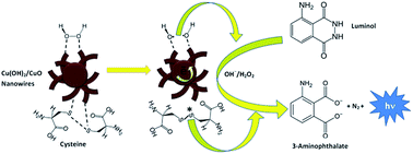
This investigation is a novel chemiluminescence (CL) reaction strategy to enhance the inherent sensitivity of the luminol–H2O2 CL system for the determination of cysteine. It was found that CL intensity of cysteine in the luminol–H2O2 system could be enhanced strongly in the presence of our synthesized copper hydroxide/copper oxide nanowires (Cu(OH)2/CuO NWs). About 3–6 fold improvement in the sensitivity was observed when cysteine was determined in the presence of colloidal Cu(OH)2/CuO NWs. Structural, morphological and optical properties of the Cu(OH)2/CuO NWs were examined by X-ray diffraction (XRD), field emission scanning electron microscopy (FE-SEM) and ultraviolet-visible (UV-Vis) spectroscopy. Fourier Transform Infrared Spectroscopy (FT-IR) studies were done to investigate the presence of organic and/or other compounds in the synthesized NWs. In order to explore the CL mechanism, UV-Vis, fluorescence, and CL spectra studies were carried out. It is suggested that the enhancement in the CL intensity of cysteine is due to the fact that luminol, H2O2 and cysteine could be adsorbed on the surface of NWs and all the electron and the energy transfer processes could be facilitated on the surface of NWs. The CL spectra showed that, the luminophor is the excited-state 3-aminophthalate anion (3-AP2−*, the oxidation product of luminol). Moreover, UV-Vis absorption spectra showed that adsorbed cysteine could be oxidized very easy on the surface of NWs and the resulting product along with O2˙− radicals generate more 3-AP2−* anions. Therefore, higher CL intensities could be produced in the presence of prepared NWs. This enhancement effect has not been seen in the CL system of luminol–H2O2–nanoparticles up to now. Based on these findings, a rapid and sensitive assay was developed for the determination of cysteine. Under the optimum conditions, the CL intensity was proportional to the concentration of cysteine in the ranges 0.8 × 10⁻⁹ to 8.0 × 10⁻⁸ mol L−1 and 8 × 10⁻⁸ to 1.0 × 10⁻⁶ mol L−1. The limit of detection was 0.69 × 10⁻⁹ mol L−1 and percent of relative standard deviations for 50 × 10−9 mol L−1 cysteine (n = 11) was 5.7%. The proposed method was applied successfully for the sensitive determination of cysteine in human plasma and synthesized injection samples.
Introduction
Metal nanoparticles (NPs) are very attractive materials owing to their high surface-to-volume ratio and high surface energy, which make their surface atoms very active. Interest in these particles stems from their unique optical, electronic, and catalytic properties, which are different from those of their bulk counterparts and hence have led to novel applications in sensors, catalysts, bioassays, biological imaging, etc.1 Chemiluminescence (CL) is an attractive means of detection; because it presents low detection limits, a wide linear working range and uses relatively simple instrumentation. For these reasons, CL has received much attention in various fields, especially in combination with separation methods, for analysis of drugs in biological samples.2–8 CL is usually researched as an effective means gaining insight into chemical reactions and widening applications associated with it.9–12 However, it is challenging today to explore novel CL reaction strategies providing new approaches to enhance the inherent sensitivity of CL techniques to widen and deepen its applications.13–15 Among a variety of new materials, nanomaterials are introduced into the CL system as the most promising to improve sensitivity and stability of CL.16–20 A recent challenging research field is the use of nanomaterial for increasing selectivity21 and as a kind of catalyst,22–24 reductant,25 luminophor26,27 or energy accepter28–30 in CL systems, due to their high surface areas, good adsorption characteristics, high activity and high selectivity. In the recent decade, analytical applications of NPs have widely been explored in CL methods.14,24,31–43 It has been reported that NPs can enhance luminol–oxidant CL signals and analytes could enhance or inhibit the CL signals from luminol–oxidant–NPs system. Cysteine (a –SH containing amino acid) have been determined using some of these methods. Table 1 summarizes some analytical characteristics of CL methods proposed for the determination of cysteine with aid of NPs. Cysteine plays a critical role in a variety of important cellular functions, such as detoxification and metabolism.44 Altered levels of cysteine have been implicated in a number of pathological conditions including Alzheimer's and Parkinson's disease.45 Cysteine in most of NP assisted CL methods, had an inhibition effect on the CL intensity.24,31,33,35,46–52 In this study, copper hydroxide/copper oxide nanowires (Cu(OH)2/CuO NWs) were prepared by a very facile co-precipitation method at low temperature as well as short time in comparison with others.53–57 In the CL investigations of our synthesized Cu(OH)2/CuO NWs, cysteine showed a strong enhancement in the CL intensity of luminol–H2O2 system. This enhancement effect has not been seen in the CL system of luminol–H2O2–NPs up to now. Therefore, the mechanism of the CL reaction was investigated in details. The proposed method was successfully used for the quantification of cysteine in synthesized injection and human plasma samples.
Table 1 Response characteristics of the proposed method in comparison with other NPs assisted CL methods for the determination of cysteine
CL system |
NPs |
Effect of cysteine on the CL system |
LDR (×10−9 mol L−1) |
LOD (×10−9 mol L−1) |
Application |
Reference |
Rhodamine 6G–Cerium(IV) |
Au–Ag alloy |
Enhancement |
4–17 000 |
2.2 |
— |
58 |
Lucigenin–KI |
Ag |
Enhancement |
Not reported |
Not reported |
— |
25 |
Lucigenin–hydrazine |
Pt |
Enhancement |
Not reported |
Not reported |
— |
59 |
Luminol–[Ag(IO6)2]7− |
Fe3O4 capped with β-cyclodextrins |
Enhancement |
8–1000 |
2.0 |
Plasma |
60 |
Luminol–AgNO3 |
Ag |
Enhancement |
Not reported |
Not reported |
— |
61 |
Ce(IV)–Na2SO3 |
Ag nanoclusters |
Quenching |
5–1000 |
2.5 |
— |
51 |
Luminol (without oxidant) |
Networked Au |
Quenching |
5–10 000 |
2.0 |
Urine, plasma |
50 |
Luminol–H2O2 |
Triangular Au |
Quenching |
50–2000 |
0.5 |
— |
46 |
Luminol–H2O2 |
Au |
Quenching |
10–70, 100–7000 |
0.82 |
Injection |
47 |
Luminol–H2O2 |
Au |
Quenching |
Not reported |
Not reported |
— |
48 |
Luminol–H2O2 |
Fe2O3 coated with 8-hydroxyquinoline |
Quenching |
2.5 × 10⁴ to 2.5 × 10⁶ |
Not reported |
— |
49 |
Luminol–H2O2 |
Ag |
Quenching |
Not reported |
Not reported |
— |
33 |
Luminol–H2O2 |
Pt |
Quenching |
Not reported |
Not reported |
— |
35 |
Luminol–H2O2 |
Au |
Quenching |
2–2000 |
0.64 |
— |
31 |
Luminol–H2O2 |
ZnO |
Quenching |
2–2000 |
0.85 |
— |
24 |
Luminol–H2O2 |
Au |
Quenching |
1–7000 |
0.29 |
— |
52 |
Luminol–H2O2 |
Cu(OH)2/CuO NWs |
Enhancement |
0.8–80, 80–1000 |
0.69 |
Plasma, synthesized injection |
Proposed method |
Experimental
Reagents
L-Cysteine hydrochloride and luminol were purchased from Sigma-Aldrich company. H2O2, acetone and sodium carbonate were purchased from Dr Mojallali company (Iran). NH4OH solution was purchased from Chem. Lab. Company and copper(II) acetate dihydrate (Cu(CH3COO)2·2H2O) was provided from Merck company. The cysteine stock solution (0.001 mol L−1) was prepared by dissolving cysteine (0.0157 g) in 100 mL deionized water. Working standard solutions were freshly diluted with deionized water. Luminol stock solution (0.01 mol L−1) was prepared by dissolving luminol (0.183 g) in 0.10 mol L−1 Na2CO3 (100 mL). Working solution of H2O2 was prepared freshly daily from 30% (w/w) H2O2. Unless other indicated, all reagents and solvents were purchased in their highest available purity and used without further purification. Millipore Milli-Q water (18 MΩ cm−1) was used in all experiments. Plasma samples were taken from the health center of Gorgan (Iran).
All the experiments involving human subjects were approved by the Golestan University's committee and they were performed in compliance with the relevant laws and institutional guidelines. Moreover, written consent was obtained from all human subjects prior to the experiment.
Experimental method for preparation of Cu(OH)2/CuO NWs
The reaction solution for the fabrication Cu(OH)2/CuO NWs was prepared as follows: 0.002 mole copper acetate dihydrate [Cu(CH3COO)2–2H2O] solution was mixed with 1.0 mL of a fresh NH4OH solution at pH = 10, then deionized water was added to the mixed solution to make a total volume of 100.0 mL in a volumetric flask. The obtained reaction solution was poured into a glass tank being used as a reaction vessel for the NWs preparation. In the next step, the glass tank was placed in a thermostat bath set at 50 °C for 2 hours. The color of the reaction solution at the start was light blue, but changed to dark green at the end of the reaction. After 2 hours, the obtained Cu(OH)2/CuO NWs were filtered, then washed several times by deionized water and dried in air at room temperature.
CL apparatus
CL analysis was applied using a 0.50 cm light path length quartz cell. The CL signal was measured with a CL analyzer with PMT (Hammamatso, model R212, Japan) using a low pass filter which its output was connected to a data processing system with a Pentium IV PC. A schematic block diagram of the used instruments is shown in Fig. S1 in the ESI.†
Procedure for CL detection
An aliquot (200 μL) of standard solution of cysteine with 400 μL of luminol and 200 μL of colloidal Cu(OH)2/CuO NWs (after 10 seconds sonication) were transferred into the 0.50 cm path light length quartz cell. Then, the cell was placed at its location in front of PMT and the program was started. After a few seconds, 200 μL of H2O2 was injected into the cell by a microsyringe and the peak-like CL emission was recorded by a computer (with interval times of 100 ms). Those data information were collected into Excel software. Maximum CL response of cysteine appeared about 0.5 seconds after injection of H2O2 solution. For obtaining the analytical signal, the background signal from the colloidal Cu(OH)2/CuO NWs solution was subtracted from maximum peak height of each sample.
Results and discussion
Characterization of Cu(OH)2/CuO NWs
The size and the surface morphology of the obtained Cu(OH)2/CuO NWs were investigated by field emission scanning electron microscopy (FE-SEM: Hitachi S-4160) under an acceleration voltage of 15 kV. The FE-SEM images of the prepared Cu(OH)2/CuO NWs at different magnifications are shown in Fig. 1a–c. The Cu(OH)2/CuO NWs with approximate diameters of 54 nm and the lengths of about 1 μm are seen in Fig. 1a. It seems that the aggregation of the NWs lead to the formation of the wicker texture structure (Fig. 1a and b).
 |
| Fig. 1 FE-SEM images of the prepared Cu(OH)2/CuO NWs at different magnifications. | |
X-ray diffraction (XRD, philips PW 1800) was performed to characterize the phase and structure of the prepared NWs using CuKa radiation (40 kV and 40 mA) at angles 2θ from 10 to 80°. Fig. 2a shows the XRD pattern of a typical of the prepared Cu(OH)2/CuO NWs.
 |
| Fig. 2 (a) XRD pattern of the prepared Cu(OH)2/CuO NWs. (b) optical absorbance spectrum of the prepared CuO/Cu(OH)2-NWrs (inset: plot of (αhν)2 versus hν of the prepared CuO/Cu(OH)2 NWs). | |
The XRD pattern revealed that the obtained Cu(OH)2/CuO NWs to be polycrystalline and combination of a orthorhombic (copper hydroxide) structure of Cu(OH)2 and a monoclinic (tenorite) crystal structure of CuO. The four strong peaks observed in XRD pattern at about 16.76, 23.84, 33.98, and 53.20° are related to (020), (021), (002), and (150) planes of Cu(OH)2 orthorhombic structure (JCPDS card no. 13-0420), respectively. Besides, two strong peaks observed at around 35.6 and 38.5° are corresponded to (
11) and (111) planes of CuO monoclinic structure (JCPDS card no. 05-0661), respectively. On the basis of the full width at half-maximum (FWHM) of (020) and (
11) peaks and applying the Debeye–Scherrer equation,62 the average nanocrystallite size of Cu(OH)2 and CuO NWs were estimated to be 13 and 28 nm, respectively.
Fig. 2b shows the optical absorbance spectrum of the prepared Cu(OH)2/CuO NWs. From the absorption data, the band gap energy of the Cu(OH)2/CuO NWs was estimated using the well-known relation for semiconductors:63 αhν = k(hν − Eg)n/2; where Eg is the band gap energy, k is a constant, and n is a constant equal to 1 or 4 for direct and indirect band gap materials, respectively. A plot of (αhν)2 versus hν (inset of Fig. 2b) is linear at the absorption edge, which means that the mode of transition in these films has a direct nature. The band gap energy, Eg, was calculated about 3.7 eV from an extrapolation of the straight-line portion of the (αhν)2 vs. hν plot to zero absorption coefficient value. The obtained band gap energy for the prepared Cu(OH)2/CuO NWs is larger than that of the bulk CuO which could be assigned to quantum confinement effects in nanosize materials.
The chemical composition of the prepared Cu(OH)2/CuO was studied by Energy Dispersive X-ray (EDX) analysis and confirmed the presence of Cu and O in the sample (Fig. S2 in the ESI†). In the obtained EDX analysis, the Au peaks are related to the signal detected from the gold coating by sputtering during FE-SEM sample preparation. In EDX analysis (Fig. S2 in the ESI†) Cu and O are the dominant elements throughout the surface of the Cu(OH)2/CuO NWs with weight percentages of 72.6%, and 27.4, respectively.
Fig. S3 in the ESI† shows the FT-IR spectrum of the Cu(OH)2/CuO NWs in the range of 400–4000 cm−1. FT-IR spectroscopy was carried out in order to investigate purity as well as the presence of organic and/or other compounds in the prepared Cu(OH)2/CuO NWs.
Hydroxides and oxides of metal NPs usually gives absorption peak in the finger print region i.e. below wavelength of 1000 cm−1 arising from inter-atomic vibrations.64 Normally the appeared peaks at 400–850 cm−1 are corresponded to lattice vibration of Cu–O, O–Cu–O, and Cu–O–Cu.65 In this study, we observed the sharp and strong peaks at 487, 564, 653, and 894 cm−1 which can be assigned to Cu–O stretching vibration mode.66 The strong and sharp absorption peaks at about 3300–3500 cm−1 and 1500 cm−1 are respectively attributed to the normal polymeric O–H stretching vibrations of Cu–OH and H–O–H bending vibrations of H2O adsorbed onto the Cu(OH)2/CuO NWs lattice.
The characteristics of luminol CL in the presence of Cu(OH)2/CuO nanowires and cysteine
The typical CL profiles are shown in Fig. 3. It can be seen without cysteine and colloidal Cu(OH)2/CuO NWs, the oxidation of luminol by H2O2 is a slow reaction and only emits weak CL (Fig. 3a). Cu(OH)2/CuO NWs (Fig. 3c) or cysteine (Fig. 3b) could enhance the CL from luminol–H2O2 system. Cysteine in presence of NWs can greatly enhance the CL intensity (Fig. 3d). The CL reaction was rapid and the CL intensity reached to maximum in 0.5 second, and then attenuated quickly to baseline.
 |
| Fig. 3 The typical CL profiles of luminol–H2O2 system in presence of (a) blank (water) (b) cysteine (c) blank (Cu(OH)2/CuO NWs) and (d) cysteine–Cu(OH)2/CuO NWs. Conditions: luminol (5.0 × 10⁻⁴ mol L−1), H2O2 (5 × 10⁻⁵ mol L−1), cysteine (1.0 × 10⁻⁷ mol L−1), Cu(OH)2/CuO NWs (0.5 mg L−1), Na2CO3 (0.1 mol L−1). For preparing of the figure, four separate peaks have been pasted together. | |
Optimization of reaction condition
The reaction conditions were optimized for the CL system to obtain higher sensitivities and lower backgrounds for cysteine in presence of NWs. Therefore, reaction parameters optimized so that both signal-to-background (S/B) and net signal (S–B) be as high as possible; (B is the background CL intensity from NWs and S is CL intensity of cysteine in presence of NWs).
For selecting suitable reaction medium, effect of 0.1 mol L−1 NaOH, 0.1 mol L−1 NaHCO3–NaOH buffer and 0.1 mol L−1 Na2CO3 were studied on CL intensity of cysteine in presence of NWs. It was found that, the background from NWs is very high in NaOH and NaHCO3–NaOH buffer solutions so that no enhancement in the CL intensity of cysteine could be observed in these media. Only in the Na2CO3 solution the background was low and cysteine had a great enhancement in the CL intensity; therefore luminol solutions were prepared in the Na2CO3 solution.
The effect of H2O2 on the CL intensity was studied in the range of 2 × 10⁻⁵ to 1.0 × 10⁻³ mol L−1. As shown in Fig. 4a, both the CL signal (from cysteine + NWs) and the background (from NWs) increased with increasing H2O2 concentration. Therefore, to obtain better S/B and S–B, concentration of 5 × 10⁻⁵ mol L−1 H2O2 was chosen for further investigations.
 |
| Fig. 4 Optimization of chemical variables; (a) H2O2 (b) luminol (c) Cu(OH)2/CuO NWs and (d) Na2CO3. Conditions: cysteine (5.0 × 10⁻⁷ mol L−1) (a) luminol (5.0 × 10⁻⁴ mol L−1), NWs (0.4 mg L−1), Na2CO3 (0.1 mol L−1) (b) H2O2 (5 × 10⁻⁵ mol L−1), NWs (0.4 mg L−1), Na2CO3 (0.1 mol L−1) (c) H2O2 (5 × 10⁻⁵ mol L−1), luminol (5.0 × 10⁻⁴ mol L−1), Na2CO3 (0.1 mol L−1) (d) H2O2 (5 × 10⁻⁵ mol L−1), luminol (5.0 × 10⁻⁴ mol L−1), NWs (1.0 mg L−1). | |
The effect of luminol concentration was investigated from 5.0 × 10⁻⁵ to 5.0 × 10⁻³ mol L−1. It was found that the CL intensity increased with the luminol concentration from 5.0 × 10⁻⁵ to 5.0 × 10⁻⁴ mol L−1 (Fig. 4b). A higher concentration of luminol produced self-absorption of the emitted radiation and decreased the CL intensity.14 The background was relatively constant in this region. Therefore, concentration of 5.0 × 10⁻⁴ mol L−1 luminol was chosen as the optimal concentration.
The effect of concentration of Cu(OH)2/CuO NWs was also investigated. As shown in Fig. 4c, the CL intensity and the background increase with increasing Cu(OH)2/CuO NWs concentration in the range of 8.0 × 10⁻³ to 8.0 mg L−1. As a compromise, 1.0 mg L−1 was chosen for further investigations.
Effect of concentration Na2CO3 on the CL intensity has been shown in Fig. 4d. Highest CL intensity and acceptable level of background were achieved at 0.1 mol L−1 Na2CO3.
Analytical features
Under optimum conditions, a long series of standard solutions of cysteine were subjected to the optimized CL method for the purpose of calibration. CL response was found to be linear in two concentration ranges from (0.8–80.0) × 10−9 and (80–1000) × 10−9 mol L−1. The correlation equation between CL intensity and concentration of cysteine in linear ranges were: ICL = 0.144CCys − 0.019 (R2 = 0.9983) and ICL = 0.078CCys + 6.868 (R2 = 0.9930), respectively (where ICL is CL intensity (mV) and CCys is cysteine concentration (×10−9 mol L−1)).
The limit of detection (LOD) was calculated as 3σ/m where σ is the standard deviation existing in 10 times determination of the blank response and m is slope of the lower calibration curve ((0.8–80.0) × 10−9 mol L−1). The LOD obtained was 0.69 × 10⁻⁹ mol L−1, indicating good detectability. The reproducibility was investigated and the percent of relative standard deviations (%RSDs) for 50 × 10−9 mol L−1 cysteine (n = 11) was 5.7%. Typical CL profiles for different concentrations including 0.0, 2.0 × 10⁻⁸, 6.0 × 10⁻⁸, 5.0 × 10⁻⁷ and 1.5 × 10⁻⁶ mol L−1 of cysteine at optimum conditions are shown in Fig. 5.
 |
| Fig. 5 Typical CL profiles of cysteine in the optimized CL system of luminol–H2O2–Cu(OH)2/CuO NWs. Concentration of cysteine: (a) 0.0 (b) 2.0 × 10⁻⁸ (c) 6.0 × 10⁻⁸ (d) 5.0 × 10⁻⁷ and (e) 1.5 × 10⁻⁶ mol L−1. | |
As can be seen in Fig. 6, determination of cysteine using Cu(OH)2/CuO NWs with respect to conventional luminol–H2O2 system (without Cu(OH)2/CuO NWs), shows significant improvement in the sensitivity (approximately 3–6 times).
 |
| Fig. 6 Comparison between CL intensity of cysteine in luminol–H2O2 system at optimum conditions (a) without Cu(OH)2/CuO NWs (b) in presence of Cu(OH)2/CuO NWs. All the CL intensities are background corrected. | |
Interference study
In order to validate of the possible analytical application of the method, interference effect of some common ions, excipients in pharmaceutical preparations and some amino acids were studied by recovering 5.0 × 10⁻⁸ mol L−1 of cysteine in presence of each substance. The tolerance of each substance was taken as the largest amount yielding an error of less than 3σ in the analytical signal of 5.0 × 10⁻⁸ mol L−1 of cysteine (σ is the standard deviation in the response obtained from 11 times determination of 5.0 × 10⁻⁸ mol L−1 of cysteine). No detectable interference found when 1000-fold K+, NO3−, Na+, Ca2+, NH4+, SO42−, Zn2+, alanine, leucine, arginine, saccharin and starch, 250-fold cystine, glucose, fructose, lactose and histidine, 100-fold tyrosine and 10-fold ascorbic acid and SO32− were added into the CL system. Concentrations more than equal amount of Fe2+, Cu2+, Co2+ and glutathione could interfere in this CL system.
According to the literatures, reference (normal) concentrations of Fe2+, Cu2+, Co2+ and glutathione in human plasma are (7–29) × 10−6 mol L−1 Fe2+,67 (1.6–2.4) × 10−6 mol L−1 Cu2+,68 (0.7–7) × 10−6 mol L−1 Co2+ (ref. 69) and (2.35–4.43) × 10−6 mol L−1 glutathione.70
After 5000-folds dilution of the plasma sample, concentration of cysteine will be in the range of (35–76) × 10−9 mol L−1 and concentrations of Fe2+, Cu2+, Co2+ and glutathione will be in the ranges of (1.4–5.8) × 10−9 mol L−1, (0.32–0.48) × 10−9 mol L−1, (0.14–1.4) × 10−9 mol L−1 and (0.47–0.89) × 10−9 mol L−1, respectively. For these interfering substances, concentration of cysteine is at least 6 to 540 times higher than the interfering substance and concentrations of interferences are about 8 to 80 times lower than their tolerance limit (5.0 × 10⁻⁸ mol L−1). Therefore, dilution factor of 5000 is adequate for eliminating the effect of interferences and there is no need to separate the interferences.
Application
This novel CL approach has been successfully used for the determination of cysteine in the synthetic injection formulation and human plasma. Synthetic injection formulation including 50 mg mL−1 L-cysteine hydrochloride prepared in our laboratory according to Loyd and Allen.71 An appropriate volume of the sample solution was further diluted with water so that the final concentration of cysteine was in the working range. However, direct determination of cysteine in plasma was not successful. Therefore, according to Rezaei et al.,60 a deproteination process was carried out by using acetone and the standard addition was carried out by spiking known amounts of cysteine standard solution to plasma samples. For the release of cysteine which is bound to protein in plasma, 10.0 μL β-mercaptoethanol was added to 2.0 mL of the plasma. After precipitation of proteins with 2 mL acetone, the protein-free supernatant was transferred into a small conical flask and evaporated to dryness under a stream of nitrogen at room temperature. Then the dry residue was diluted 10
000-folds. The results are given in Table 2. As can be seen, these values are in the normal range of cysteine in the human plasma (174–378 × 10−6 mol L−1 (ref. 72)).
Table 2 Results for the determination of cysteine in the injection and human plasma
Sample |
Added (mol L−1) |
Founda (mol L−1) |
Recovery (%) |
Mean values of three replications. |
Injection |
— |
(3.07 ± 0.25) × 10−7 |
— |
0.5 × 10⁻⁷ |
(3.56 ± 0.29) × 10−7 |
98.0 |
1.0 × 10⁻⁷ |
(4.11 ± 0.37) × 10−7 |
104.0 |
4.0 × 10⁻⁷ |
(6.89 ± 0.45) × 10−7 |
95.5 |
Plasma 1 |
— |
(341.3 ± 29) × 10−6 |
— |
Plasma 2 |
— |
(211.7 ± 27) × 10−6 |
— |
Plasma 3 |
— |
(308.5 ± 19) × 10−6 |
— |
The obtained results from analyzing of synthesized cysteine injection were also certified using the reference method described in the British Pharmacopoeia (BP).73 Moreover, two new plasma samples were analyzed using the proposed method and a medical laboratory analysis. Statistical analysis (student t-test and the variance ratio F-test) of the results acquired using the BP and CL methods showed no significant difference between the performance of two methods as regards to accuracy and precision. The results are presented in Table 3.
Table 3 Analysis of three samples using the proposed method and the reference method
Sample |
Analytical results (×10−6 mol L−1) |
t-Testd |
F-Teste |
CL method |
Reference method |
Mean values of four replications. British Pharmacopoeia method: based on a redox titration using potassium iodide (excess)/iodine (0.05 mol L−1) as reactant and sodium thiosulfate (0.1 mol L−1) as titrant. Clinical lab analysis was based on a HPLC method with pre column derivatization using ortho-phthalaldehyde (OPA) and UV detection at 333 nm (the results are without replication). Student t-test calculated, theoretical value = 3.182 (P = 0.05). F-Test calculated, theoretical value = 9.28 (P = 0.05). |
Injection |
3.22 ± 0.19 a |
3.11 ± 0.44a,b |
0.65 |
5.36 |
Plasma 4 |
155.2 ± 24.2 |
171.8c |
— |
— |
Plasma 5 |
213.9 ± 14.3 |
228.4c |
— |
— |
Possible CL mechanism
Some CL pathways might be investigated for the luminol–H2O2–Cu(OH)2/CuO NWs-cysteine CL system, involving well known excited-stat 3-aminophthalate anion (3-AP2−*),74 CL from NPs26,27 and oxidation products of analyte.75 In order to explore the possible mechanism of the CL system of luminol–H2O2 in presence of Cu(OH)2/CuO NWs and cysteine, some experiments performed and following results were obtained.
The CL spectrum was scanned with a spectrofluorimeter (Spectrolab, model Spectro-96) using batch mode, a fast scan (15
000 nm min−1) and with turned off excitation lamp. The CL spectra were acquired as shown in Fig. 7 for luminol–H2O2 (spectrum a), luminol–H2O2–cysteine (spectrum b), luminol–H2O2–Cu(OH)2/CuO NWs (spectrum c) and luminol–H2O2–Cu(OH)2/CuO NWs-cysteine (spectrum d). It was clearly indicated that the maximum emission for all mixtures was ∼425 nm which it is the same as fluorescence maximum of luminol (inset in Fig. 7), revealing that the luminophor for the CL system was still the 3-AP2−* with CL emission at 425 nm.36,76,77 Addition of cysteine and Cu(OH)2/CuO NWs did not lead to the generation of a new luminophor in this CL system. Moreover, UV-Vis spectra taken from luminol–cysteine–H2O2 (Fig. S4b in the ESI†) and luminol–cysteine–H2O2–Cu(OH)2/CuO NWs (Fig. S4a in the ESI†) solutions are similar to luminol–H2O2 spectrum (Fig. S4c in the ESI†) and did not reveal any extra peak. Therefore, UV-Vis spectra confirm the results from CL spectra.
 |
| Fig. 7 CL spectra of (a) luminol–H2O2 (b) luminol–H2O2–cysteine (c) luminol–H2O2–Cu(OH)2/CuO NWs (d) luminol–H2O2–Cu(OH)2/CuO NWs–cysteine. Insect is the fluorescence spectrum of luminol. Conditions: cysteine (5.0 × 10⁻⁷ mol L−1), luminol (5.0 × 10⁻⁴ mol L−1), NWs (8.0 mg L−1), Na2CO3 (0.1 mol L−1), H2O2 (5 × 10⁻⁵ mol L−1). | |
Luminol with H2O2 in alkaline solution only produce weak CL in the absence of a catalyst (Fig. 3a). Some oxygen-related radicals, such as hydroxyl radical (OH˙), superoxide radical anion (O2˙−) and other radical derivatives, have been reported to be involved in the luminol oxidation processes.14,33
The strong CL activity of the NPs might be ascribed to mechanisms facilitating the active oxygen intermediate generation and electron-transfer processes on the surface of the NPs.31,35,50,51 Colloidal NPs as a catalyst could transfer electrons to H2O2 to cause them decompose in weakly alkaline solutions.14,24,78 Therefore, we suggest that the O–O bond of H2O2 might be rapidly broken up into to OH˙ radicals by the catalytic action of Cu(OH)2/CuO NWs. The redox potential33 and the shape46 of NPs are important for their catalysis ability in luminol–H2O2 CL system. The resulting OH˙ radicals might be stabilized at the surface of NPs via partial electron exchange interactions.24,31 Then the OH˙ radicals would react with luminol anions and HO2− to facilitate the formation of luminol radical (L˙−) and O2˙−.24,79,80 Production of L˙− was supposed to be the rate-determining step of luminol CL.81 The further reaction between L˙− and O2˙− radicals on the surface of Cu(OH)2/CuO NWs would take place to produce 3-AP2−* anions, the key intermediate of light emission, leading to the enhanced CL.
As can be seen in Fig. S4 in the ESI† (spectrum a and b), maximum absorption of luminol at 350 nm is decreased in the presence of Cu(OH)2/CuO NWs more than that of absence of Cu(OH)2/CuO NWs. As mentioned above, this is due to facilitating production of OH˙ radicals on the surface of Cu(OH)2/CuO NW lead to increasing production of L˙−. It also might be due to adsorption of luminol on the surface of Cu(OH)2/CuO NWs and electron transfer processes between L˙− and O2˙− radicals31,46,82 on the surface of Cu(OH)2/CuO NWs. In order to evaluate the possibility of adsorption of luminol on the Cu(OH)2/CuO NWs, time course curve for luminol at 350 nm was obtained in presence of Cu(OH)2/CuO NWs. As shown in Fig. S5 in the ESI,† concentration of luminol in the solution is decreasing slightly in the presence of Cu(OH)2/CuO NWs that it might be due to adsorption of luminol on the surface of Cu(OH)2/CuO NWs.
OH˙ radicals can readily attack thiol-containing compounds (RSH) to form sulfide radical anion (RS˙).83 It is proposed that RSH could ionize to produce sulfide anion (RS−), which could react with RS˙ to form disulfide radical anion (RSSR˙−).84 It was also reported that RSSR˙− could transfer their unpaired electron to oxygen (O2) to produce O2˙−and RSSR.85
It is also reported that O2˙− and OH˙ radicals can react with cysteine to produce the excited oxidized cysteine (RSSR*) molecule (cystine). When RSSR* returns to the ground state, it can transfer the energy to the ground state of 3-AP2− ion, the oxidation product of luminol, and produce more 3-AP2−* ions. The 3-AP2−* ions return to the ground state and CL intensity enhances.78 Therefore, very high enhancement effect of cysteine in the CL system of luminol–H2O2–Cu(OH)2/CuO NWs could be attributed to production of more O2˙− and RSSR* at the surface of Cu(OH)2/CuO NWs. Cysteine has an affinity to be adsorbed on the copper surface86,87 and oxidation of the cysteine on the NPs of copper occurs at higher rates kinetically and at lower potentials thermodynamically.87
In our investigations we found that cysteine also could be adsorbed on the surface of Cu(OH)2/CuO NWs where luminol and OH˙ radicals are also adsorbed and all electron and energy transfer process could be facilitated by Cu(OH)2/CuO NWs.
Fig. 8 shows the UV-Vis absorption spectra of Cu(OH)2/CuO NWs (spectrum a) cysteine (spectrum b), and cysteine–Cu(OH)2/CuO NWs (spectrum c) solutions. No strong absorption was observed in the spectrum of Cu(OH)2/CuO NWs. An enhancement, broadening and red shift (5 nm) in the band of cysteine was observed in the presence of Cu(OH)2/CuO NWs. These results preliminary suggest that cysteine could interact with Cu(OH)2/CuO NWs. New peak in spectrum c from 350 to 200 nm could be due to autoxidation of cysteine and production of S–S band (maximum 240–260 nm (ref. 88)) on the surface of Cu(OH)2/CuO NWs. The autoxidation of thiol compounds such as cysteine by dissolved oxygen in the presence of Cu(II) has been extensively investigated, and reactive oxygen species such as H2O2,89 O2˙− (ref. 90) and OH˙ radicals91 have been shown to be produced in the reaction. A luminol CL method has also been proposed for the determination of some thiol compounds in the absence of oxidant.92
 |
| Fig. 8 UV-Vis absorption spectra of (a) Cu(OH)2/CuO NWs (b) cysteine (c) cysteine–Cu(OH)2/CuO NWs. Conditions: NWs (8.0 mg L−1), cysteine (2.5 × 10⁻⁴ mol L−1). | |
According to the above discussion, the following mechanism (shown in Scheme 1 and Fig. 9) is proposing for the CL reaction of luminol–H2O2–Cu(OH)2/CuO NWs–cysteine.
 |
| Scheme 1 The mechanism proposed for the CL system of luminol–H2O2–Cu(OH)2/CuO NWs-cysteine.14,24,31,33,35,59,78 | |
 |
| Fig. 9 Schematic illustration of possible mechanism. | |
1 The luminophor is 3-AP2−* (the oxidation product of luminol).
2 Luminol, H2O2 and cysteine could be adsorbed on the surface of Cu(OH)2/CuO NWs and all the electron and energy transfer process could be facilitated on the surface of Cu(OH)2/CuO NWs.
3 Adsorbed cysteine could be oxidized very easy on the surface of Cu(OH)2/CuO NWs and resulting RSSR* and O2˙− aid to produce more 3-AP2−* anions.
Conclusions
In this study, first, we prepared Cu(OH)2/CuO NWs by a very facile co-precipitation method at low temperature as well as short time. Then the prepared Cu(OH)2/CuO NWs were used as catalyst in the CL system of luminol–H2O2. We suggest that very high enhancement in the CL intensity of cysteine in the system of luminol–H2O2–Cu(OH)2/CuO NWs may be attributed to production of more O2˙− and RSSR* at the surface of these NWs. Moreover in our investigations we found that cysteine could be adsorbed on the NWs and adsorbed cysteine could be oxidized very easy on the surface of NWs where luminol and OH˙ radicals are also adsorbed. Therefore, all electron and energy transfer process could be facilitated on the surface of Cu(OH)2/CuO NWs. Using these NWs an analytical application was proposed for the sensitive determination of cysteine in human plasma and injections.
Acknowledgements
We are grateful to the campus of Golestan University for supporting of this research.
Notes and references
- N. Li, D. Liu and H. Cui, Anal. Bioanal. Chem., 2014, 406, 5561–5571 CrossRef CAS PubMed.
- H. Wu, M. Chen, Y. Fan, F. Elsebaei and Y. Zhu, Talanta, 2012, 88, 222–229 CrossRef CAS PubMed.
- E. Nalewajko-Sieliwoniuk, J. Malejko, M. Mozolewska, E. Wołyniec and J. Nazaruk, Talanta, 2015, 133, 38–44 CrossRef CAS PubMed.
- Y. Sun, Z. Zhang and X. Zhang, Spectrochim. Acta, Part A, 2013, 105, 171–175 CrossRef CAS PubMed.
- Z. Lin, X. Sun, W. Hu, Y. Yin and G. Chen, Electrophoresis, 2014, 35, 993–999 CrossRef CAS PubMed.
- Y. Liu, Y. Liu, M. Zhou, K. Huang, J. Cao, H. Wang and Y. Chen, J. Chromatogr. A, 2014, 1340, 128–133 CrossRef CAS PubMed.
- Y. m. Liu, L. Mei, Y. y. Liu, M. Zhou, K. j. Huang, Y. h. Chen and S. w. Ren, Electrophoresis, 2014, 35, 972–977 CrossRef CAS PubMed.
- J. L. Adcock, J. M. Terry, C. J. Barrow, N. W. Barnett, D. C. Olson and P. S. Francis, Drug Test. Anal., 2011, 3, 139–144 CrossRef CAS PubMed.
- R. Freeman, X. Liu and I. Willner, J. Am. Chem. Soc., 2011, 133, 11597–11604 CrossRef CAS PubMed.
- A. Roda and M. Guardigli, Anal. Bioanal. Chem., 2012, 402, 69–76 CrossRef CAS PubMed.
- S. Deng and H. Ju, Analyst, 2013, 138, 43–61 RSC.
- J. L. Adcock, N. W. Barnett, C. J. Barrow and P. S. Francis, Anal. Chim. Acta, 2014, 807, 9–28 CrossRef CAS PubMed.
- Z. Lin, X. Dou, H. Li, Q. Chen and J.-M. Lin, Microchim. Acta, 2014, 181, 805–811 CrossRef CAS.
- W. Chen, L. Hong, A.-L. Liu, J.-Q. Liu, X.-H. Lin and X.-H. Xia, Talanta, 2012, 99, 643–648 CrossRef CAS PubMed.
- Y. J. Chao, L. X. Xie and W. Cao, Key Eng. Mater., 2014, 575, 249–252 Search PubMed.
- Y. Zhou, H. Chen, N. Ogawa and J.-M. Lin, J. Lumin., 2011, 131, 1991–1997 CrossRef CAS.
- S. He, W. Shi, X. Zhang, J. Li and Y. Huang, Talanta, 2010, 82, 377–383 CrossRef CAS PubMed.
- Y. Fan, W. Shi, X. Zhang and Y. Huang, J. Mater. Chem. A, 2014, 2, 2482–2486 CAS.
- J. Xie, X. Zhang, H. Jiang, S. Wang, H. Liu and Y. Huang, RSC Adv., 2014, 4, 26046–26049 RSC.
- Z. Wang, F. Liu and C. Lu, Biosens. Bioelectron., 2012, 38, 284–288 CrossRef CAS PubMed.
- G. Guan, L. Yang, Q. Mei, K. Zhang, Z. Zhang and M.-Y. Han, Anal. Chem., 2012, 84, 9492–9497 CrossRef CAS PubMed.
- Y. Qi, F.-R. Xiu and B. Li, Anal. Biochem., 2014, 449, 1–8 CrossRef CAS PubMed.
- S. Li, S. Tao, F. Wang, J. Hong and X. Wei, Microchim. Acta, 2010, 169, 73–78 CrossRef CAS.
- S.-F. Li, X.-M. Zhang, W.-X. Du, Y.-H. Ni and X.-W. Wei, J. Phys. Chem. C, 2008, 113, 1046–1051 Search PubMed.
- J.-Z. Guo and H. Cui, J. Phys. Chem. C, 2007, 111, 12254–12259 Search PubMed.
- D. Lee, S. Khaja, J. C. Velasquez-Castano, M. Dasari, C. Sun, J. Petros, W. R. Taylor and N. Murthy, Nat. Mater., 2007, 6, 765–769 CrossRef CAS PubMed.
- Z. Wang, J. Li, B. Liu, J. Hu, X. Yao and J. Li, J. Phys. Chem. B, 2005, 109, 23304–23311 CrossRef CAS PubMed.
- X. Huang and J. Ren, Anal. Chim. Acta, 2011, 686, 115–120 CrossRef CAS PubMed.
- G. Qin, S. Zhao, Y. Huang, J. Jiang and F. Ye, Anal. Chem., 2012, 84, 2708–2712 CrossRef CAS PubMed.
- J.-M. Lin and M. Liu, J. Phys. Chem. B, 2008, 112, 7850–7855 CrossRef CAS PubMed.
- Z.-F. Zhang, H. Cui, C.-Z. Lai and L.-J. Liu, Anal. Chem., 2005, 77, 3324–3329 CrossRef CAS PubMed.
- S. Zhang, H. Zhong and C. Ding, Anal. Chem., 2008, 80, 7206–7212 CrossRef CAS PubMed.
- J.-Z. Guo, H. Cui, W. Zhou and W. Wang, J. Photochem. Photobiol., A, 2008, 193, 89–96 CrossRef CAS.
- W. Shi, X. Zhang, S. He and Y. Huang, Chem. Commun., 2011, 47, 10785–10787 RSC.
- S. L. Xu and H. Cui, Luminescence, 2007, 22, 77–87 CrossRef CAS PubMed.
- S. Li, X. Li, J. Xu and X. Wei, Talanta, 2008, 75, 32–37 CrossRef CAS PubMed.
- A. Safavi, G. Absalan and F. Bamdad, Anal. Chim. Acta, 2008, 610, 243–248 CrossRef CAS PubMed.
- F. Wang, C. Ma, X. Zeng, C. Li, Y. Deng and N. He, J. Biomed. Nanotechnol., 2012, 8, 786–790 CrossRef CAS PubMed.
- J. Abolhasani, J. Hassanzadeh and E. Ghorbani-Kalhor, Luminescence, 2015, 31, 247–254 CrossRef PubMed.
- S. M. Wabaidur, S. M. Alam, Z. A. Alothman, M. R. Siddiqui, M. Naushad and A. A. Alqadami, Bioprocess Biosyst. Eng., 2015, 1–8 Search PubMed.
- M. J. Chaichi and M. Ehsani, J. Fluoresc., 2015, 1–10 Search PubMed.
- J. Du, Y. Wang and W. Zhang, Spectrochim. Acta, Part A, 2015, 149, 698–702 CrossRef CAS PubMed.
- C. Y. Wang, Y. X. Qi and X. Q. Liu, Adv. Mater. Res., 2014, 1033, 533–536 Search PubMed.
- Z. P. Li, X. R. Duan, C. H. Liu and B. A. Du, Anal. Biochem., 2006, 351, 18–25 CrossRef CAS PubMed.
- M. T. Heafield, S. Fearn, G. B. Steventon, R. H. Waring, A. C. Williams and S. G. Sturman, Neurosci. Lett., 1990, 110, 216–220 CrossRef CAS PubMed.
- Q. Li, F. Liu, C. Lu and J.-M. Lin, J. Phys. Chem. C, 2011, 115, 10964–10970 Search PubMed.
- W. Liu, J. Luo, Y. Guo, J. Kou, B. Li and Z. Zhang, Talanta, 2014, 120, 336–341 CrossRef CAS PubMed.
- C. Lu, Q. Li, S. Chen, L. Zhao and Z. Zheng, Talanta, 2011, 85, 476–481 CrossRef CAS PubMed.
- T. Triantis, K. Papadopoulos, E. Yannakopoulou, D. Dimotikali, J. Hrbáč and R. Zbořil, Chem. Eng. J., 2008, 144, 483–488 CrossRef CAS.
- L. Zhang, B. Lu and C. Lu, Analyst, 2013, 138, 850–855 RSC.
- X. Yu, Q. Wang, X. Liu and X. Luo, Microchim. Acta, 2012, 179, 323–328 CrossRef CAS.
- P. Yang, Y. Chen, Q. Zhu, F. Wang, L. Wang and Y. Li, Microchim. Acta, 2008, 163, 263–269 CrossRef CAS.
- S. B. Khan, M. Faisal, M. M. Rahman, I. Abdel-Latif, A. A. Ismail, K. Akhtar, A. Al-Hajry, A. M. Asiri and K. A. Alamry, New J. Chem., 2013, 37, 1098–1104 RSC.
- R. Sathyamoorthy and K. Mageshwari, Phys. E, 2013, 47, 157–161 CrossRef CAS.
- D. Gao, J. Zhang, J. Zhu, J. Qi, Z. Zhang, W. Sui, H. Shi and D. Xue, Nanoscale Res. Lett., 2010, 5, 769–772 CrossRef CAS PubMed.
- J. Xiang, J. Tu, L. Zhang, Y. Zhou, X. Wang and S. Shi, Electrochim. Acta, 2010, 55, 1820–1824 CrossRef CAS.
- H. Siddiqui, M. S. Qureshi and F. Z. Haque, Optik, 2014, 125, 4663–4667 CrossRef CAS.
- S.-F. Li, X.-M. Zhang, Z.-J. Yao, R. Yu, F. Huang and X.-W. Wei, J. Phys. Chem. C, 2009, 113, 15586–15592 CAS.
- B. Liu, Y. He, C. Duan, N. Li and H. Cui, J. Photochem. Photobiol., A, 2011, 217, 62–67 CrossRef CAS.
- B. Rezaei, A. A. Ensafi, L. Zarei and P. Kameli, Luminescence, 2012, 27, 390–397 CrossRef CAS PubMed.
- S. Li, H. Sun, D. Wang, J. Hong, S. Tao, H. Yu, X. Wang and X. Wei, Luminescence, 2012, 27, 211–216 CrossRef CAS PubMed.
- A. Goudarzi, G. M. Aval, R. Sahraei and H. Ahmadpoor, Thin Solid Films, 2008, 516, 4953–4957 CrossRef CAS.
- A. Goudarzi, A. D. Namghi and C.-S. Ha, RSC Adv., 2014, 4, 59764–59771 RSC.
- D. Astruc, F. Lu and J. R. Aranzaes, Angew. Chem., Int. Ed., 2005, 44, 7852–7872 CrossRef CAS PubMed.
- X.-Y. Yu, R.-X. Xu, C. Gao, T. Luo, Y. Jia, J.-H. Liu and X.-J. Huang, ACS Appl. Mater. Interfaces, 2012, 4, 1954–1962 CAS.
- L.-J. Chen, G.-S. Li and L.-P. Li, J. Therm. Anal. Calorim., 2008, 91, 581–587 CrossRef CAS.
- L. Gomella and S. Haist, Clinician’s pocket reference, 2007 Search PubMed.
- D. Longo, A. Fauci, D. Kasper and S. Hauser, Harrison's Principles of Internal Medicine, McGraw-Hill Professional, 18th edn, 2011 Search PubMed.
- J. D. Kopple and S. G. Massry, Kopple and Massry's nutritional management of renal disease, Lippincott Williams & Wilkins, 2004 Search PubMed.
- F. Michelet, R. Gueguen, P. Leroy, M. Wellman, A. Nicolas and G. Siest, Clin. Chem., 1995, 41, 1509–1517 CAS.
- V. Loyd and J. Allen, US Pharm., 2011, 36, 41–42 Search PubMed.
- S. P. Stabler, P. D. Marcell, E. R. Podell and R. H. Allen, Anal. Biochem., 1987, 162, 185–196 CrossRef CAS PubMed.
- British Pharmacopoeia, British Pharmacopoeia Commission, London, 2009 Search PubMed.
- J. Lind, The chemiluminescence of luminol in aqueous solutions, a pulse radiolytic study, Royal Inst. of Technology, Dept. of Nuclear Chemistry, Stockholm, Sweden, 1980 Search PubMed.
- A. áVan den Borre and G. áVan der Weken, Analyst, 1995, 120, 463–466 RSC.
- B. Haghighi and S. Bozorgzadeh, Microchem. J., 2010, 95, 192–197 CrossRef CAS.
- Y. Li, W. Niu and J. Lu, Talanta, 2007, 71, 1124–1129 CrossRef CAS PubMed.
- Y. Li, C. Zhu and L. Wang, Microchim. Acta, 2005, 150, 95–99 CrossRef CAS.
- G. Merenyi and J. S. Lind, J. Am. Chem. Soc., 1980, 102, 5830–5835 CrossRef CAS.
- A. L. Rose and T. D. Waite, Anal. Chem., 2001, 73, 5909–5920 CrossRef CAS PubMed.
- T. G. Burdo and W. R. Seitz, Anal. Chem., 1975, 47, 1639–1643 CrossRef.
- B. Rezaei, A. A. Ensafi and L. Zarei, Spectrochim. Acta, Part A, 2012, 90, 223–229 CrossRef CAS PubMed.
- V. Mišík, I. T. Mak, R. E. Stafford and W. B. Weglicki, Free Radicals Biol. Med., 1993, 15, 611–619 CrossRef.
- C. C. Winterbourn, Free Radicals Biol. Med., 1993, 14, 85–90 CrossRef CAS PubMed.
- L. Packer and E. Cadenas, Biothiols in health and disease, Marcel Dekker Inc., 1995 Search PubMed.
- E. Mateo Marti, C. Methivier and C. Pradier, Langmuir, 2004, 20, 10223–10230 CrossRef CAS PubMed.
- H. Heli, M. Hajjizadeh, A. Jabbari and A. Moosavi-Movahedi, Anal. Biochem., 2009, 388, 81–90 CrossRef CAS PubMed.
- F. X. Schmid, eLS, 2001 Search PubMed.
- A. Hanaki and H. Kamide, Bull. Chem. Soc. Jpn., 1983, 56, 2065–2068 CrossRef CAS.
- H. P. Misra, J. Biol. Chem., 1974, 249, 2151–2155 CAS.
- A. V. Kachur, C. J. Koch and J. E. Biaglow, Free Radical Res., 1999, 31, 23–34 CrossRef CAS PubMed.
- H. Li and J. Du, Anal. Lett., 2009, 42, 2131–2140 CrossRef CAS.
Footnote |
† Electronic supplementary information (ESI) available. See DOI: 10.1039/c5ra21085b |
|
This journal is © The Royal Society of Chemistry 2016 |
Click here to see how this site uses Cookies. View our privacy policy here.