Thermally sensitive conductive hydrogel using amphiphilic crosslinker self-assembled carbon nanotube to enhance neurite outgrowth and promote spinal cord regeneration

Lili Sang abc, Yuqing Liub, Wenxi Huaa, Kaige Xub, Guobao Wanga, Wen Zhongd, Leyu Wanga, Shuchai Xuc, Malcolm M. Q. Xing*b and Xiaozhong Qiu*a
aGuangdong Provincial Key Laboratory of Construction and Detection in Tissue Engineering, Southern Medical University, Guangdong, Guangzhou 510515, China. E-mail: qqiuxzh@163.com
bDepartment of Mechanical Engineering and Manitoba Institute of Child Health, University of Manitoba, Winnipeg, MB R3T 2N2, Canada. E-mail: malcolm.xing@umanitoba.ca
cThe Second Affiliated Hospital of Guangzhou University of Traditional Chinese Medicine, Guangdong, Guangzhou 510120, China
dDepartment of Biosystem Engineering, Faculty of Engineering, Department of Medical Microbiology, Faculty of Medicine, University of Manitoba, Winnipeg, Manitoba R3T 2N2, Canada

Received 7th October 2015 , Accepted 22nd February 2016

First published on 23rd February 2016


Abstract

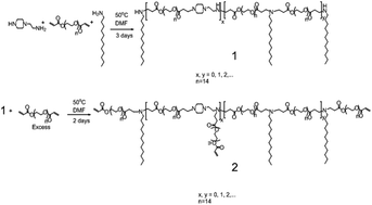
Spinal cord injury leads to severe sensory or motor damage in the human body. Efforts have been made to activate the nerve function by trying physical and biochemical strategies. Carbon nanotubes as conductive materials have been used to transmit electrical signals to improve cell–cell communication and cross-talk, besides providing an extracellular scaffold for neurons. We reported a thermally sensitive hydrogel using copolymerization of n-isopropylacrylamide, the oligomeric amphiphilic crosslinker of polyethylene glycol diacrylate–dodecylamine–1-(2-aminoethyl)piperazine (PEGDA–DD–AEP), and single-walled carbon nanotubes. We hypothesized that carbon nanotubes in the gel could improve neurite outgrowth and nerve regeneration. In order to overcome the aggregation issue of carbon nanotubes, the hydrophobic chains of the amphiphilic crosslinker were used to stabilize the nanotubes. The carbon nanotube–poly(n-isopropylacrylamide) (PNIPAAM) hydrogel was injectable and improved the electrical conductivity. We found that the hydrogel may have potential to promote the growth of SH-SY5Y cells, with significant neurite outgrowth while electrical stimulation was given. In a spinal cord injury model, creating a 1 mm × 1 mm × 1 mm cavity at C7, we found that the hydrogel promoted nerve tissue regeneration and reduced the formation of scar tissue. Therefore, the hydrogel may be a potential repairing biomaterial for neuron network reconstruction and spinal cord regeneration.


Introduction

Spinal cord injury (SCI) leads to severe sensory or motor damage in the human body.1 Difficulty in achieving functional recovery results in overwhelming medical cost and psychological burden, which necessitates the mitigation of neuronal cell loss and repair of nerves.2 Multiple approaches have been undertaken for neurite outgrowth and nerve regeneration with the strategies of autografts, allografts, and xenografts in nerve tissue engineering.3 Though autografts are generally considered to be the gold standard, many limitations, such as donor site morbidity, insufficient donor tissue, etc., attract intense research attention to other alternative sources.4

Neurons are electrically excitable and can directionally transmit electrical signals. For fulfilling the aforementioned function, electrically conductive materials have been synthesized to be intended as a substrate for neuron growth. Those polymers, such as polyaniline (PANI),5 polypyrrole (PPY),4,6 poly(3,4-ethylenedioxythiophene) (PEDOT)7,8 and their derivatives, have been tested and verified for their functions in neuron activation, except not being confirmed for biocompatibility, and acid dopants are unlikely to boost these polymers’ conductivity.9

Inorganic carbon nanotubes (CNTs) already receive extensive attention in potential biomedical applications because of their outstanding properties in electronics, strength, and photonics.14 For instance, CNTs as scaffolds promote nerve and cardiac reconstruction15–18 by attracting stem cells to defective tissues and minimizing cellular apoptosis there by providing and building an electrical signaling network between cells and injured and healthy tissues.19,20 Specifically, many reports have demonstrated that CNTs enhance the neurite outgrowth of various cell types.21–23 For example, Roman et al. reported that single-walled carbon nanotube-functionalized polyethylene glycol (SWNT-PEG) had efficacy by enhancing the neurite outgrowth of hippocampal neurons and giving functional recovery of hind limb locomotion after SCI.20 However, easy hydrophobic aggregation and difficult uniform dispersal limit their application.14

To this end, we fabricated a SWNT-based thermosensitive hydrogel using a multifunctional cross-linker and N-isopropylacrylamide (NIPAAM). By including dodecylamine in the amphiphilic crosslinker, the long hydrophobic alkyl chain can stabilize the SWNT. Thermally sensitive hydrogels experience morphology alteration, sol–gel transition or chemical bonding/debonding in response to temperature change.10 Among them, PNIPAAM shows sol-to-gel transition properties. Its lower critical solution temperature (LCST) of about 32 °C (ref. 2, 11–13) inspires lots of bioengineering applications in carriers of drugs and stem cells. We hypothesize that the SWNT/PNIPAAM hydrogel can promote neuron regeneration after SCI. We first investigated the morphology of the human neuroblastoma cell line (SH-SY5Y cells) in the gel and studied the effect of the electrically conductive property of the SWNT-PNIAAM hydrogel on neuron growth. Finally, we utilized the injectable SWNT–PNIPAAM hydrogel in a rat model with SCI to determine its potential efficacy in nerve repair in vivo (Scheme 1).


image file: c5ra20780k-s1.tif
Scheme 1 Schematic illustration of temperature-sensitive hydrogel with amphiphilic crosslinker and carbon nanotubes for the treatment spinal cord injury (APS: ammonium persulfate; TEMED: tetramethylethylenediamine).

Materials and methods

Materials

Polyethylene glycol diacrylate (PEG-DA, Mw = 700, Sigma Aldrich), dodecylamine (DDA, TCI, 99%), 1-(2-aminoethyl)piperazine (AEP, Aldrich), dimethylformamide (DMF, Sigma Aldrich), DD water (ultrapure water, 18.2 MΩ, from a Direct-Q®3 instrument). NIPAAM was ordered from Sigma and SWNT was obtained from USA Nano (TA, USA). All reagents were used as received. The Human SH-SY5Y neuroblastoma cell line was obtained from the ATCC (Manassas, VA, USA). Dulbeccos Modified Eagle Medium (high-glucose DMEM), 0.25% trypsin/1 mM EDTA, Fetal Bovine Serum (FBS) and phosphate buffered saline (PBS) were all purchased from Gibco BRL (Gaithersburg, MD, USA). Cell Counter Kit-8 (CCK-8) was ordered from Dojindo Laboratory (Japan). The Cell Lysis Buffer and BCA Protein Assay Kit were provided by Keygen Biotech (China). Goat anti-rabbit β-tublin antibody (ME, 1[thin space (1/6-em)]:[thin space (1/6-em)]1000) was purchased from Sigma (USA). An Olympus inverted microscope (Olympus Optical, Tokyo, Japan) was used to obtain light and fluorescence photographs. An ATFxxB Series Function generator was ordered from the ATTEN company (Hongkong, China). Platinum electrode (99.99%, 0.0707 mm2 in area) was obtained from Three Precision Alloy Corporation (China).

Synthesis of PEG-DDA-AEP oligomer crosslinker

0.63 g polyethylene glycol diacrylate (Mw = 700, 0.9 mmol), 0.1483 g dodecylamine (0.8 mmol), 0.0517 g 1-(2-aminoethyl)piperazine (0.4 mmol), and 4 ml DMF were added into a 25 ml single-neck flask capped with a rubber septum. The flask was heated to 50 °C in an oil bath, and the solution was stirred with a magnetic stirrer bar. After 72 h, a 500 μl aliquot (product 1, intermediate product) was collected from the flask using a pipette, and dried at room temperature under vacuum for 48 h for NMR characterization without further purification. Another 3.15 g polyethylene glycol diacrylate (4.5 mmol) was dissolved in 1 ml DMF, dropped into the flask and mixed with the solution homogeneously. Then, the reaction was continued at 50 °C for another 48 h. After reaction, the solution was terminated by cooling in cold water, and the product was purified by dialysis against DD water in a pre-wetted dialysis tube (MWCO = 1000) for 72 h.15,24,25 After dialysis, the solvent was removed using a rotary evaporator, and the oligomer crosslinker (product 2), a kind of light brown viscous oil, was recovered under vacuum at room temperature with an overall yield of 80% (1.10 g). 1H NMR (ppm) of product 1 (intermediate product): δ 0.86 (t, –CH3), δ 1.15–1.50 (s, –NH–(CH2)10CH2CH3), δ 2.30–2.50 (m, –NH–CH2–CH2–COO–), δ 2.50–2.65 (m, –NH–CH2–(CH2)10–CH3), δ 2.60–2.80 (m, –NH–CH2–CH2–COO–, –(CH2)2N–CH2–CH2–NH–, –(CH2)2N–CH2–CH2–NH2), δ 2.80–2.95 (m, –N(CH2)2–(CH2)2N–), δ 3.50–3.8 (s, [–CH2–CH2–O–]n), δ 4.10–4.35 (m, –COO–CH2–CH2–O–). 1H NMR (ppm) of product 2 (PEG-DDA-AEP crosslinker): δ 0.86 (t, –CH3), δ 1.15–1.50 (s, –NH–CH2–(CH2)10CH3), δ 2.30–2.50 (m, –NH–CH2–CH2–COO–), δ 2.50–2.65 (m, –NH–CH2–(CH2)10–CH3), δ 2.60–2.80 (m, –NH–CH2–CH2–COO–, –(CH2)2N–CH2–CH2–NH–, –(CH2)2N–CH2–CH2–NH2), δ 2.80–2.95 (m, –N(CH2)2–(CH2)2N–), δ 3.50–3.8 (m, [–CH2–CH2–O–]n), δ 4.10–4.35 (m, –COO–CH2–CH2–O–), 5.75–6.50 (m, –CH[double bond, length as m-dash]CH2).

NMR characterization

1H NMR experiments were conducted on a Bruker Avance 300 MHz NMR Spectrometer at room temperature. The samples were dissolved in chloroform-d at a concentration of 20 mg ml−1, and the relaxation delay (d1) was set as 5 s.

Hydrogel synthesis

After being lyophilized in a freeze-dyer, a 20 mg ml−1 solution of the crosslinker was prepared in ddH2O and SWNTs were added to obtain a concentration of 0.2 mg ml−1 of nanotubes. The solution was sonicated for 15 × 2 min in an ice bath to get uniformly dispersed SWNTs. 1 ml of this solution was added to 9 ml aqueous N-isopropylacrylamide (80 mg ml−1 in H2O) and then 150 μl ammonium persulfate (200 mg ml−1) and 10 μl N,N,N′,N′-tetramethylethylenediamine were added in an ice bath. After 2 hours, the mixture was dialyzed in ddH2O for 72 h and the resultant product lyophilized in a freeze-dyer for 72 h to acquire the polymer.

Electrical conductivity test

All the electrical conductivity tests were carried out using a Multifunction Digital Four-probe Tester (Suzhou Jingge Electronic Co., Ltd).

Transmission electron microscopy

3 μl of SWNT-hydrogel solution at room temperature was dropped onto a copper grid for TEM sample preparation to determine the dispersion of SWCNTs in the solution. A Hitachi TEM system (H-7650) was used for the measurements.

Cell culture

Human SH-SY5Y neuroblastoma cells (ATCC, Manassas, VA, USA) were maintained in high-glucose DMEM supplemented with 10% FBS and 100 U ml−1 penicillin–streptomycin at 5% CO2, 37 °C and subcultured every the other day when cell confluence was approximately 80%.

The viability of the SH-SY5Y cells on the SWNT–PNIPAAM

20 μl hydrogel was spread on a 96-well plate with temperature under 25 °C. The plate uniformly coated with hydrogel solution was transferred into the humidified incubator (37 °C) to form a gel on the dish. Cells digested from 75 cm2 culture flasks were seeded on the shrunk hydrogel with a density of 5000 cells per well. After 30 min at 37 °C, additional high-glucose DMEM was added for cell culture. They were divided into three groups: SWNT–PNIPAAM hydrogel (n = 6), PNIPAAM hydrogel (n = 6), and control group (n = 6). The viability of the cells was detected by CCK-8 analysis after 24 h and 48 h. The optical density (OD) value was measured at 450 nm using a microplate reader (Bio-Rad 680, Hercules, CA, USA) to test the cell viability. Each experiment was repeated independently three times.

The electrical stimulation of SH-SY5Y cells encapsulated in SWNT–PNIPAAM hydrogel

Cells were digested from 75 cm2 culture flasks and centrifuged at 800 rpm, 37 °C for 5 min. Then, 60 μl soluble hydrogel and 2 × 10⁵ cells were mixed uniformly at 25 °C, and the mixture drawn in straight lines on the cover slips of a 24-well plate. The plate was placed in the incubator (37 °C) for sol–gel transition. After 10 min, high-glucose DMEM was added. Three groups were designated as follows: cells loaded into SWNT–PNIPAAM hydrogel (n = 4, 3D), cells loaded into PNIPAAM hydrogel (n = 4, 3D), and cells seeded on the plate only (n = 4, 2D). Each group was then divided into 2 subgroups: electrical stimulation (ES) and nonstimulation (NS). After the initial 24 h culture, the cells were electrically stimulated with a rectangular wave (frequency 20 Hz, potential 100 mV) for 2 h. For electrical stimulation, two platinum wires served as the anode and cathode. After exposure to constant electrical stimulation, the cells were cultured for an additional 24 h (a total of 48 h from the start of the experiment).

Cell morphology observation

After the electrically stimulated cells were incubated for another 24 h, the cover slips were gently washed with PBS (37 °C) 3 times and fixed with 4% paraformaldehyde overnight. After leaving overnight, the cover slips were lightly washed with PBS (37 °C) 3 times. Then, PBS was exchanged with 0.2% Triton X-100/PBS for 10 min. After washing with PBS at 37 °C, BSA was added. After 30 min, the cover slips were then incubated with the primary antibody (β-tublin, 1[thin space (1/6-em)]:[thin space (1/6-em)]1000, Sigma, USA) overnight (4 °C). The next day, the cover slips were incubated with the goat anti-rabbit secondary antibody (1[thin space (1/6-em)]:[thin space (1/6-em)]1000, Sigma, USA), after washing with PBS at room temperature, for 1 h. Finally, DAPI was used for counterstain mounting at room temperature (RT). The morphology of the samples was recorded with an Olympus BX-51 inverted phase-contrast microscope (Olympus Optical Co., Ltd., Tokyo, Japan).

Neurite length measurements

The length of the individual neurites for each cell was measured using the NIH IMAGE software. Length was defined as the straight-line distance from the tip of the neurite to the junction between the cell body and neurite base. In the rare case of branched neurites, the length of the longest branch was selected. For each experiment, three duplicate wells were observed under a fluorescence microscope. For each well, approximately 9 images were taken randomly by scanning the wells from left to right and top to bottom. Experiments were repeated on at least two separate days.

The post-SCI repair evaluation in rats

The rats were purchased from the Animal Experiment Center, Southern Medical University (Guangzhou, China). The animal experimental procedures were carried out in the Animal Experiment Center, Southern Medical University and approved by the Ethics Committee of Southern Medical University. NIH guidelines for laboratory animal care and safety were strictly followed. All efforts were made to minimize the suffering of the animals. A total of 24 adult female SD rats (180–200 g) were subjects of this study. All the surgery was done under deep anesthesia with 10% chloral hydrate (0.3 ml/100 g). The nape area was shaved and disinfected with povidone iodine. A dorsal column lesion of the spinal cord was applied at the cervical 7 (C7) level. Briefly, after the anesthetised rats were fixed prone, the dorsal soft tissues were cut open step by step and a longitudinal incision of the skin and muscles was made at C7. Then, a small dural and leptomeningeal incision was made following dorsal laminectomy, and then an approximate 1 mm × 1 mm × 1 mm cavity was made with lidocaine for local anesthesia on the right dorsal spinal cord. Following this, the cavities in the different groups were injected with the SWNT–PNIPAAM hydrogel (group D, n = 6), PNIPAAM hydrogel only (group C, n = 6) and with nothing for the sham group (group B, n = 6). The untreated rats were taken as the controls (group A, n = 5). Dural and other soft tissues were sutured layer by layer with 11–0 and 3–0 silk thread, respectively. Two months later, the center and adjacent regions were excised for H&E staining. The process of making paraffin sections and H&E staining is as follows: the excised tissues were fixed in neutral buffered formalin, dehydrated in alcohol from low to high concentration, made transparent with dimethyl benzene, embedded in melted paraffin, sectioned with 5 μm thickness, then stained using hemotoxylin and eosin. The stained sections were dehydrated and made transparent again. Finally, the sections could be utilized for imaging after mounting with resin. The sections for fluorescence immunoassay and DAPI staining were made as in above method.

Weight test and neurological evaluation (Cheerio test)

The weight test and neurological test were performed every other day from the operation day. The Cheerio test is used to assess forelimb function by observing animals eating cereal of a consistent size (about 14 mm in diameter) according to the scale of neurological evaluation (Table 1).
Table 1 Scale of neurological evaluationa
Score Qualifying parameters
a Supportive contact = no treat manipulation. Coordinated contact = manipulation. Occasional = <50% of the time. Frequent = >50% of the time but <95% of the time. Consistent = 95–100% of the time.
0 No ipsilateral forelimb usage/contact with treat
1 Supportive dorsal forelimb contact with treat, non-elevated forelimbs, drops
2 Supportive dorsal forelimb contact with treat, occasional–frequent elevated (flexed) forelimbs, drops
3 Supportive dorsal–dorsal/palmar forelimb contact, frequent–consistent elevated (flexed) forelimbs, drops
4 Supportive dorsal–dorsal/palmar forelimb contact, frequent–consistent elevated (flexed) forelimbs, drops–no drops
5 Supportive dorsal–dorsal/palmar forelimb contact, frequent–consistent elevated (flexed) forelimbs, no drops
6 Supportive-coordinated dorsal–dorsal/palmar forelimb contact, frequent–consistent elevated (flexed) forelimbs, no drops
7 Coordinated dorsal/palmar forelimb contact, consistent elevated (flexed) forelimbs, no drops
8 Coordinated palmar forelimb contact, consistent elevated (flexed) forelimbs, no drops


Two independent examiners observed the forelimb movements of the rats and scored the motor and sensory function according to the detailed scale ranging from 0 (no movement) to 8 (normal movement). A minimum of 3 trials was assessed for each rat.

Statistics

Statistical analysis was performed using the Statistical Package for Social Science (SPSS Software, Minitab Inc., USA). Data were expressed as average ± standard error (SEM). Statistical differences were assessed by one-way ANOVA followed by Bonferroni post hoc test for multiple comparisons. P-Values less than 0.05 were deemed as statistically significant.

Results and discussion

PEG-DDA-AEP crosslinker and hydrogel synthesis

The amphiphilic PEG-DDA-AEP oligomer crosslinker was synthesized via two steps of Michael addition condensation polymerization in one pot, as shown in Fig. 1.
image file: c5ra20780k-f1.tif
Fig. 1 Synthesis of the crosslinker PEG-DDA-AEP.

In this Michael addition polymerization, the reactivity of the nucleophilic groups (Michael donors) is according to the following sequence: secondary amine on the ring of piperazine (–NH(CH2)2–, 2° amine on ring) > primary amine (NH2–, 1° amine) ≫ secondary amine (–NH–, 2° amine, formed during reaction). Therefore, in the first step of the reaction, 1.8 mmol acrylate would prefer to first react with 0.4 mmol 2°amine on the ring and 1.2 mmol 1° amine, and then react with 1.2 mmol 2° amine, which formed after 1° amine addition.26,27 After reacting at 50 °C for 3 days, the sample was collected and dried for NMR characterization without further purification. As shown in Fig. 2, the 3 peaks at 6.42 ppm, 6.13 ppm and 5.82 ppm, which are assigned to the three protons on –CH–CH2, disappeared completely after reaction, indicating the completion of the Michael addition polymerization. Due to the equal consumption of Michael donors and acceptors and the different reactivity of the three types of amine, all 2° amines on the rings and 1° amines were consumed and 0.2 mmol 2° amine reacted as well. So, the oligomer (1) formed is as shown in the scheme, with the two ends of the oligomer capped by secondary amine functional groups formed from DDA, and 1.0 mmol secondary amine was left after reaction (a small amount of PEG-DA reacted with the secondary amine on the aminoethyl piperizine unit to form a branch chain, but was capped by DDA as well). To achieve the amphiphilic crosslinker, the chain of compound 1 was extended by reacting with an excess of PEG-DA via the second step of the Michael addition reaction in the same pot. 5 equivalents of PEG-DA were added into the reaction to react under the same conditions for another 48 h, and all excess PEG-DA was removed by dialysis again with DD water after reaction. As shown in Fig. 2c, the peaks belonging to the protons of the acrylate groups appeared again, indicating the successful addition of PEG-DA. In both Fig. 2b and c, the amount of PEG in the oligomer can be calculated by comparing the peak intensity at 3.6 ppm with the peak intensity at 0.86 ppm. The former is from the protons on the PEG backbone, and the latter is assigned to the protons on the methyl groups of DDA (–CH3). The calculated molar ratios of PEG to DDA are 1.0 and 2.16 respectively in products 1 and 2. Given the DDA amount is the same as its feeding amount (0.8 mmol), 0.928 mmol PEG-DA conjugated with the oligomer during the addition reaction, which is comparable to the amount of secondary amine (1.0 mmol). Therefore, it can be concluded that almost all secondary amine groups are reacted with PEG-DA, and all aminoethyl piperazine units formed branch PEG-acrylate chains (Fig. 2). As shown in Fig. 3A, the final gel undergoes a sol–gel transition from room temperature to 37 °C. We then tested the surface conductivity of the hydrogel at 37 °C with 0.01 s m−1. The TEM micrograph shows the gel morphology and carbon nanotubes (Fig. 3B).


image file: c5ra20780k-f2.tif
Fig. 2 1H NMR spectra of (A) polyethylene glycol diacrylate (PEGDA, Mw = 700), (B) product 1, the intermediate product, and (C) product 2, PEG-DDA-AEP crosslinker.

image file: c5ra20780k-f3.tif
Fig. 3 Sol–gel transition (A) and TEM micrograph (B) of SWNT-hydrogel (scale bar = 40 nm).

Cell viability on SWNT–PNIPAAM hydrogel

SH-SY5Y cells were seeded on the SWNT–PNIPAAM hydrogel to evaluate its cytotoxicity. In the process of the operation, the temperature was controlled at 37 °C. It was about 2 h after adding the CCK-8 agent that the microplate reader (Bio-Rad 680, Hercules, CA, USA) was used to detect the optical density (OD, 450 nm). As shown in Fig. 4, the absorbances with the SWNT–PNIPAAM hydrogel were 0.10 ± 0.001 (24 h) and 0.15 ± 0.010 (48 h), significantly higher than those with the PNIPAAM hydrogel, which were 0.09 ± 0.003 (24 h) and 0.13 ± 0.008 (48 h) (Fig. 4). It was found that the absorbance of the SWNT–PNIPAAM group was significantly higher than that of the PNIPAAM group.
image file: c5ra20780k-f4.tif
Fig. 4 The cytotoxicity of SWNT–PNIPAAM and PNIPAAM hydrogels towards SH-SY5Y cells by CCK-8 assay after 24 h and 48 h. The absorbance of the SWNT–PNIPAAM group was significantly higher than that of PNIPAAM group both after 24 h and 48 h (error bars = average ± standard deviations, *p < 0.05).

Suitable scaffolding materials to support cells on implantation must be safe and the matrix must therefore provide an appropriate spatial microenvironment for cell proliferation, differentiation and axon extension.28 CNTs have been reported to be biocompatible platforms for neuronal growth and differentiation.21,22 In this work, it was found that the absorbance of the SWNT–PNIPAAM group was significantly higher than that of PNIPAAM group. Such data may indicate that SWNT may have potential to promote the growth of SH-SY5Y cells and also have promise for nerve tissue engineering.

ES enhances neurite outgrowth of 3D SH-SY5Y cells loaded in SWNT–PNIPAAM hydrogel

Cho and Borgens23 demonstrated the effect of electrical stimulation on pheochromocytoma cells through a CNT–collagen composite with 5% SWNT loading. They found that electrical stimulation promoted cell adhesion and proliferation, and enhanced neurite outgrowth. Currently, the immortalized and proliferative SH-SY5Y cells are one of the most commonly applied cell lines in neuroscience and neuroblastoma research.29 Herein, we hypothesized that ES could promote the outgrowth of SH-SY5Y cells encapsulated in the 3D SWNT–PNIPAAM hydrogel. We tuned the parameters of ES in our study according to the pioneering work of Schmidt4 in conductive materials. Therefore, after constant ES (rectangular wave: 20 Hz, 100 mV) for 2 h, cells were incubated for an additional 24 h (a total of 48 h from the start of the experiment), as shown in Fig. 5.
image file: c5ra20780k-f5.tif
Fig. 5 Illustration of the electrical stimulation of cells. (a) 60 μl hydrogel and 2 × 10⁵ cells were mixed uniformly together on ice (<32 °C). (b) The mixture was drawn in straight lines on the cover slips of a 24-well plate. (c) Electrodes were added. (d) The parameters of the electrical stimulator were frequency (20 Hz), potential (100 mV), wave shape (rectangular wave), the period of stimulation (for 2 h after the cells were cultured for 24 h), and the electrode (platinum wire electrode).

Our study investigated the effects of ES on the morphologies of 3D encapsulated SH-SY5Y cells compared with 2D. SH-SY5Y cells (2D), PNIPAAM hydrogel (3D), and SWNT–PNIPAAM hydrogel (3D) are shown in Fig. 6 (from top to bottom) for comparison. To evaluate the morphologies of SH-SY5Y cells after ES, a fluorescence immunoassay was performed by staining the neuron scaffolding protein (β-tublin, red) and cellular nuclear staining (DAPI, blue). The morphologies of the SH-SY5Y cells showed no significant differences in the 2D groups (Fig. 6a–f) and the 3D PNIPAAM hydrogel groups (Fig. 6g–l). However, the SWNT–PNIPAAM hydrogel enhanced neurite outgrowth of the 3D SH-SY5Y cells with ES (Fig. 6m–o) compared with NS (Fig. 6p–r). The 3D SH-SY5Y cell morphologies of the SWNT–PNIPAAM hydrogel group after ES were significantly different from the other 3D groups.


image file: c5ra20780k-f6.tif
Fig. 6 Immunofluorescence staining of β-tublin (red) and DAPI (blue) of SH-SY5Y cells with electrical stimulation (ES) and non-stimulation (NS). (A) The effects of ES on 2D groups, 3D SWNT–PNIPAAM hydrogel groups and 3D PNIPAAM hydrogel groups. (B) The effects of NS on 2D groups, 3D SWNT–PNIPAAM hydrogel groups and 3D PNIPAAM hydrogel groups. The morphologies of the SH-SY5Y cells showed no significant differences in 2D groups (a–f) and 3D PNIPAAM hydrogel groups (g–l). However, the SWNT–PNIPAAM hydrogel enhanced neurite outgrowth of the 3D SH-SY5Y cells with ES (m–o) compared with NS (p–r) (scale bar = 40 μm).

The neurite distribution was recorded as a frequency histogram. The median neurite length (12.59 μm) and average length (14.24 μm) with ES were much longer than those with NS, which were 8.04 μm and 7.98 μm, respectively. The distribution of the neurite lengths for the SWNT–PNIPAAM hydrogel (ES, NS) are shown in Fig. 7. We found that ES not only enhanced the neurite sprouting, but made the dividing cells gather and form multinucleate cells, which come out with amazing interfacial connections with each other. We calculated the relative value between the neurite number and the cell number to find the value with ES of 0.62 was obviously much larger than that with NS, which was 0.04. The morphologies of the SH-SY5Y cells in the NS SWNT–PNIPAAM hydrogel (3D) group, ES PNIPAAM hydrogel (3D) group and NS PNIPAAM hydrogel (3D) group showed no significant differences. Similarly, there were also no significant differences between the ES 2D group and NS 2D group.


image file: c5ra20780k-f7.tif
Fig. 7 The merged images of DIC and β-tublin/DAPI, and neurite length histograms. (A) SH-SY5Y cells loaded in SWNT–PNIPAAM hydrogel with ES and NS. The application of ES significantly enhanced neurite lengths of SH-SY5Y cells and promoted connection formation among the neurons. (B) The average and median lengths of SH-SY5Y cells in SWNT–PNIPAAM hydrogel group with ES were significantly larger than those with NS (scale bar = 20 μm).

Recently, CNTs and their composites have played a valuable role in nanomedicine and tissue engineering due to their outstanding electrical properties and other excellent properties. In 2005, Lovat et al. suggested that CNTs may enhance neural signal transmission due to their high electrical conductivity.30 The presence of SWNTs in a hydrogel can similarly improve the conductivity of the hydrogel. In this study, we confirmed the effect of the electrical conductivity of the SWNT–PNIPAAM hydrogel on SH-SY5Y cells. The ES significantly increased the number of neurites and largely enhanced neurite outgrowth compared with NS through the SWNT-PNIPAAM hydrogel. The study demonstrated that the PNIPAAM hydrogel loaded with SWNTs (3D) enhanced neurite sprouting.

SWNT–PNIPAAM promoting the regeneration of the spinal cord and the functional recovery post-spinal cord injury (SCI)

The images showed that the neurons migrated into the injury site (Fig. 8d and 9j–l) and formed interactions and connections between the hydrogel and neurons. The implantation of SWNT–PNIPAAM into the cavity of dorsal horn (DH) provided an obvious improvement for the spinal cord injury, while group B formed scar tissue (Fig. 8b and 9d–f) and group C developed barrier film formation, resisting repair of the injury (Fig. 8c and 9g–i). Therefore it became obvious that the copolymers were highly biocompatible in vivo compared with the PNIPAAM hydrogel and possessed the potential capacity to serve as an extraordinary extracellular matrix (ECM) for nervous tissue engineering. We also tracked the function scores and body weight (in grams) (Fig. 8b) of the animals. Though all the animals presented moderate functional defects compared with the normal group, animals in group D had better functional recovery scores (6.50 ± 0.55) of the forelimb locomotion after two months compared with group C (5.67 ± 0.52) and the group B (5.67 ± 0.82). The animals of all the groups exhibited gradual improvements in body weight. However, the weights (in grams) of group D (299 ± 15) were significantly higher than those of group C (272 ± 21), group B (266 ± 17) and group A (279 ± 12) two months post-SCI. The better functional recovery and the higher body weight of group D indicated that the copolymers induced no overt toxicity to the animal body and the whole physical condition was enhanced by the injectable SWNT–PNIPAAM hydrogel.
image file: c5ra20780k-f8.tif
Fig. 8 HE staining of the dorsal column lesion of C7 sections and monitoring of animals by weight and Cheerio test two months post-injury. (A) Group A (a) was the normal control; group B (b) remained untreated and formed scar tissue; group C (c) was implanted with PNIPAAM hydrogel and developed barrier film formation, resisting the repair of the injury; and group D (d) was grafted with SWNT–PNIPAAM hydrogel and showed enhanced neuron migration into the injury site. (B) Group D had higher body weights (in grams) and better scores of Cheerio test compared with other groups (scale bar = 200 μm; error bars = average ± standard deviation, n = 6).

image file: c5ra20780k-f9.tif
Fig. 9 Immunofluorescence staining of β-tublin (red) and DAPI (blue) of the lesion sections two months post-injury. (a–c) Group A was the normal control. (d–f) Group B remained untreated and formed scar tissue. (g–i) Group C was implanted with PNIPAAM hydrogel and developed barrier film formation, resisting the repair of the injury. (j–l) Group D was grafted with SWNT–PNIPAAM hydrogel and showed enhanced neuron migration into the injury site (scale bar = 200 μm).

Of the multiple temperature-stimulated hydrogels, the PNIPAAM hydrogel has been extensively explored and used for drug delivery systems and tissue engineering because of the smart LCST. Hydrogels have many advantages as cellular scaffolds, for example having similar mechanical properties to soft tissue, which results in low interfacial tension and makes neurons able to move across the tissue-implant interface, as well as allowing diffusion of oxygen, nutrients and waste.31 The injectable PNIPAAM hydrogel has been deployed in the nervous system, and provided support for neurons implanted within the matrix. Meanwhile, the SWNT–PNIPAAM hydrogel has better electrical conductive capacity, making the biocopolymer more appropriate in implantations for SCI. Previous study showed that the better result for repairing damaged axons post-SCI was rejoining the lesion area.32 However, it was so hard to achieve functional recovery due to neuron loss and scar tissue forming in the lesion site. Mattson and coworkers21 reported that the molecular functionalized CNTs could not only enhance nerve cell growth but also reduce scar formation. Possible therapies are directed to neuron regeneration and promoting the recovery of morphology of the injured spinal cord.33 In our report, the copolymer scaffold of SWNT–PNIPAAM hydrogel was designed to function as an injectable matrix for nerve tissue applications, and the migrated neurons of the native tissue repaired the injury site two months post-injury.

Conclusion

We developed an injectable carbon nanotube-based temperature-sensitive hydrogel. By developing an amphiphilic crosslinker to stabilize the conductive carbon nanotubes, a uniform hydrogel was obtained with conductive properties. We studied the SWNT–PNIPAAM hydrogel for its potential role in the growth of SH-SY5Y cells and found it can enhance neurite outgrowth when adding electrical stimulation in vitro. Finally, in a SCI model, we found that the SWNT–PNIPAAM hydrogel not only promoted nerve tissue regeneration, but also reduced the formation of nerve tissue scars in vivo.

Acknowledgements

This work was supported by the National Natural Science Foundation of China (31371000 and 51428301) and Guangdong Natural Science Foundation (s2013010016032). M. X. would like to thank the support from the NSERC Discovery Grant, CIHR-RPP and China 863 Project.

References

  1. J. Ma, L. N. Novikov, M. Wiberg and J. O. Kellerth, Exp. Brain Res., 2001, 139, 216 CrossRef CAS PubMed.
  2. N. Comolli, B. Neuhuber, I. Fischer and A. Lowman, Acta Biomater., 2009, 5, 1046 CrossRef CAS PubMed.
  3. H. Cheng, Y. C. Huang, P. T. Chang and Y. Y. Huang, Biochem. Biophys. Res. Commun., 2007, 357, 938 CrossRef CAS PubMed.
  4. C. E. Schmidt, V. R. Shastri, J. P. Vacanti and R. Langer, Proc. Natl. Acad. Sci. U. S. A., 1997, 94, 8948 CrossRef CAS.
  5. V. Guarino, M. A. Alvarez-Perez, A. Borriello, T. Napolitano and L. Ambrosio, Adv. Healthcare Mater., 2013, 2, 218 CrossRef CAS PubMed.
  6. H. Xu, J. M. Holzwarth, Y. Yan, P. Xu, H. Zheng and Y. Yin, Biomaterials, 2014, 35, 225 CrossRef CAS PubMed.
  7. Y. Furukawa, A. Shimada, K. Kato, H. Iwata and K. Totimitsu, Biochim. Biophys. Acta, 2013, 1830, 4329 CrossRef CAS PubMed.
  8. X. Luo, C. L. Weaver, D. D. Zhou, R. Greenberg and X. T. Cui, Biomaterials, 2011, 32, 5551 CrossRef CAS PubMed.
  9. S. Jafarzadeh, P. M. Claesson, P. E. Sundell, J. Pan and E. Thormann, ACS Appl. Mater. Interfaces, 2014, 6, 19168 CAS.
  10. Y. Qiu and K. Park, Adv. Drug Delivery Rev., 2001, 53, 321 CrossRef CAS PubMed.
  11. J. Y. Wu, S. Q. Liu, P. W. Heng and Y. Y. Yang, J. Controlled Release, 2005, 102, 361 CrossRef CAS PubMed.
  12. S. B. Turturro, M. J. Guthrie, A. A. Appel, P. W. Drapalab, E. M. Breya and V. H. Pérez-Lunab, Biomaterials, 2011, 32, 3620 CrossRef CAS PubMed.
  13. L. Klouda and A. G. Mikos, Eur. J. Pharm. Biopharm., 2008, 68, 34 CrossRef CAS PubMed.
  14. E. L. Hopley, S. Salmasi, D. M. Kalaskar and A. M. Seifalian, Biotechnol. Adv., 2014, 32, 1000 CrossRef CAS PubMed.
  15. X. Li, J. Zhou, Z. Liu, J. Chen, S. Lü and H. Sun, Biomaterials, 2014, 35, 5679 CrossRef CAS PubMed.
  16. S. Yasmeen, M. K. Lo, S. Bajracharya and M. Roldo, Langmuir, 2014, 30, 12977 CrossRef CAS PubMed.
  17. A. Fraczek-Szczypta, Mater. Sci. Eng., C, 2014, 34, 35 CrossRef CAS PubMed.
  18. A. Fabbro, M. Prato and L. Ballerini, Adv. Drug Delivery Rev., 2013, 65, 2034 CrossRef CAS PubMed.
  19. Z. Liu, S. Tabakman, K. Welsher and H. Dai, Nano Res., 2009, 2, 85 CrossRef CAS PubMed.
  20. J. A. Roman, T. L. Niedzielko, R. C. Haddon, V. Parpura and C. L. Floyd, J. Neurotrauma, 2011, 28, 2349 CrossRef PubMed.
  21. M. P. Mattson, R. C. Haddon and A. M. Rao, J. Mol. Neurosci., 2000, 14, 175 CrossRef CAS PubMed.
  22. M. C. Serrano, S. Nardecchia, C. Garcia-Rama, M. L. Ferrer, J. E. Collazos-Castrob and M. C. Gutiérrez, Biomaterials, 2014, 35, 1543 CrossRef CAS PubMed.
  23. Y. Cho and R. B. Borgens, J. Biomed. Mater. Res., 2010, 95, 510 CrossRef PubMed.
  24. J. Chen, J. Ouyang, J. Kong, W. Zhong and M. M. Xing, ACS Appl. Mater. Interfaces, 2013, 5, 3108 CAS.
  25. J. Chen, X. Qiu, J. Ouyang, J. Kong, W. Zhong and M. M. Xing, Biomacromolecules, 2011, 12, 3601 CrossRef CAS PubMed.
  26. D. Wu, Y. Liu, C. He and T. S. Chung, Macromolecules, 2004, 37, 6763 CrossRef CAS.
  27. C. Y. Hong, Y. Z. You, D. C. Wu, Y. Liu and C. Y. Pan, J. Am. Chem. Soc., 2007, 129, 5354 CrossRef CAS PubMed.
  28. K. E. Crompton, J. D. Goud, R. V. Bellamkonda, T. R. Gengenbach, D. I. Finkelstein, M. K. Horne and J. S. Forsythe, Biomaterials, 2007, 28, 441 CrossRef CAS PubMed.
  29. J. I. Forster, S. Köglsberger, C. Trefois, O. Boyd, A. S. Baumuratov, L. Buck, R. Balling and P. M. Antony, J. Biomol. Screening, 2016, 6 Search PubMed.
  30. V. Lovat, D. Pantarotto, L. Lagostena, B. Cacciari, M. Grandolfo and M. Righi, Nano Lett., 2005, 5, 1107 CrossRef CAS PubMed.
  31. B. D. Ratner and A. S. Hoffman, ed. J. D. Andrade, ACS, Washington, DC, 1976, p. 1.
  32. H. Hu, Y. C. Ni, V. Montana, R. C. Haddon and V. Parpura, Nano Lett., 2004, 4, 507 CrossRef CAS PubMed.
  33. C. D. Galindo, B. G. González, E. Salinas, D. C. Vallejo, I. H. Jasso, E. Bautista and J. L. Quintanar, Neural Regener. Res., 2015, 10, 1819 CrossRef PubMed.

Footnote

Equal contribution.

This journal is © The Royal Society of Chemistry 2016
Click here to see how this site uses Cookies. View our privacy policy here.