Comparative study of dG affinity vs. DNA methylation modulating properties of side chain derivatives of procainamide: insight into its DNA hypomethylating effect

R. L. Gawadea, D. K. Chakravartyb, J. Debguptac, E. Sangtania, S. Narwadeb, R. G. Gonnadea, V. G. Puranik*a and D. D. Deobagkar*b
aCentre for Materials Characterisation, CSIR-National Chemical Laboratory, Dr. Homi Bhabha Road, Pune 411008, India
bDepartment of Zoology, Centre for Advanced Studies, Savitribai Phule Pune University, Pune 411007, India. E-mail: deepti@bioinfo.net.in
cPhysical and Materials Chemistry Division, CSIR-National Chemical Laboratory, Dr. Homi Bhabha Road, Pune 411008, India

Received 28th September 2015 , Accepted 4th January 2016

First published on 7th January 2016


Abstract

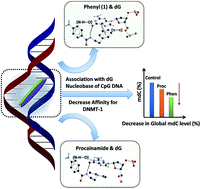
Procainamide derivatives have been synthesized to investigate the role of side chains in modulating the DNA methylation level in cancer cells and gain insight into its mechanism of action. The synthesized derivatives comprised of flexible (dimethyl), constrained (pyrrolidine, piperidine, morpholine) and planar aromatic (pyridine, phenyl) side chain motifs. The affinity of procainamide and its derivatives towards the deoxyguanosine (dG) base in neutral form has been assessed by performing Differential Pulse Voltammetry (DPV) under physiological conditions. Further, molecular docking with hemimethylated CpG rich DNA acquired from an active mDNMT-1-DNA (PDB ID-4DA4) crystal structure, reveals their preferential non-covalent interaction with dG nucleobase in the intercalation cavity of the minor groove. Differential affinity of the derivatives to dG base in neutral and bound forms (DNA) is correlated with their DNA methylation modulating properties at sub-lethal concentrations. Among all the derivatives, a compound with an aromatic phenyl side chain (1) has shown a highest binding affinity for dG nucleobase in neutral form as well as for partially denatured CpG rich DNA which is attributed to the formation of π⋯π stacking interaction in addition to N–H⋯O hydrogen bonding with the pyrimidine ring of dG base. It also shows the highest cytotoxicity and global hypomethylation at a sub-lethal level in the MCF-7 cancer cell line compared to other derivatives and procainamide. A docking study has also illustrated the plausible structural basis of DNA methylation modulating a property of procainamide. Strong association of procainamide with dG bases of partially denatured CpG rich DNA via H-bonding and other non-covalent interactions may alter the active topology of DNA required by the DNA-binding regulatory proteins (e.g. DNMT-1) which is validated by a DNMT-1 inhibition assay. This systematic investigation leads to a new potent alternative to procainamide being found and gives a plausible insight into the DNA hypomethylating effect of procainamide.


Introduction

Epigenetic regulation such as DNA methylation and histone modification plays a major role in embryonic development, cellular differentiation, suppression of repetitive elements, X-chromosome inactivation, and long-term memory.1 Furthermore, it regulates gene expression by reversible and heritable changes in DNA without altering the nucleotide sequence of the genome. DNA methylation is a key epigenetic regulatory process that predominantly occurs in CpG rich DNA clusters called “CpG islands” present at the promoter regions of genes.2 The process involves reversible modification of DNA by covalent addition of the methyl group at the 5-carbon position of the cytidine. Cancer cells show significantly altered DNA methylation profiles compared to normal cells that are used as diagnostic and prognostic markers for many cancer types.3 Global hypomethylation observed in cancer cells is responsible for inducing genomic instability owing to under-methylation of endogenous retroviral elements, oncogenes, and heterochromatic DNA repeats. However, aberrant hypermethylation within promoter regions of tumor suppressor genes is accountable for uncontrolled growth.3

The strategy of utilizing DNA hypomethylating agents (DHAs) for reducing aberrant methylation level of regulatory genes is used to treat cancer and neurological disorders.4 Nucleoside analogs such as 5-azacytidine, 5-aza-2′-deoxycytidine, zebularine, and 5-fluoro-2′-deoxycytidine have shown to reduce aberrant methylation level in cancer cells by inhibiting the activity of DNA methyltransferase (DNMT) enzymes.5 However, their clinical side effects and genotoxicity remained a great concern. Non-nucleoside methylation modulators are the promising alternative due to their low genotoxicity and mutagenicity. The non-nucleoside DNA methylation modulators are broadly categorized into three major classes based on their site of action and binding modes. The first one is the catalytic/allosteric site (protein) binding compounds that interfere with the catalytic activity of the DNMTs6 whereas the second one is CpG rich DNA binding compounds that form complex with DNA and reduces their affinity for DNMT proteins.7 The third category of compounds compete with DNA substrate and block the DNA-binding pocket of DNMT proteins.8 Recently FDA approved local anaesthetic drug procaine, and antiarrhythmic drug procainamide have been explored as DNA hypomethylating agents (DHA) against numerous malignancies as well as type-II diabetes both in vitro and in vivo.9 A striking feature of their action is their ability to modulate DNA methylation level at sub-lethal concentration.10 Non-genotoxicity, low cytotoxicity, and protective effect make them suitable anticancer agents for the combinatorial chemotherapeutic approach.11 In combination with the pharmacologically active scaffold of other DNMT inhibitors or co-administration of procaine and procainamide with known anticancer drugs has shown higher potency against many cancer types.12 However, the exact mechanism of their DNA methylation modulating activity so far is not clearly understood. The first report of their interaction with genomic DNA was noticed when patients undergoing prolonged therapy of procainamide and procaine showed the presence of antinuclear antibodies in the serum and 30% of them developed systemic lupus erythematosus syndrome.13 Pathogenesis of procainamide was linked to stabilization of immunogenic CpG rich Z-DNA and preferential affinity towards CpG rich DNA sequences.14 Link between CpG rich DNA binding affinity and DNA methylation modulating property of procainamide was first established by Lee et al.15 It has been suggested that procainamide inhibits binding of DNMT-1 to hemimethylated DNA leading to suppression of its catalytic activity. Based on these reports procainamide was categorized as class II demethylating agent. However, recently reported docking studies16 with constructed homology models also suggested procainamide may form strong association with amino acids (Arg174) of DNA binding pocket of DNMT-1 protein that may reduce its affinity for 2′-deoxycytidine of target hemimethylated DNA consequently may serve as class III methylation modulator.

One way to investigate the mechanistic basis of bioactive compounds is to design derivatives and study their structure–activity relationship.17 This approach not only provides the insight into the mechanism but also offers lead for improving the therapeutic value of the drugs. However, it is necessary to identify pharmacophore moiety within the drug molecule whose presence is essential to trigger the desired biological response. Our investigation attempts the similar approach. Evidence reported earlier for procaine18 has been used as clues to choose selective derivatives of procainamide. Procaine exhibits the strong association with the guanine base compared to other nucleobases mainly due to strong hydrogen bonding between 4-amino benzoic acid backbone and guanine base. Furthermore, constrained side chain derivatives of procaine reveal higher demethylating activity18 compared to their flexible counterparts. On the similar note, side chain derivatives of procainamide may also possess differential DNA hypomethylating activity if 4-aminobenzamide (anchoring site) remains unmodified.

Here we report the synthesis of procainamide derivatives comprising flexible (dimethyl), constrained (pyrrolidine, piperidine, morpholine) and planar aromatic side chain motifs (pyridine, phenyl) without modifying 4-amino benzamide backbone. Diethyl side-chain motif of procainamide is also flexible due to free rotation of both the ethyl moieties covalently linked to tertiary nitrogen. The differential affinity of procainamide and its derivatives towards dG nucleobase in the neutral and bound state have been investigated using DPV and molecular docking study respectively. Further, we explored growth inhibitory properties of these compounds towards MCF-7 cancer cell lines, quantified global DNA methylation level at a sub-lethal concentration and their effect on DNMT1 activity in vitro to evaluate cytotoxicity and suitability as DHA. It can be seen that the differences in the affinity towards dG nucleobase and CpG rich DNA, cytotoxic effect, global hypomethylation at sub-lethal level (MCF-7 cancer cell line) are correlated with the structural features of side chain motifs. This comparative study has helped in deriving the relation between dG affinity and DNA methylation modulating activity of procainamide and its derivatives. Based on the study we have also hypothesized the plausible mechanism by which procainamide may act as DHA in cancer cells.

Result and discussion

Chemistry

Procainamide derivatives were prepared as per the reported procedure (Fig. S1, ESI).19 4-Nitro benzoyl chloride was added to primary amines in the presence of triethylamine base to form corresponding 4-nitro carboxamides. Further reduction of the nitro group in the presence of catalytic amount of Pd/C and H2 yielded compounds 1–6. The compounds were named based on their side chain. They were further characterized by 1H NMR and HR-MS (Fig. S2–S13, ESI). Good quality crystals were grown for single crystal X-ray diffraction. The purity of the compounds was checked by HPLC (Fig. S14–S19, ESI) and further used for biological studies.

Crystallography

Molecular conformations in crystal structures and solution state share some analogy. Comparative analysis of crystal structures of isostructural compounds provides valuable information about their preferred geometry and torsional constraints of rotatable bonds within pharmacophore.20 Pure compounds were crystallized from dichloromethane/petroleum ether solvent mixture by a slow evaporation method. Crystal structure analysis was carried out using Bruker Smart Apex II X-ray diffractometer. Detailed information about crystallographic data and ORTEPs diagrams are provided in ESI (Table S1, Fig. S20, ESI). Atomic coordinates of procainamide were retrieved from Cambridge Structural Database (CSD, CCDC number 694545).21 Based on reported studies with procaine18 the 4-aminobenzamide of procainamide may play a pivotal role as backbone moiety of pharmacophore.18 Similarly alkyl chain (–CH2–CH2– hinge) may play a significant role in providing flexibility to side chains (binder) (Fig. 1) and eventually responsible for the structural variation. Structure overlay (Fig. 2a) and torsion angle τ1 (C1–C7–N–C8), τ2 (C7–N–C8–C9), and τ3 (N–C8–C9–R) are tabulated in (Fig. 2b, Table 1). As expected the torsional difference across 4-aminobenzamide backbone showed less change whereas torsional variation across side-chains displayed marked deviation.
image file: c5ra20012a-f1.tif
Fig. 1 Description of pharmacophore features of procainamide and molecular structures of its side chain motif derivatives 1–6.

image file: c5ra20012a-f2.tif
Fig. 2 (a) Structure overlay of procainamide and its derivatives and (b) scheme displaying different sites of torsion in the compounds.
Table 1 Torsion angles (τ1τ3) of compounds 1–6 and procainamide extracted from their crystal structures
Compounds τ1 (°) τ2 (°) τ3 (°)
Phenyl (1) 171.71 94.6 179.08
Pyrrolidine (2) 172.68 −168.93 −51.77
Dimethyl (3) −175.34 −139.39 −61.58
Morpholine (4) −172.43 −101.03 −164.72
Piperidine (5) −170.0 −105.96 −167.33
Pyridine (6) 173.80 84.44 71.66
Procainamide 166.14 88.18 173.24


Electrochemical studies

Electroactivity of nucleobases has been used for their quantification in biological samples as well as to study their interaction with other chemical species.22 DPV study and DFT calculation reported earlier18 for procaine and DNA bases reveal that binding affinity of procaine towards DNA bases is in the order G > A > T > C. The strongest binding affinity for guanine base in neutral form is attributed to co-planar C–H⋯N and N–H⋯O hydrogen bonding between 4-amino benzoic acid of procaine and pyrimidine moiety of guanine. Similarly, procainamide may also exhibit the strong association with guanine due to the presence of 4-aminobenzamide moiety. The binding affinity for cytosine base was found to be much lower than other bases. CpG rich DNA is built by the alternate arrangement of guanine and cytosine bases linked via phosphate-deoxyribose backbone. In view of this, binding affinity towards guanine was employed as a parameter for structure–activity correlation. We envisaged that the affinity study with deoxyguanosine (dG) instead of guanine base may give broader view and significant variation for deriving structure–activity relation based on the fact that guanine conjugated with deoxyribose sugar residue provides multiple interaction sites. Redox electrochemical behaviour of guanine is depicted in Fig. 3. To investigate differential affinity of procainamide and its derivatives (1–6) to dG nucleobase base in a neutral form, DPV was performed using CHI 900b potentiostat with a pulse amplitude of 50 mV, pulse width of 0.2 s and a pulse period of 0.5 s (for detailed procedure see page S28, ESI). Fig. 4 shows the differential pulse voltammograms of pure dG as well as dG in the presence of procainamide and compounds 1–6. When the potential of the GC electrode, kept in a solution of dG, is made more positive as compared to the open circuit potential (OCP), a clear peak is observed at 0.79 V (vs. SCE, 0.1 M KCl) (there is no peak in case of buffer solution alone, Fig. S21) due to the oxidation of dG as shown in the reaction mechanism in Fig. 3. Moreover, when the same concentration of dG is used in all the experiments (dG to compound ratio: 4), the peak current density of dG oxidation drops in the presence of carboxamide ligands (1–6 and procainamide) (Fig. 4). The results indicate binding of added ligands to dG and hence number of free dG molecules depletes in the solution. As a result, peak current decreases because the current is directly proportional to the concentration of the active species in the solution. It invariably suggests that the drop in peak current of dG is due to the interactions of the procainamide and compounds 1–6 with dG. However, the extent of the drop in peak current is differentially related to structural features of side-chain motifs of ligands. Phenyl substituent is found to cause a maximum drop in peak current of dG (Fig. 4b) and thus more efficiently binds to dG. The effective binding affinity of procainamide derivatives (according to Fig. 4b) follow the order, phenyl (1) > pyridine (6) ≈ piperidine (5) > morpholine (4) > procainamide > dimethyl (3) ≈ pyrrolidine (2). The somewhat broad peak observed prior to the dG peak is due to the oxidation of compound 1–6 and procainamide that is demonstrated by control experiment for phenyl (1) (Fig. S21). A broad peak observed at 0.6 V (vs. SCE, 0.1 M KCl) prior to dG oxidation peak, attributed to the oxidation of phenyl (1) to form radical cation. The peak that appears after dG peak corresponds the oxidative decomposition/cleavage of the compound–dG complex on the application of a more positive potential. It is supported by the fact that both dG, as well as phenyl derivative alone, do not show such peak after the oxidation potential of dG. Such peak is observed only in case of mixed solutions of dG and compound under study. Careful observation of these peaks reveals that the phenyl–dG complex requires much higher potential than any other compound–dG complex and thereby it is most stable. This result indirectly indicates the possibility of strong binding of phenyl with dG.
image file: c5ra20012a-f3.tif
Fig. 3 Scheme displaying redox behaviour of guanine base.

image file: c5ra20012a-f4.tif
Fig. 4 (a) Differential pulse voltammograms of dG in the presence of compounds 1–6 and procainamide; glassy carbon, Pt-foil and standard calomel electrode (SCE, 0.1 M KCl) are used as working, counter and reference electrodes respectively (pulse amplitude of 50 mV, pulse width of 0.2 s and a pulse period of 0.5 s); systematic drop in peak current density (j) in presence of different compounds suggest their differential binding affinity to dG; (b) relative drop in peak current density (j) of dG with respect to peak current of pure dG (j0) (j is normalized with respect to j0).

Molecular docking study

Many experimental evidences were reported for association of procainamide with CpG rich DNA. However, two reports provided important clues about their selective affinity. First report revealed shift in the midpoint of transition is observed during salt-induced B–Z transition of poly CpG DNA in the presence of procainamide14 while the second report suggested it shows specific affinity for hemimethylated-DNMT-1 bound complex over unmethylated-DNMT-1 bound counterpart.15 Cellular processes that involve dynamic topological variation of DNA such as maintenance methylation, B–Z transition, replication, DNA modification and repair share common structural features. Partial denaturation of DNA and flipping out of bases at the target site is observed during these events.23 We predicted procainamide may show more affinity for partially denatured CpG rich DNA and driving force is provided by its affinity for the dG base. To validate our hypothesis we used molecular docking study.

Docking study provides a good estimate of sequence specificity and intercalation mechanism of DNA-binding drugs.24 Due to the significant variation observed for minor groove size within the crystal structures of AT-rich and GC-rich DNA sequences,25 it is advisable to use crystal coordinates instead of modelled DNA for carrying out docking study with minor groove binding agents. The atomic coordinates of CpG rich hemimethylated DNA (hmDNA) were extracted from RCSB Protein Data Bank (PDB ID:4DA4) and used for docking study.26 Structural analysis reveals that the H-bonding donor/acceptor groups of dG bases are exposed by partial denaturation of DNA. Further, non-conventional pyrimidine: pyrimidine base pairing between dG bases and flipping out of nucleobases creates small intercalation cavity at the target site.

Docking was performed with Schrodinger Maestro Suite and flexible docking method.27 Detailed procedure and docking protocol are provided in ESI (pg. S29, ESI). Post-docking minimization and residue-wise interaction parameters were calculated. Most important step for performing docking simulation is to generate all possible bioactive conformers of the compounds and to choose reliable docked poses for comparative analysis. Atomic coordinates of compounds 1–6 and CSD database survey28 (Fig. S22, Table S2, ESI) suggested permissible variation for torsion angle τ1 that lies within the range of 160–180°. Accordingly the filtering of the docked poses has been carried out where ligand conformation matched with our cut-off value. Further, conformers with lowest glide scores were used for comparative studies. Structural overlay and torsional parameters of ligand conformations extracted from docked poses are provided in ESI (Fig. S23, Table S3).

Docking analysis with hmDNA reveals that all the compounds are docked inside the intercalation cavity of the DNA minor groove (Fig. 5a) and have formed a strong association with a dG base of target site except the dimethyl derivative. Carboxamide N–H proton of 4-aminobenzamide backbone formed strong N–H⋯O hydrogen bonding interaction with the carbonyl oxygen C[double bond, length as m-dash]O of dG base whereas N–H proton of para-amino group formed a N–H⋯O hydrogen bonding with O acceptor atom of phosphodiester linkage of DNA (Fig. 5b–h). Although all the derivatives except the dimethyl (3) showed similar binding features, variation in their H-bonding geometries with the dG base was noticed (Fig. 6a). Glide score (docking score) was used to evaluate binding affinity for entire hmDNA whereas, H-bonding interaction length, H-bonding energy and van der Waals energy were taken into account for assessing the binding affinity for dG at the target site. Based on the glide score, affinity trend for hmDNA was found to be in the order, phenyl (1) > pyridine (6) > pyrrolidine (2) > piperidine (5) > morpholine (4) > procainamide > dimethyl (3) H-bonding energy score (E1) between ligands and dG residue revealed phenyl (1), procainamide, and pyridine (6) derivatives form strong hydrogen bonding interaction with dG nucleobase (Fig. 6a). Moreover, van der Waals interaction energy scores (E2) suggests that all the derivatives except dimethyl (5) are strongly associated via van der Waals forces with dG nucleobase in addition to their H-bonding interactions. Docked postures of phenyl and pyridine showed the formation of parallel displaced π-stacking interaction between benzene/pyridine moieties with pyrimidine ring of the dG base. However, the geometrical parameters for stacking interactions (Cg⋯Cg distance for phenyl 3.588 Å and pyridine 3.613 Å, the dihedral angle between aromatic rings α for phenyl 15.85° and for pyridine 18.67°) suggests benzene ring of phenyl (1) formed comparatively stronger π⋯π interaction than pyridine ring of compound 6 (Fig. 6b and S24, ESI).


image file: c5ra20012a-f5.tif
Fig. 5 (a) Compilation of docked poses showed most favourable binding site lies within intercalation cavity. Detailed view of individual docked poses of (b) phenyl (c) pyrrolidine (d) dimethyl (e) morpholine (f) piperidine (g) pyridine (h) procainamide with hmDNA, showing strong hydrogen-bonding association with dG nucleobase (except dimethyl) and O-acceptor of phosphodiester linkage at the target site.

image file: c5ra20012a-f6.tif
Fig. 6 (a) Tabulated glide score, affinity parameters to dG residue H-bond distance, hydrogen bonding energy score E1, van der Waals association energy score E2 of docked poses of procainamide and derivatives (1–6) with hmDNA and (b) structural depiction of association between phenyl (1) and dG base at the target site via H-bonding and aromatic π⋯π stacking interaction.

The molecular docking study reveals that the side-chain topology of compounds play a pivotal role for binding to CpG rich hmDNA. The strength of the binding affinity increases as the rigidity and bulkiness of side chain motif increases. The comparative analysis showed that hydrogen bonding between 4-aminobenzamide backbone and dG nucleobase in the intercalation cavity allowed all the derivatives to dock inside the minor groove intercalation cavity but the strength of their association within target site is further determined by other non-covalent interactions between side chain and target base. The aromatic side chain phenyl (1) showed the highest affinity due to the formation of π⋯π stacking interaction with the dG base (Fig. 6b).

Cytotoxicity study

Cytotoxicity towards MCF-7 breast cancer cell lines was investigated to evaluate the differential cytotoxic effect of procainamide and its derivatives (1–6). Five different concentrations of compounds from 500 μM to 100 μM at 24 h, 48 h, and 72 h time points were used to treat the cell line. Detailed procedure and protocol are provided in ESI (pg. S32). The results depicted in ESI showed that all the derivatives (1–6) and procainamide were non-cytotoxic at 24 h and 48 h time points (Fig. S25 and 26, ESI). Notably, even after 72 h and 100 μM concentration of compounds did not exhibit obvious cytotoxicity and viability ranges from 80% to 100% (Fig. S27, ESI). However, at 500 μM concentration and 72 h time point, significant variation in their cytotoxicity was observed (Fig. 7). Phenyl (1) showed almost 50% cell death with the highest cytotoxicity whereas pyridine (6) and dimethyl (3) derivatives possess moderate cytotoxicity. Procainamide, pyrrolidine (2), piperidine (5), and morpholine (4) containing derivatives showed the absence of significant cell death even at the highest concentration. The trend observed for growth inhibitory properties towards breast cancer cell line is in the order, phenyl (1) > dimethyl (3) > pyridine (6) > pyrrolidine (2) > piperidine (5) > morpholine (4) > procainamide.
image file: c5ra20012a-f7.tif
Fig. 7 A comparison of cell viability assay (data in %) after 72 h of treatment with 500 μM conc. of procainamide and its derivatives (1–6) towards MCF-7 cancer cell line.

Global methylation quantification

Procainamide and its derivatives (1–6) showed variation in their affinity towards the dG base in neutral form, partially denatured CpG rich DNA and cytotoxicity towards breast cancer cell line. However, their cytotoxic effect alone may not infer their ability to modulate DNA methylation level and candidature as DHA. Secondly, procainamide is promising combinatorial drug that reduces aberrant methylation at the sub-lethal level and increases vulnerability of cancer cells for strong anticancer drugs.11,12 Relative shift in the global methylation level in MCF-7 cancer cells upon treatment with sub-lethal concentration of compounds is used as a parameter for structure–activity correlation. In recent years, many techniques and methods have been reported for quantifying global methylation levels. These methods are luminometric, electrophoretic, pyro-sequencing, HPLC and colorimetric-based assays.29 For our study ELISA based methylated DNA quantification kit (colorimetric) from Abcam was used for determination of % 5-methyl-2′-deoxycytidine content (% mdC) in treated and untreated MCF-7 cancer cells. The detailed experimental procedure is provided in ESI (Fig. S36, ESI). The C1 and C2 were the two negative controls used for this study. Control C2 was used as 0.5% DMSO to eliminate the error of demethylating effect of DMSO solvent.30 5-Azacytidine (5 μM) was used as a positive control. Methylation level in case of negative control DNA samples, C1 (no DMSO) and C2 (0.5% DMSO) ranged from 5.37% to 5.68% which is similar to one reported in published literature.31 Positive control 5-azacytidine showed highest decrease in global methylation level whereas trend in the decrease in methylation level for procainamide and its derivatives (1–6) followed the order as phenyl (1) > piperidine (5) > procainamide > dimethyl (3) > morpholine (4) ≈ pyridine (6) > pyrrolidine (2) (Fig. 8). The highest decrease in the methylation level was observed for phenyl (1) containing derivative whereas the lowest decrease was seen for the pyrrolidine (2) derivative. Previous reports have shown that slight modulation in global methylation level is attributed to significant change at the gene regulation level,32 which suggested phenyl derivative can be used as DHA at sub-lethal concentration.
image file: c5ra20012a-f8.tif
Fig. 8 A comparison of global methylation quantification (in %) after 72 h of treatment with 100 μM conc. of procainamide, its derivatives (1–6) and controls (C1 = blank, C2 = 0.5% DMSO) towards MCF-7 cancer cell lines.

DNMT-1 inhibition assay

DNMT-1 inhibitory activity of compound 1 (comparatively higher active) and procainamide were assessed using DNA methyltransferase 1 activity/inhibitor screening assay core kit (P-3006A). Detailed procedure is provided in the ESI (page 37). Both the compounds showed inhibition of DNMT-1 activity in in vitro. However, no significant difference was observed which may be attributed to their similar mode of action (Fig. 9).
image file: c5ra20012a-f9.tif
Fig. 9 A comparison of in vitro DNMT-1 inhibitory activity (%) of compound 1 (phenyl) and procainamide at 100 μM conc. (DMSO 0.01%).

Conclusions

Four activity parameters namely, dG affinity (neutral state), CpG rich binding affinity, cytotoxicity to MCF-7 cancer cell lines, % mdC level (global) showed significant variation with respect to topology of side-chain motifs of procainamide and its derivatives. DPV study revealed that aromatic side chains containing, phenyl (1) and pyridine (6) derivatives showed strong association with the dG base in a neutral form whereas flexible side chain containing, dimethyl (3) and procainamide derivatives showed the least association. Constrained heterocyclic side chain containing morpholine (4) and piperidine (5) derivatives showed intermediate association with dG base except pyrrolidine (2). DPV results demonstrated good correlation with docking study with partially denatured hmDNA. Phenyl (1) and pyridine (6) derivatives formed π⋯π stacking interaction with the dG base that manifested their strong association with CpG rich hmDNA. Conversely, flexible dimethyl (3) and procainamide showed the least affinity. Based on the glide score, constrained heterocyclic side chain derivatives possessed intermediate (between aromatic and flexible side chain derivatives) binding affinity. Combined results signify that the aromatic side chains possess a higher affinity towards CpG rich DNA due to their strong intermolecular association with dG nucleobase.

Cytotoxicity data demonstrated good agreement with DPV/docking study. It is further used to evaluate the sub-lethal concentration of procainamide and derivatives 1–6. Interestingly all the derivatives of procainamide have shown the decrease in % mdC level in comparison with control in MCF-7 cancer cells at the sub-lethal concentration which may be attributed to their affinity towards CpG rich DNA especially to dG base. Moreover, phenyl (1) showed a higher demethylating effect compared to other derivatives (2–6) including its parent analogue procainamide. The increased demethylating property is attributed to its strong association with dG nucleobase via hydrogen bonding and π⋯π stacking interaction within the CpG rich minor groove of DNA. Procainamide has been shown to induce structural changes in circular supercoiled plasmid DNA and alter tertiary topology of DNA.14 On the similar note strong association of phenyl (1) with CpG rich DNA may alter its active conformation required by regulatory proteins such as DNMT-1 and may consequently inhibit protein–DNA binding which is validated by in vitro DNMT-1 inhibition assay. Thus, our study provides mechanistic insight into the plausible mode of methylation modulating activity of procainamide and its analogues. Our systematic investigation also provides a rapid method of screening for finding other non-nucleoside DNA binding (CpG rich) demethylating agents as shown in Fig. 10. The screening may involve four primary steps before going to in vivo trial. Step one will involve the rapid evaluation of binding affinity of compounds for a dG base in a neutral form. The extent of affinity will be determined by the drop in the peak current. Step two comprise of docking simulation study with hmDNA to evaluate the affinity to partially denatured CpG rich DNA. The third step will evaluate cytotoxicity and sub-lethal concentration at in vitro. Finally increase or decrease in the global methylation level in cancer cells will decide the candidature of the compounds as DHA. Currently in our lab we are exploring sulphonamide based novel methylation modulators due to the additional conformational flexibility of 4-amino benzene sulfonamide compare to 4-amino benzamide backbone.


image file: c5ra20012a-f10.tif
Fig. 10 Cartoon illustration of protocol for the rapid method of screening for non-nucleosidal CpG DNA binding methylation modulators.

Experimental

Experimental details of compounds characterization, crystallographic, electrochemical, docking, cytotoxicity, global methylation quantification study and DNMT-1 inhibition assay are provided in ESI file (ESI).

Acknowledgements

RLG thanks CSIR for fellowship. DK thanks CSIR-NCL for funding. This work was made possible by financial support from CSIR (Origin) and UGC UPE II grant.

Notes and references

  1. (a) J. C. Rice and C. D. Allis, Curr. Opin. Cell Biol., 2001, 13, 263–273 CrossRef PubMed; (b) R. Jaenisch and A. Bird, Nat. Genet., 2003, 33, 245–254 CrossRef CAS PubMed; (c) W. Reik, Nature, 2007, 447, 425–432 CrossRef CAS PubMed; (d) P. A. Jones and D. Takai, Science, 2001, 293, 1068–1070 CrossRef CAS PubMed; (e) F. Mohn and D. Schübeler, Trends Genet., 2009, 25, 129–136 CrossRef CAS PubMed; (f) R. K. Slotkin and R. Martienssen, Nat. Rev. Genet., 2007, 8, 272–285 CrossRef CAS PubMed; (g) W. Reik and A. Lewis, Nat. Rev. Genet., 2005, 6, 403–410 CrossRef CAS PubMed; (h) S. N. Rajpathak and D. D Deobagkar, Curr. Pharm. Des., 2014, 20, 1778–1785 CrossRef CAS PubMed; (i) S. N. Rajpathak, S. K. Vellarikkal, A. Patowary, V. Scaria, S. Sivasubbu and D. D. Deobagkar, PLoS One, 2014, 9, e100076 Search PubMed; (j) A. Kelkar and D. Deobagkar, Epigenetics, 2010, 5, 612–618 CrossRef CAS PubMed; (k) N. Tsankova, W. Renthal, A. Kumar and E. J. Nestler, Nat. Rev. Neurosci., 2007, 8, 355–367 CrossRef CAS PubMed.
  2. A. M. Deaton and A. Bird, Genes Dev., 2011, 25, 1010–1022 CrossRef CAS PubMed.
  3. (a) M. Ehrlich, Oncogene, 2002, 21, 5400–5413 CrossRef CAS PubMed; (b) M. Ehrlich, Epigenomics, 2009, 1, 239–259 CrossRef CAS PubMed; (c) R. J. W. O'Neill, M. J. O'Neill and J. A. M. Graves, Nature, 1998, 393, 68–72 CrossRef PubMed; (d) S. P. Kabekkodu, S. Bhat, R. Radhakrishnan, A. Aithal, R. Mascarenhas, D. Pandey, L. Rai, P. Kushtagi, G. P. Mundyat and K. Satyamoorthy, J. Biol. Chem., 2014, 289, 10637–10649 CrossRef CAS PubMed; (e) P. M. Das and R. Singal, J. Clin. Oncol., 2004, 22, 4632–4642 CrossRef CAS PubMed; (f) Q. W. Chen, X. Y. Zhu, Y. Y. Li and Z. Q. Meng, Oncol. Rep., 2014, 31, 523–532 CAS; (g) M. Esteller, Oncogene, 2002, 21, 5427–5440 CrossRef CAS PubMed.
  4. (a) J. P. Issa, Clin. Cancer Res., 2007, 13, 1634–1637 CrossRef CAS PubMed; (b) F. Lyko and R. Brown, J. Natl. Cancer Inst., 2005, 97, 1498–1506 CrossRef CAS PubMed; (c) C. B. Yoo and P. A. Jones, Nat. Rev. Drug Discovery, 2006, 5, 37–50 CrossRef CAS PubMed; (d) X. Yang, F. Lay, H. Han and P. A. Jones, Trends Pharmacol. Sci., 2010, 31, 536–546 CrossRef CAS PubMed; (e) S. Heerboth, K. Lapinska, N. Snyder, M. Leary, S. Rollinson and S. Sarkar, Genet. Epigenet., 2014, 6, 9–19 CAS.
  5. (a) J. K. Christman, Oncogene, 2002, 21, 5483–5495 CrossRef CAS PubMed; (b) C. Stresemann and F. Lyko, Int. J. Cancer, 2008, 123, 8–13 CrossRef CAS PubMed; (c) J. C. Cheng, C. B. Matsen, F. A. Gonzales, W. Ye, S. Greer, V. E. Marquez, P. A. Jones and E. U. Selker, J. Natl. Cancer Inst., 2003, 95, 399–409 CrossRef CAS PubMed; (d) Q. Zhao, J. Fan, W. Hong, L. Li and M. Wu, SpringerPlus, 2012, 1, 65 CrossRef PubMed.
  6. (a) S. Castellano, D. Kuck, M. Viviano, J. Yoo, F. LÓpez-Vallejo, P. Conti, L. Tamborini, A. Pinto, J. L. Medina-Franco and G. Sbardella, J. Med. Chem., 2011, 54, 7663–7677 CrossRef CAS PubMed; (b) M. Z. Fang, Y. Wang, N. Ai, Z. Hou, Y. Sun, H. Lu, W. Welsh and C. S. Yang, Cancer Res., 2003, 63, 7563–7570 CAS; (c) D. Kuch, L. Schermelleh, S. Manetto, H. Leonhardt and T. Carell, Angew. Chem., Int. Ed., 2008, 47, 1515–1518 CrossRef CAS PubMed; (d) Z. Liu, S. Liu, Z. Xie, R. E. Pavlovicz, J. Wu, P. Chen, J. Aimiuwu, J. Pang, D. Bhasin, P. Neviani, J. R. Fuchs, C. Plass, P. K. Li, C. Li, T. H. M. Huang, L. C. Wu, L. Rush, H. Wang, D. Perrotti, G. Marcucci and K. K. Chan, J. Pharmacol. Exp. Ther., 2009, 329, 505–514 CrossRef CAS PubMed; (e) T. Suzuki, R. Tanaka, S. Hamada, H. Nakagawa and N. Miyata, Bioorg. Med. Chem. Lett., 2010, 20, 1124–1127 CrossRef CAS PubMed.
  7. C. Gros, L. Fleury, V. Nahoum, C. Faux, S. Valente, D. Labella, F. Cantagrel, E. Rilova, M. A. Bouhlel, M. H. David-Cordonnier, I. Dufau, F. Ausseil, A. Mai, L. Mourey, L. Lacroix and P. B. Arimondo, J. Biol. Chem., 2015, 290, 6293–6302 CrossRef CAS PubMed.
  8. R. L. Fagan, D. E. Cryderman, L. Kopelovich, L. L. Wallrath and C. Brenner, J. Biol. Chem., 2013, 288, 23858–23867 CrossRef PubMed.
  9. (a) Z. Gao, Z. Xu, M. S. Hung, Y. C. Lin, T. Wang, M. Gong, X. Zhi, D. M. Jablons and L. You, Oncol. Rep., 2009, 22, 1479–1484 CAS; (b) X. Lin, K. Asgari, M. J. Putzi, W. R. Gage, X. Yu, B. S. Cornblatt, A. Kumar, S. Piantadosi, T. L. DeWeese, A. M. De Marzo and W. G. Nelson, Cancer Res., 2001, 61, 8611–8616 CAS; (c) B. Segura-Pacheco, C. Trejo-Becerril, E. Perez-Cardenas, L. Taja-Chayeb, I. Mariscal, A. Chavez, C. Acuña, A. M. Salazar, M. Lizano and A. Dueñas-Gonzalez, Clin. Cancer Res., 2003, 9, 1596–1603 CAS; (d) M. Tada, F. Imazeki, K. Fukai, A. Sakamoto, M. Arai, R. Mikata, T. Tokuhisa and O. Yokosuka, Hepatology International, 2007, 1, 355–364 CrossRef PubMed; (e) A. Villar-Garea, M. F. Fraga, J. Espada and M. Esteller, Cancer Res., 2003, 63, 4984–4989 CAS; (f) W. F. El-Hadidy, A. R. Mohamed and H. F. Mannaa, Alexandria Journal of Medicine, 2015, 51, 65–71 CrossRef.
  10. Y. S. Lin, A. Y. Shaw, S. G. Wang, C. C. Hsu, I. W. Teng, M. J. Tseng, T. H. Huang, C. S. Chen, Y. W. Leu and S. H. Hsiao, J. Biomed. Sci., 2011, 18, 3 CrossRef CAS PubMed.
  11. (a) C. Fenoglio, E. Boncompagni, B. Chiavarina, S. Cafaggi, M. Cilli and M. Viale, Anticancer Res., 2005, 25, 4123–4128 CAS; (b) M. Viale, I. Pastrone, C. Pellecchia, M. O. Vannozzi, S. Cafaggi and M. Esposito, Anti-Cancer Drugs, 1998, 9, 457–463 CrossRef CAS PubMed; (c) M. Viale, M. O. Vannozzi, I. Pastrone, M. A. MariggiÓ, A. Zicca, A. Cadoni, S. Cafaggi, G. Tolino, G. Lunardi, D. Civalleri, W. E. Lindup and M. Esposito, J. Pharmacol. Exp. Ther., 2000, 293, 829–836 CAS; (d) J. G. Zhang, L. F. Zhong, M. Zhang and Y. X. Xia, Arch. Toxicol., 1992, 66, 354–358 CrossRef CAS PubMed; (e) A. Zicca, S. Cafaggi, M. A. MariggiÓ, M. O. Vannozzi, M. Ottone, V. Bocchini, G. Caviglioli and M. Viale, Eur. J. Pharmacol., 2002, 442, 265–272 CrossRef CAS PubMed.
  12. (a) O. Altundag, K. Altundag and M. Gunduz, Med. Hypotheses, 2004, 63, 684–687 CrossRef CAS PubMed; (b) L. Halby, C. Champion, C. Sénamaud-Beaufort, S. Ajjan, T. Drujon, A. Rajavelu, A. Ceccaldi, R. Jurkowska, O. Lequin, W. G. Nelson, A. Guy, A. Jeltsch, D. Guianvarc'h, C. Ferroud and P. B. Arimondo, ChemBioChem, 2012, 13, 157–165 CrossRef CAS PubMed.
  13. M. M. Reidenberg and D. E. Drayer, Angiology, 1986, 37, 968–971 CAS.
  14. (a) T. J. Thomas and R. P. Messner, Arthritis Rheum., 1986, 29, 638–645 CrossRef CAS PubMed; (b) W. Zacharias and W. J. Koopman, Arthritis Rheum., 1990, 33, 366–374 CrossRef CAS PubMed.
  15. B. H. Lee, S. Yegnasubramanian, X. Lin and W. G. Nelson, J. Biol. Chem., 2005, 280, 40749–40756 CrossRef CAS PubMed.
  16. N. Singh, A. DueÇas-Gonzlez, F. Lyko and J. L. Medina-Franco, ChemMedChem, 2009, 4, 792–799 CrossRef CAS PubMed.
  17. J. D. McKinney, A. Richard, C. Waller, M. C. Newman and F. Gerberick, Toxicol. Sci., 2000, 56, 8–17 CrossRef CAS PubMed.
  18. (a) S. Castellano, D. Kuck, M. Sala, E. Novellino, F. Lyko and G. Sbardella, J. Med. Chem., 2008, 51, 2321–2325 CrossRef CAS PubMed; (b) G. Ping, G. Lv, S. Gutmann, C. Chen, R. Zhang and X. Wang, J. Nanosci. Nanotechnol., 2006, 6, 91–94 CAS; (c) G. Lv, Z. Chen, J. Zheng, F. Wei, H. Jiang, R. Zhang and X. Wang, J. Mol. Struct.: THEOCHEM, 2010, 939, 44–52 CrossRef CAS.
  19. H. R. Lawrence, M. P. Martin, Y. Luo, R. Pireddu, H. Yang, H. Gevariya, S. Ozcan, J. Y. Zhu, R. Kendig, M. Rodriguez, R. Elias, J. Q. Cheng, S. M. Sebti, E. Schonbrunn and N. J. Lawrence, J. Med. Chem., 2012, 55, 7392–7416 CrossRef CAS PubMed.
  20. (a) K. A. Brameld, B. Kuhn, D. C. Reuter and M. Stahl, J. Chem. Inf. Model., 2008, 48, 1–24 CrossRef CAS PubMed; (b) H. F. G. Velec, H. Gohlke and G. Klebe, J. Med. Chem., 2005, 48, 6296–6303 CrossRef CAS PubMed.
  21. O. M. Peeters, N. M. Blaton, C. J. De Ranter, O. Denisoff and L. Molle, Cryst. Struct. Commun., 1980, 9, 851–856 CAS.
  22. (a) H. E. S. Kara, Bioelectrochemistry, 2014, 99, 17–23 CrossRef PubMed; (b) R. M. Shubietah, A. Z. A. Zuhri and A. G. Fogg, Fresenius' J. Anal. Chem., 1994, 348, 754–757 CrossRef CAS; (c) E. Palecek and M. Bartosik, Chem. Rev., 2012, 112, 3427–3481 CrossRef CAS PubMed; (d) N. Li, L. Guo, J. Jiang and X. Yang, Biophys. Chem., 2004, 111, 259–265 CrossRef CAS PubMed.
  23. S. KlimaŠauskas, Z. LiutkeviČiŪtĖ and D. DaujotytĖ, Biophysics and the Challenges of Emerging Threats, Springer, Netherlands, 2009, ch. Biophysical Approaches To Study DNA Base Flipping, pp. 51–64 Search PubMed.
  24. (a) C. G. Ricci and P. A. Netz, J. Chem. Inf. Model., 2009, 49, 1925–1935 CrossRef CAS PubMed; (b) H. K. Srivastava, M. Chourasia, D. Kumar and G. N. Sastry, J. Chem. Inf. Model., 2011, 51, 558–571 CrossRef CAS PubMed.
  25. (a) E. P. Bishop, R. Rohs, S. C. J. Parker, S. M. West, P. Liu, R. S. Mann, B. Honig and T. D. Tullius, ACS Chem. Biol., 2011, 6, 1314–1320 CrossRef CAS PubMed; (b) S. C. J. Parker and T. D. Tullius, Curr. Opin. Struct. Biol., 2011, 21, 342–347 CrossRef CAS PubMed.
  26. J. Song, M. Teplova, S. Ishibe-Murakami and D. J. Patel, Science, 2012, 335, 709–712 CrossRef CAS PubMed.
  27. (a) R. A. Friesner, J. L. Banks, R. B. Murphy, T. A. Halgren, J. J. Klicic, D. T. Mainz, M. P. Repasky, E. H. Knoll, M. Shelley, J. K. Perry, D. E. Shaw, P. Francis and P. S. Shenkin, J. Med. Chem., 2004, 47, 1739–1749 CrossRef CAS PubMed; (b) Schrödinger Suite 2011 Protein Preparation Wizard; Epik version 2.3, Schrödinger, LLC, New York, NY, 2012; Impact version 5.8, Schrödinger, LLC, New York, NY, 2012; Prime version 3.1, Schrödinger, LLC, New York, NY, 2012. Suite 2012: LigPrep, version 2.5, Schrödinger, LLC, New York, NY, 2012.
  28. F. H. Allen, Acta Crystallogr., Sect. B: Struct. Sci., 2002, 58, 380–388 CrossRef.
  29. (a) S. Lisanti, W. A. W. Omar, B. Tomaszewski, S. De Prins, G. Jacobs, G. Koppen, J. C. Mathers and S. A. S. Langie, PLoS One, 2013, 8, e79044 Search PubMed; (b) A. Kelkar and D. Deobagkar, Epigenetics, 2009, 4, 415–420 CrossRef CAS PubMed.
  30. M. Iwatani, K. Ikegami, Y. Kremenska, N. Hattori, S. Tanaka, S. Yagi and K. Shiota, Stem Cells, 2006, 24, 2549–2556 CrossRef CAS PubMed.
  31. M. Batchman, S. Uribe-Lewis, X. Yang, M. Williams, A. Murrell and S. Balasubramanian, Nat. Chem., 2014, 6, 1049–1055 CrossRef PubMed.
  32. S. Shpyleva, S. Ivanovsky, A. de Conti, S. Melnyk, V. Tryndyak, F. A. Beland, S. J. James and I. P. Pogribny, PLoS One, 2014, 9, e113712 Search PubMed.

Footnote

Electronic supplementary information (ESI) available. CCDC 1425934–1425938 and 1442290. For ESI and crystallographic data in CIF or other electronic format see DOI: 10.1039/c5ra20012a

This journal is © The Royal Society of Chemistry 2016
Click here to see how this site uses Cookies. View our privacy policy here.