Mesoporous transition metal dichalcogenide ME2 (M = Mo, W; E = S, Se) with 2-D layered crystallinity as anode materials for lithium ion batteries

Yoon Yun Leea, Gwi Ok Parkbc, Yun Seok Choic, Jeong Kuk Shon c, Jeongbae Yoonb, Kyoung Ho Kimc, Won-Sub Yoon*b, Hansu Kim*d and Ji Man Kim*abc
aSKKU Advanced Institute of Nanotechnology (SAINT), Sungkyunkwan University, Suwon 16419, Republic of Korea
bDepartment of Energy Science, Sungkyunkwan University, Suwon 16419, Republic of Korea. E-mail: wsyoon@skku.edu
cDepartment of Chemistry, Sungkyunkwan University, Suwon 16419, Republic of Korea. E-mail: jimankim@skku.edu; Fax: +82 31 299 4174; Tel: +82 31 290 5930
dDepartment of Energy Engineering, Hanyang University, Seoul 04763, Republic of Korea. E-mail: khansu@hanyang.ac.kr

Received 25th September 2015 , Accepted 27th January 2016

First published on 29th January 2016


Abstract

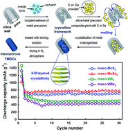
Mesoporous transition metal dichalcogenides (TMDCs), composed of group VI metals (Mo and W) and chalcogens (S and Se), with 2-D layered crystalline frameworks and 3-D pore structures were successfully prepared via a melting-infiltration assisted nano-replication method using a mesoporous template KIT-6 with cubic Ia3d symmetry. Combined analysis using X-ray diffraction, N2 adsorption–desorption and electron microscopy indicated that the mesoporous TMDCs, thus obtained, exhibited high surface areas (87–105 m2 g−1), large pore volumes (0.21–0.25 cm3 g−1) and well-defined mesopores about 20 nm in diameters. The mesoporous TMDCs showed outstanding rate capabilities up to 2C as well as high reversible lithium storage capacities (MoS2 710 mA h g−1; MoSe2 744 mA h g−1; WS2 501 mA h g−1; WSe2 427 mA h g−1) without a remarkable fading of capacity.


1. Introduction

Due to their high energy and power densities, lithium ion batteries (LIBs) are becoming the most important power sources for portable devices and electric vehicles (EVs).1–4 However, commercial LIBs (comprised of lithium metal oxide cathode and carbonaceous anode) are not as sufficient as high power sources for EVs, because of their low lithium storage capacities caused by one or less electron transfer reactions of electrode materials (e.g. six carbon atoms in graphite react with only one Li+).5–7 In order to accomplish sufficiently high energy and power densities, it is necessary to develop novel electrode materials that can exhibit multi-electron transfer reactions with high lithium storage capacity.6,7

Carbonaceous materials are the mostly used anode material for LIBs due to their low working voltage, low cost, good stability and long cycle life.7–10 Because of low theoretical capacity (LiC6, 372 mA h g−1), novel materials such as alloys, metal oxides, metal phosphides and metal sulphides have been investigated as alternative anode materials for LIB anode.4,11–24 Unfortunately, these materials have some limits in their practical application such as low coulombic efficiency, high irreversible capacity, high cost, unstable solid electrolyte interphase (SEI) layer formation, and short life cycle.7

Transition metal dichalcogenides (TMDCs), composed of group IV–VI transition metals and chalcogens (S, Se and Te), with graphite-like layered crystalline frameworks are expected to have great potential in nano-electronics, sensing, catalysis, energy storage and harvesting.25–32 In particular, due to the interesting intercalation chemistry caused by their layered crystal structures, group VI metal (Mo and W) based TMDCs have been investigated as anode materials for LIBs.33–38 Indeed, TMDCs are being regarded as promising candidates for LIB anodes because of high reversible capacity and an excellent rate capability.39

Mesoporous materials, exhibiting well-defined mesopore sizes and nano-sized wall-thicknesses, have some advantages as electrode materials for LIBs: (1) the buffer effect by their cavities (mesopores) for volume change during the charge–discharge processes, (2) controllable mesopore size and connectivity that offer a short diffusion pathway for the electrolyte and transport of lithium ions into the surface of the material, (3) shortening of the solid-state diffusion path for the lithium ion into the material by their nano-sized framework (usually <10 nm), and (4) large electrochemically active surface area between the electrode material and the electrolyte. Accordingly, mesoporous materials with inorganic frameworks such as carbons and metal oxides have been widely investigated as electrode material for LIBs.40–53 However, due to the lack of suitable precursors, a major challenge for preparing high quality mesoporous TMDCs is to find a suitable and facile synthesis route. There are only a few examples of mesoporous TMDCs, MoS2, MoSe2 WS2 and WSe2, were synthesized via the nano-replication method, which is well-known for preparing ordered mesoporous materials, using 2-D hexagonal P6mm SBA-15 as a hard-template with direct or in situ generated H2S and H2Se gases as reducing agents.54–59 To the best of our knowledge, there have been no reports on applications of ordered mesoporous TMDCs with 3-D pore channel systems for LIB electrodes.

In the present work, mesoporous TMDCs with 3-D pore channel systems and crystalline frameworks of MoS2, MoSe2, WS2 and WSe2 were successfully prepared via the nano-replication method with a melting-infiltration of chalcogen (S or Se) powders using the 3-D bicontinuous cubic Ia3d KIT-6 silica template (Fig. 1). The mesoporous TMDCs, exhibiting ordered 3-D mesostructures with layered crystalline framework structures, show high surface areas, large pore volumes, high reversible electrochemical lithium storage capacities, and outstanding rate capabilities up to 2C.


image file: c5ra19799f-f1.tif
Fig. 1 A schematic illustration of melting-infiltration assisted nano-replication method for the synthesis of mesoporous TMDCs.

2. Experimental

2.1 Preparation of KIT-6 template

Ordered mesoporous silica (OMS) template KIT-6 exhibiting 3-D bicontinuous cubic Ia3d mesostructures was prepared using Pluronic® P-123 block copolymer as the template and tetraethyl orthosilicate (TEOS) as the silica source. The Pluronic® P-123 block copolymer was dissolved in n-butanol solvent, and distilled water was added to the mixture at room temperature. The solutions were kept in a water bath at 35 °C, and hydrochloric acid (HCl) was added into the solution followed by addition of tetraethyl orthosilicate (TEOS). After that, the solutions were vigorously stirred for 24 h in a water bath at 35 °C. The precipitated products were filtered, dried, and washed with distilled water after further hydrothermal treatment at 100 °C for 24 h. Finally, products were dried at 80 °C for 12 h and calcined in air at 550 °C for 3 h.

2.2 Synthesis of mesoporous TMDCs

As shown in Fig. 1, mesoporous TMDCs of MoS2, MoSe2, WS2 and WSe2 with developed layered crystallinity were obtained via the nano-replication method with a melting-infiltration of chalcogen (S or Se) powders approach using bicontinuous cubic Ia3d structured KIT-6 silica as a hard-template. Phosphomolybdic acid hydrate (H3PMo12O40·xH2O, Sigma-Aldrich, ACS reagent), phosphotungstic acid hydrate (H3PW12O40·xH2O, Aldrich, reagent grade), sulphur (S, Sigma-Aldrich, purum p.a., ≥99.5%) and selenium (Se, Aldrich, ∼100 mesh, 99.99% trace metal basis) were used as received without pre-treatment. For preparation of the mesoporous MoS2 and MoSe2, 0.85 g and 0.54 g of H3PMo12O40·xH2O, respectively, were dissolved in 3.0 g of doubly distilled water solvent, and each solution was impregnated into the mesopore of 3.0 g of KIT-6 silica template by the incipient-wetness method. After evaporation of water solvent, 0.36 g of sulphur and 0.56 g of selenium powders were mixed with respective silica/precursor composites. The mixtures were kept at 160 or 240 °C for 12 hours in an autoclave for the melting infiltration of elemental sulphur or selenium within mesopores of the silica template containing transition metal precursors. Then, the composites were heated at 800 °C for 3 hours under an H2 atmosphere for the crystallization of metal chalcogenides. The silica templates were almost perfectly etched by using 2 M NaOH aqueous solution. For the synthesis of the mesoporous WS2 and WSe2, 0.90 g and 0.65 g of H3PW12O40·xH2O were used with 0.23 g of sulphur and 0.42 g of selenium, respectively, as precursors, and other procedures were made in the same way as the synthesis of MoS2 and MoSe2.

2.3 Characterization

Low-angle X-ray diffraction (XRD) patterns of the mesoporous silica KIT-6 were measured in transmittance mode using the Rigaku D/MAX-2500 equipped with Cu Kα radiation at an acceleration voltage of 40 kV and a current of 300 mA, and low- and wide-angle XRD patterns of the mesoporous TMDCs were obtained in reflection mode using a Rigaku Ultima IV equipped with Cu Kα at 40 kV and 30 mA. Raman spectrums were obtained by using WITEC α-300 Raman spectrometer with Ar ion laser of 532 nm at room temperature. N2 adsorption–desorption isotherms were measured by the Micromeritics TriStar II series at liquid N2 temperature. All of the samples were completely dried under vacuum at 100 °C for 24 h before taking measurements. The specific BET surface area was calculated from the adsorption branches in the range of p/p0 = 0.05–0.20, and pore diameters were obtained using the BJH method. Scanning electron microscopy (SEM) images were taken using a JEOL JSM-7100F FE-SEM operating at an accelerating voltage of 15 kV. Transmission electron microscopy (TEM) images were obtained using a JEOL JEM 3010 at an acceleration voltage of 300 kV.

The electrochemical performances were characterized using a WBCS3000K8 battery test instrument (Won-A Tech powered by Zive Lab) at room temperature. The electrochemical lithium storage behaviours of the mesoporous TMDCs of ME2 were observed using the half-cell test. In order to prepare the working electrode, active materials (mesoporous ME2), conductive additives (Super P® carbon black, TimCal Graphite & Carbon) and binder (polyamide-imide polymers, PAI, Solvay, Torlon® 4000T) dissolved in N-methylpyrrolidone (NMP, 1-methyl-2-pyrrolidone, C5H9NO, Sigma-Aldrich, biotech. grade, ≥99.5%) at a typical weight ratio of 70[thin space (1/6-em)]:[thin space (1/6-em)]15[thin space (1/6-em)]:[thin space (1/6-em)]15 were casted on a copper (Cu) foil current collector. Afterwards, the electrodes were dried in a vacuum oven at 200 °C for 2 h, and CR2032 coin type cells were employed in the battery tests, with lithium foil serving as counter electrode and a porous Celgard 2400 polypropylene microporous membrane as separator. The electrolyte was 1.3 M LiPF6 dissolved in ethyl carbonate (EC) and diethyl carbonate (DEC) mixture, in a volume ratio of 30[thin space (1/6-em)]:[thin space (1/6-em)]70.

3. Result and discussion

3.1 Characterization of mesoporous TMDCs

Mesoporous TMDCs ME2 (M = Mo, W; E = S, Se) were obtained by the melting infiltration-assisted nano-replication route as depicted in Fig. 1. After loading metal precursors into the highly ordered mesoporous silica template (KIT-6, bicontinuous cubic Ia3d, see Fig. S1), elemental S and Se powders were impregnated through the melting infiltration method within mesopores. As shown in Fig. S2, diffraction peaks of crystalline sulphur powders completely disappear after the melting infiltration process, implying faithful dispersion of sulphur elements within mesopore of silica template.

Low-angle XRD patterns with two distinct peaks at around 0.65° and 1.10°, which correspond to the (110) and (211) plane of the mesoporous TMDCs, are shown in Fig. 2A. The presence of the (110) diffraction peak presents mesostructural transformation from cubic Ia3d of the KIT-6 silica template to tetragonal I41/a or lower symmetry of the TMDC replica after the template removal process. Despite the use of the same KIT-6 silica as a hard-template, the peak positions of the mesoporous TMDCs are slightly different from each other, which may be due to the low ordering of the mesostructures of resulting materials.


image file: c5ra19799f-f2.tif
Fig. 2 (A) Low- and (B) wide-angle XRD patterns of the mesoporous (a) MoS2, (b) MoSe2, (c) WS2, and (d) WSe2.

The wide-angle XRD patterns of the mesoporous TMDCs in Fig. 2B show Bragg peaks corresponding to (002), (100), (102), (103), (105), (110), (200) and (203) reflection planes in the range of 10–80°, which can be indexed to the hexagonal P63/mmc 2H-syn structures as defined in JCPDS cards (PDF no. 87-2416 for MoS2; 87-2419 for MoSe2; 87-2417 for WS2; and 87-2418 for WSe2). Broad peaks in Fig. 2B may be attributed to the small grain sizes and nano-sized frameworks. The peak positions of the mesoporous TMDCs are almost identical, and could not be distinguished because of the equal ionic radii of Mo and W (115 pm) in hexagonal 2H-syn crystal phase. However, MoS2/MoSe2 and WS2/WSe2 are distinguishable in the XRD patterns, because the ionic radius of S (100 pm) in 2H-syn crystal structure differs from that of Se (115 pm). Note that impurity peaks could not be observed, suggesting that impregnated precursors were completely converted into the crystalline TMDC framework. The (002) peak positions of the mesoporous MoS2 (14.01°), MoSe2 (13.55°), WS2 (14.19°) and WSe2 (13.70°) were shifted to a lower angle region compared to the JCPDS values (PDF no. 87-2416 MoS2 14.12°; 87-2416 MoSe2 13.68°; 87-2416 WS2 14.32°; and 87-2416 WSe2 13.73°), revealing a slight expansion in the interlayer distance of the (002) plane (MoS2: mesoporous 0.632 nm, JCPDS 0.626 nm; MoSe2: mesoporous 0.653 nm, JCPDS 0.647 nm; WS2: mesoporous 0.624 nm, JCPDS 0.618 nm; WSe2: mesoporous 0.646 nm, JCPDS 0.644 nm). The expanded interlayer distance of the mesoporous TMDCs could be attributed to the twisted or nano-sized crystal structure.54 Raman spectroscopy studies also confirmed the 2H-syn layered crystal structure of mesoporous TMDCs with distinguishable peaks which corresponds to 380 and 405 cm−1 for the E11g and A1g of the MoS2, 240 and 287 cm−1 for the A1g and E11g of the MoSe2, 347 and 417 cm−1 for the E11g and A1g of the WS2, and 249 cm−1 for the A1g of the WSe2.60–64

The N2 sorption isotherms of the mesoporous TMDCs in Fig. 3A show typical type IV isotherms with H1 hysteresis loop in the range of p/p0 = 0.8–1.0, which are typical characteristics of mesoporous materials. The corresponding BJH pore size distribution curves calculated from the adsorption branches (Fig. 3B) show large pores of 16–22 nm. These large pore sizes of the mesoporous TMDCs support the phase transformation from cubic Ia3d to tetragonal I41/a or lower symmetry, which was expected from the low-angle XRD patterns (Fig. 2A). The mesoporous TMDCs also exhibit high specific BET surface areas and large total pore volumes as shown in Table 1.


image file: c5ra19799f-f3.tif
Fig. 3 N2 adsorption–desorption isotherms (A), and the corresponding pore size distribution (B) of the mesoporous (a) MoS2, (b) MoSe2, (c) WS2, and (d) WSe2.
Table 1 Physical properties of the mesoporous TMDCs
Material SBET (m2 g−1) Vtotal (cm3 g−1) Dp (nm) d002a (Å) d002b (Å)
a Calculated from the wide-angle XRD patterns in Fig. 2B.b Reference values given in the JCPDS cards.
(a) Meso-MoS2 87.3 0.25 21 6.32 6.26
(b) Meso-MoSe2 96.9 0.27 16 6.53 6.47
(c) Meso-WS2 97.4 0.23 19 6.24 6.18
(d) Meso-WSe2 105.0 0.21 22 6.46 6.44


As shown in the SEM images of Fig. 4A(a–d), the particle size of several micrometres and overall particle morphologies of mesoporous TMDCs are very similar to those of the KIT-6 silica template (see Fig. S1). The detailed porosities of the materials also can be confirmed by high resolution SEM (HR-SEM) studies as depicted in Fig. 4B(a–d). The structural ordering in several nano-meter scales of the mesoporous MoS2 is presented in Fig. 4C(a). However, other materials exhibit disordered pore structures (Fig. 4C(b–d)). All of the mesoporous TMDCs display developed layered crystallinities as shown in Fig. 4D(a–d). The interlayer distances of the mesoporous MoS2, MoSe2, WS2 and WSe2 are 0.63 nm, 0.65 nm, 0.62 nm and 0.65 nm, respectively, which correspond to the interlayer distances between the E–M–E (E = S, Se; M = Mo, W) tri-layers of the hexagonal P63/mmc 2H-syn crystal structure. These values are well matched with calculated values from the wide-angle XRD patterns in Fig. 2B.


image file: c5ra19799f-f4.tif
Fig. 4 (A) SEM, (B) HR-SEM, (C) TEM and (D) HR-TEM images of the mesoporous (a) MoS2, (b) MoSe2, (c) WS2, and (d) WSe2.

In order to compare electrochemical lithium storage behaviour of bulk and mesoporous TMDCs, bulk MoS2 was purchased from Sigma-Aldrich. Fig. S5 shows that bulk MoS2 also exhibits the hexagonal P63/mmc crystal phase, particle size of several micrometres with 2-D sheet-like morphologies and a thickness of tens of nanometres from the SEM images in Fig. S5(c and d), and layered crystallinities (d002 = 0.63 nm) as shown in Fig. S5(f).

3.2 Electrochemical properties of the mesoporous TMDCs

The electrochemical properties of the electrode materials were studied using cyclic voltammetry (CV) and galvanostatic discharge–charge tests. Fig. 5a shows cyclic voltammogram of the mesoporous MoS2 electrode at a scan rate of 0.05 mV s−1 in the range of 0.01–3.00 V for the first five cycles. At the first cycle, two reduction peaks appeared at 1.05 and 0.25 V. The first reduction peak at 1.05 V could be assigned to lithium insertion into the interlayer of MoS2, accompanied by phase transition from the 2H to 1T structure of LixMoS2, i.e., from trigonal prismatic to octahedral molybdenum coordination.55,65–67 The second reduction peak at 0.25 V could be ascribed to the reduction of LixMoS2 to metallic Mo and Li2S by conversion reaction. Two remarkable oxidation peaks appeared at 1.60 and 2.26 V, and the first oxidation peak may be related to partial oxidation of metallic Mo to MoS2. However, the secondary oxidation peak at 2.26 V is not clearly defined yet. In the case of nano-structured MoS2, the oxidation peak around this potential range is attributed to formation of MoS2.34,66–68 In the cases of metal oxides and sulphides, electrochemical polarizations between the discharge–charge processes are typically ca. 1.0 and 0.6 V, respectively.69,70 Y.-S. Hu et al. suggested that the oxidation peak around 2.30 V is related to formation of sulphur rather than MoS2, because if this oxidation peak represents the oxidation of Mo to MoS2, polarization between the discharge and charge curves would be around 1.87 V, which is much higher than those commonly observed at transition metal oxides electrode.55 At the second cycle, a new reduction peak appeared at 1.81 V and this peak shifted to 1.85 V for the 5th cycle, and this also supports the hypothesis that the oxidation peak at 2.26 V of the first cycle is related to the formation of sulphur.
image file: c5ra19799f-f5.tif
Fig. 5 (a and b) CV curves and (c and d) discharge–charge profiles of the (aand c) mesoporous MoS2 and (b and d) bulk MoS2.

The bulk MoS2 electrode shows two reduction peaks at 0.85 V and 0.38 V for the first CV experiment as shown in Fig. 5b. The first reduction peak corresponds to lithium intercalation and the second one is related to conversion of LixMoS2 to Mo and Li2S. However, in contrast to the mesoporous MoS2, only one oxidation peak appeared at 2.31 V, which might be assigned to oxidation of Mo to MoS2. From this cyclic voltammogram, one can reasonably expect that the oxidation of Mo and Li2S to form MoS2 simultaneously occurred in this one oxidation peak at 2.31 V. In the second cycle, a new reduction peak also appeared in the case of bulk MoS2 at 1.88 V, and this peak shifted to 1.41 V in the 5th cycle. The reduction peak at 0.85 V observed in the first cycle was shifted to 1.10 V in the second cycle, which corresponds to lithium intercalation into the MoS2 layer, and intensities gradually decrease with increases in cycle number.

Fig. 5c shows voltage profiles of the mesoporous MoS2 electrode at a constant current rate of 0.1C (1C = 670 mA h g−1 for MoS2) in the range of 0.01–3.00 V. The first voltage plateau at 1.05 V might correspond to the reduction peak at 1.05 V of the cyclic voltammogram at the first cycle (Fig. 5a), and the capacity of this plateau is about 231.7 mA h g−1 (1.38 mol of Li+ uptake). The second plateau around 0.50 V might be associated with the second reduction peak observed at 0.25 V in the cyclic voltammogram, and the capacity at this plateau (0.5–0.43 V) is 735.7 mA h g−1 corresponding to 4.4 mol of Li+ uptake into the mesoporous MoS2 electrode. The sloped region below 0.43 V has a capacity of 434.9 mA h g−1, which might be attributed to the formation of an SEI layer and/or lithium storage within the interfaces between two distinct phases of metallic Mo and Li2S.71 The voltage profile at the first cycle showed a reversible capacity of about 710 mA h g−1 with an initial efficiency of 60.5%, which is higher than the theoretical capacity of MoS2 based on the conversion mechanism (670 mA h g−1). The low initial efficiency of the mesoporous MoS2 electrode may be related to the formation of an SEI layer and/or the lithium storage reaction at the interface between Mo and Li2S. Bulk MoS2 showed a reversible capacity of about 828 mA h g−1 with an initial efficiency of 88.4%, which is also higher than that of the meso-MoS2. However, after the second discharge, no remarkable plateaus were observed and the reversible capacities dramatically decreased to 307.2 mA h g−1 after 30 cycles.

Fig. 6A and B display cycling performance and coulombic efficiency of bulk and mesoporous MoS2 electrodes. Bulk material exhibits unstable coulombic efficiencies during cycles, which are quite different from those of the mesoporous MoS2 material. Compared to the bulk MoS2, the present mesoporous MoS2 electrode exhibits higher reversible specific capacity after the 30th cycle without capacity fading with almost constant coulombic efficiency of 99.3% up to the 30th cycle. This improved electrochemical lithium storage performance of the meso-MoS2 might be attributed to its unique mesostructure.


image file: c5ra19799f-f6.tif
Fig. 6 (A) Cycling performance and (B) coulombic efficiency at 0.1C of the (a) bulk MoS2 and (b) mesoporous MoS2.

Fig. 7 shows the cyclic voltammogram and voltage profiles of the mesoporous MoSe2, WS2 and WSe2 electrode during 30 cycles. The mesoporous MoSe2 electrode shows similar cyclic voltammogram to that of MoS2 (Fig. 7A). The first (0.69 V) and second (0.29 V) reduction peaks could be assigned to Li+ insertion and the conversion reaction to form Mo and Li2Se. Two representative oxidation peaks at 1.44 and 2.18 V could not be assigned exactly, but might be associated with the electrochemical oxidation of Mo to MoSe2 and Li2Se to Se, respectively. It should be noted that a new reduction peak appeared at 1.79 V (shifted to 1.88 V at the 5th cycle) at subsequent CV cycles, but was absent in the first cycle. This newly observed reduction peak might be coupled with the oxidation peak of 2.18 V (shifted to 2.21 V at 5th cycle) at the first cycle, which is related to the formation of Se rather than MoSe2. These redox peaks in the cyclic voltammogram of mesoporous MoSe2 are highly reversible in the following cycles.


image file: c5ra19799f-f7.tif
Fig. 7 (a–c) CV curves and (d–f) discharge–charge profiles of the mesoporous (a and d) MoSe2, (b and e) WS2, and (c and f) WSe2.

Fig. 7b and c show the cyclic voltammogram of the mesoporous WS2 and WSe2 electrode, respectively, which are very similar to that of the mesoporous MoS2 electrode. In the initial cycle of mesoporous WS2, a reduction peak at 0.42 V is ascribed to the Li+ intercalation, accompanied by the decomposition of the non-aqueous electrolyte.56 At the first cycle, two remarkable oxidation peaks appeared at 1.72 and 2.35 V, which are associated with the oxidation of W to WS2, and Li2S to S, respectively. In the following cycles, new cathodic peaks could be also observed at 1.89 and 1.28 V, which shifted to lower potential compared to the first cycle. The intensities of the reduction peaks at 1.89 and 1.28 V slightly increase with cycling. The oxidation peak of 2.35 V would be related to the formation of S, likewise in case of mesoporous MoS2. The cyclic voltammogram of mesoporous WSe2 at the first cycle also shows two reduction peaks at 1.54 and 0.68 V, and two oxidation peaks at 1.08 and 2.12 V, respectively. The reduction peaks could be assigned to lithium insertion and conversion reaction containing SEI layer formation, and oxidation peaks might be related to formation of WSe2 and Se species. The cyclic voltammograms of mesoporous WS2 and WSe2 after the second cycle almost overlap, indicating excellent electrochemical reversibility of mesoporous electrodes.

Fig. 7d shows voltage profiles of the mesoporous MoSe2 electrode at a constant current rate of 0.1C (1C = 422.3 mA h g−1 for MoS2) in the range of 0.01–3.00 V. The voltage profile at the first cycle showed a reversible capacity of about 728 mA h g−1 with initial efficiency of 71.6%, which is higher than the theoretical capacity of MoS2 based on the conversion mechanism (422.3 mA h g−1). As mentioned earlier, the low initial efficiency of the mesoporous MoSe2 electrode may also be attributed to electrochemical reaction at the low potential region.

Mesoporous WS2 and WSe2 electrodes show very similar voltage profiles to those of homologues containing Mo. As shown in Fig. 7e and f, the reversible capacities of WS2 and WSe2 is 674.8 and 561.1 mA h g−1, respectively. These reversible capacities are much higher than the theoretical capacities of WS2 and WSe2 based on the conversion reaction mechanism (WS2 432.3; and WSe2 313.7 mA h g−1, based on conversion reaction). However, the initial coulombic efficiencies of W-containing mesoporous TMDCs are much lower than those of Mo-containing homologues (46.1% for WS2 and 42.4% for WSe2). These low coulombic efficiencies might be attributed to the initial sloped region at low potential range of the first discharge profiles shown in Fig. 5c and 7d–f. In order to know precisely the main reason for the lower initial coulombic efficiency of W-containing mesoporous oxide materials, several spectroscopy studies are now underway.

Fig. 8 shows cycling performance and coulombic efficiencies of mesoporous TMDC electrodes. As might be expected, mesoporous TMDC electrodes show highly stable capacity retention during 30 cycles. These excellent cycle performances originate from well-ordered mesoscale pores in the material, which act as a mechanical buffer to sustain the volume changes of the electrode material during cycling.


image file: c5ra19799f-f8.tif
Fig. 8 (A) Cycling performance and (B) coulombic efficiency at 0.1C of the mesoporous (a) MoS2, (b) MoSe2, (c) WS2, and (d) WSe2.

The rate capabilities of the mesoporous TMDCs electrodes from 0.1 to 2C are given in Fig. 9. All mesoporous electrodes show very stable cycling performance at each applied current density, but the molybdenum-based electrodes exhibit much higher rate capability compared to tungsten-based electrodes in all regions. In particular, mesoporous MoSe2 retained, even under a rate of 2C, about 82% of the capacity at a 0.1C rate. Excellent electrochemical performance including high capacities, highly stable cycle performances, and outstanding rate capabilities of the mesoporous TMDCs electrodes might be attributed to their high surface area, suitable pore sizes for diffusion of electrolyte, high activity, high electric and ionic conductivity, and short pathways for lithium insertion due to their nano-sized frameworks.


image file: c5ra19799f-f9.tif
Fig. 9 (A) Rate capabilities and (B) capacity retention from 0.1 to 2C of the mesoporous (a) MoS2, (b) MoSe2, (c) WS2, and (d) WSe2.

4. Conclusions

In conclusion, mesoporous transition metal dichalcogenide materials with good crystallinities were successfully obtained by the melting-infiltration assisted impregnation and nano-replication method. The mesoporous MoS2, MoSe2, WS2 and WSe2 electrodes achieved high reversible capacities and exhibited excellent high rate capabilities up to 2C. High electrochemical performances of the present mesoporous electrodes may mainly come from high specific areas, large pore volumes, uniform pore sizes, and short pathways for lithium ion diffusion due to the nano-sized frameworks. These results imply that the mesoporous transition metal dichalcogenide materials could be promising for high power lithium ion batteries. We also expect that this simple synthesis strategy may be very useful for preparing mesoporous metal sulphides and selenides.

Acknowledgements

This work was supported by the Energy Efficiency and Resources Core Technology program (No. 20132020000260) of the Korea Institute of Energy Technology Evaluation and Planning (KETEP), granted financial resources from the Ministry of Trade, Industry and Energy. We also thanks the partial support from the Degree and Research Center (DRC) Program (2014) through the National Research Council of Science & Technology (NST) from the Ministry of Science, ICT and Future Planning.

Notes and references

  1. P. Poizot, S. Laruelle, S. Grugeon, L. Dupont and J. M. Tarascon, Nature, 2000, 407, 496–499 CrossRef CAS PubMed.
  2. K. Zaghib, P. Charest, A. Guerfi, J. Shim, M. Perrier and K. Striebel, J. Power Sources, 2004, 134, 124–129 CrossRef CAS.
  3. M. Armand and J. M. Tarascon, Nature, 2008, 451, 652–657 CrossRef CAS PubMed.
  4. P. G. Bruce, B. Scrosati and J.-M. Tarascon, Angew. Chem., Int. Ed., 2008, 47, 2930–2946 CrossRef CAS PubMed.
  5. B. L. Ellis, K. T. Lee and L. F. Nazar, Chem. Mater., 2010, 22, 691–714 CrossRef CAS.
  6. M. M. Thackeray, C. Wolverton and E. D. Isaacs, Energy Environ. Sci., 2012, 5, 7854–7863 CAS.
  7. S. Goriparti, E. Miele, F. De Angelis, E. Di Fabrizio, R. Proietti Zaccaria and C. Capiglia, J. Power Sources, 2014, 257, 421–443 CrossRef CAS.
  8. H. Fujimoto, K. Tokumitsu, A. Mabuchi, N. Chinnasamy and T. Kasuh, J. Power Sources, 2010, 195, 7452–7456 CrossRef CAS.
  9. J. Hou, Y. Shao, M. W. Ellis, R. B. Moore and B. Yi, Phys. Chem. Chem. Phys., 2011, 13, 15384–15402 RSC.
  10. J. Yang, X.-Y. Zhou, J. Li, Y.-L. Zou and J.-J. Tang, Mater. Chem. Phys., 2012, 135, 445–450 CrossRef CAS.
  11. J. Yang, Y. Takeda, N. Imanishi, C. Capiglia, J. Y. Xie and O. Yamamoto, Solid State Ionics, 2002, 152–153, 125–129 CrossRef CAS.
  12. C. K. Chan, X. F. Zhang and Y. Cui, Nano Lett., 2007, 8, 307–309 CrossRef PubMed.
  13. S. Boyanov, K. Annou, C. Villevieille, M. Pelosi, D. Zitoun and L. Monconduit, Ionics, 2008, 14, 183–190 CrossRef CAS.
  14. C.-M. Park, J.-H. Kim, H. Kim and H.-J. Sohn, Chem. Soc. Rev., 2010, 39, 3115–3141 RSC.
  15. L. Ji, Z. Lin, M. Alcoutlabi and X. Zhang, Energy Environ. Sci., 2011, 4, 2682–2699 CAS.
  16. T. Song, H. Cheng, H. Choi, J.-H. Lee, H. Han, D. H. Lee, D. S. Yoo, M.-S. Kwon, J.-M. Choi, S. G. Doo, H. Chang, J. Xiao, Y. Huang, W. I. Park, Y.-C. Chung, H. Kim, J. A. Rogers and U. Paik, ACS Nano, 2011, 6, 303–309 CrossRef PubMed.
  17. J. R. Szczech and S. Jin, Energy Environ. Sci., 2011, 4, 56–72 CAS.
  18. A. M. Chockla, K. C. Klavetter, C. B. Mullins and B. A. Korgel, ACS Appl. Mater. Interfaces, 2012, 4, 4658–4664 CAS.
  19. M. Ge, J. Rong, X. Fang and C. Zhou, Nano Lett., 2012, 12, 2318–2323 CrossRef CAS PubMed.
  20. J. Jiang, Y. Li, J. Liu, X. Huang, C. Yuan and X. W. Lou, Adv. Mater., 2012, 24, 5166–5180 CrossRef CAS PubMed.
  21. C.-H. Lai, M.-Y. Lu and L.-J. Chen, J. Mater. Chem., 2012, 22, 19–30 RSC.
  22. Z. Wang, L. Zhou and X. W. Lou, Adv. Mater., 2012, 24, 1903–1911 CrossRef CAS PubMed.
  23. N. G. Rudawski, B. R. Yates, M. R. Holzworth, K. S. Jones, R. G. Elliman and A. A. Volinsky, J. Power Sources, 2013, 223, 336–340 CrossRef CAS.
  24. K. Zhuo, M.-G. Jeong and C.-H. Chung, J. Power Sources, 2013, 244, 601–605 CrossRef CAS.
  25. J. D. Benck, Z. Chen, L. Y. Kuritzky, A. J. Forman and T. F. Jaramillo, ACS Catal., 2012, 2, 1916–1923 CrossRef CAS.
  26. S. Tongay, J. Zhou, C. Ataca, K. Lo, T. S. Matthews, J. Li, J. C. Grossman and J. Wu, Nano Lett., 2012, 12, 5576–5580 CrossRef CAS PubMed.
  27. Q. H. Wang, K. Kalantar-Zadeh, A. Kis, J. N. Coleman and M. S. Strano, Nat. Nanotechnol., 2012, 7, 699–712 CrossRef CAS PubMed.
  28. C.-T. Li, C.-P. Lee, Y.-Y. Li, M.-H. Yeh and K.-C. Ho, J. Mater. Chem. A, 2013, 1, 14888–14896 CAS.
  29. C. Lin, X. Zhu, J. Feng, C. Wu, S. Hu, J. Peng, Y. Guo, L. Peng, J. Zhao, J. Huang, J. Yang and Y. Xie, J. Am. Chem. Soc., 2013, 135, 5144–5151 CrossRef CAS PubMed.
  30. F. K. Perkins, A. L. Friedman, E. Cobas, P. M. Campbell, G. G. Jernigan and B. T. Jonker, Nano Lett., 2013, 13, 668–673 CrossRef CAS PubMed.
  31. M. Sup Choi, G.-H. Lee, Y.-J. Yu, D.-Y. Lee, S. Hwan Lee, P. Kim, J. Hone and W. Jong Yoo, Nat. Commun., 2013, 4, 1624 CrossRef PubMed.
  32. W. Wu, L. Wang, Y. Li, F. Zhang, L. Lin, S. Niu, D. Chenet, X. Zhang, Y. Hao, T. F. Heinz, J. Hone and Z. L. Wang, Nature, 2014, 514, 470–474 CrossRef CAS PubMed.
  33. G. X. Wang, S. Bewlay, J. Yao, H. K. Liu and S. X. Dou, Electrochem. Solid-State Lett., 2004, 7, A321–A323 CrossRef CAS.
  34. C. Feng, J. Ma, H. Li, R. Zeng, Z. Guo and H. Liu, Mater. Res. Bull., 2009, 44, 1811–1815 CrossRef CAS.
  35. R. Bhandavat, L. David and G. Singh, J. Phys. Chem. Lett., 2012, 3, 1523–1530 CrossRef CAS PubMed.
  36. X. Fang, C. Hua, X. Guo, Y. Hu, Z. Wang, X. Gao, F. Wu, J. Wang and L. Chen, Electrochim. Acta, 2012, 81, 155–160 CrossRef CAS.
  37. X. Fang, C. Hua, C. Wu, X. Wang, L. Shen, Q. Kong, J. Wang, Y. Hu, Z. Wang and L. Chen, Chem.–Eur. J., 2013, 19, 5694–5700 CrossRef CAS PubMed.
  38. Y. Liao, K.-S. Park, P. Singh, W. Li and J. B. Goodenough, J. Power Sources, 2014, 245, 27–32 CrossRef CAS.
  39. T. Stephenson, Z. Li, B. Olsen and D. Mitlin, Energy Environ. Sci., 2014, 7, 209–231 CAS.
  40. H. Zhou, S. Zhu, M. Hibino, I. Honma and M. Ichihara, Adv. Mater., 2003, 15, 2107–2111 CrossRef CAS.
  41. F. Jiao, K. M. Shaju and P. G. Bruce, Angew. Chem., Int. Ed., 2005, 44, 6550–6553 CrossRef CAS PubMed.
  42. J.-Y. Luo, J.-J. Zhang and Y.-Y. Xia, Chem. Mater., 2006, 18, 5618–5623 CrossRef CAS.
  43. F. Jiao, J. Bao and P. G. Bruce, Electrochem. Solid-State Lett., 2007, 10, A264–A266 CrossRef CAS.
  44. F. Jiao and P. G. Bruce, Adv. Mater., 2007, 19, 657–660 CrossRef CAS.
  45. K. M. Shaju, F. Jiao, A. Debart and P. G. Bruce, Phys. Chem. Chem. Phys., 2007, 9, 1837–1842 RSC.
  46. F. Jiao, J. Bao, A. H. Hill and P. G. Bruce, Angew. Chem., Int. Ed., 2008, 47, 9711–9716 CrossRef CAS PubMed.
  47. Y. Ren, A. R. Armstrong, F. Jiao and P. G. Bruce, J. Am. Chem. Soc., 2009, 132, 996–1004 CrossRef PubMed.
  48. Y. Shi, B. Guo, S. A. Corr, Q. Shi, Y.-S. Hu, K. R. Heier, L. Chen, R. Seshadri and G. D. Stucky, Nano Lett., 2009, 9, 4215–4220 CrossRef CAS PubMed.
  49. J. K. Shon, H. Kim, S. S. Kong, S. H. Hwang, T. H. Han, J. M. Kim, C. Pak, S. Doo and H. Chang, J. Mater. Chem., 2009, 19, 6727–6732 RSC.
  50. W. Yue, C. Randorn, P. S. Attidekou, Z. Su, J. T. S. Irvine and W. Zhou, Adv. Funct. Mater., 2009, 19, 2826–2833 CrossRef CAS.
  51. W. Yue, X. Xu, J. T. S. Irvine, P. S. Attidekou, C. Liu, H. He, D. Zhao and W. Zhou, Chem. Mater., 2009, 21, 2540–2546 CrossRef CAS.
  52. Y. Ren, L. J. Hardwick and P. G. Bruce, Angew. Chem., Int. Ed., 2010, 49, 2570–2574 CrossRef CAS PubMed.
  53. D. Saikia, T.-H. Wang, C.-J. Chou, J. Fang, L.-D. Tsai and H.-M. Kao, RSC Adv., 2015, 5, 42922–42930 RSC.
  54. Y. Shi, Y. Wan, R. Liu, B. Tu and D. Zhao, J. Am. Chem. Soc., 2007, 129, 9522–9531 CrossRef CAS PubMed.
  55. X. Fang, X. Yu, S. Liao, Y. Shi, Y.-S. Hu, Z. Wang, G. D. Stucky and L. Chen, Microporous Mesoporous Mater., 2012, 151, 418–423 CrossRef CAS.
  56. H. Liu, D. Su, G. Wang and S. Z. Qiao, J. Mater. Chem., 2012, 22, 17437–17440 RSC.
  57. H. Liu, D. Su, R. Zhou, B. Sun, G. Wang and S. Z. Qiao, Adv. Energy Mater., 2012, 2, 970–975 CrossRef CAS.
  58. Y. Shi, C. Hua, B. Li, X. Fang, C. Yao, Y. Zhang, Y.-S. Hu, Z. Wang, L. Chen, D. Zhao and G. D. Stucky, Adv. Funct. Mater., 2013, 23, 1832–1838 CrossRef CAS.
  59. F. Chen, J. Wang, B. Li, C. Yao, H. Bao and Y. Shi, Mater. Lett., 2014, 136, 191–194 CrossRef CAS.
  60. J. Kibsgaard, Z. Chen, B. N. Reinecke and T. F. Jaramillo, Nat. Mater., 2012, 11, 963–969 CrossRef CAS PubMed.
  61. P. Tonndorf, R. Schmidt, P. Böttger, X. Zhang, J. Börner, A. Liebig, M. Albrecht, C. Kloc, O. Gordan, D. R. T. Zahn, S. Michaelis de Vasconcellos and R. Bratschitsch, Opt. Express, 2013, 21, 4908–4916 CrossRef CAS PubMed.
  62. W. Zhao, Z. Ghorannevis, K. K. Amara, J. R. Pang, M. Toh, X. Zhang, C. Kloc, P. H. Tan and G. Eda, Nanoscale, 2013, 5, 9677–9683 RSC.
  63. M. Shanmugam, R. Jacobs-Gedrim, E. S. Song and B. Yu, Nanoscale, 2014, 6, 12682–12689 RSC.
  64. S.-Y. Chen, C. Zheng, M. S. Fuhrer and J. Yan, Nano Lett., 2015, 15, 2526–2532 CrossRef CAS PubMed.
  65. E. Benavente, M. A. Santa Ana, F. Mendizábal and G. González, Coord. Chem. Rev., 2002, 224, 87–109 CrossRef CAS.
  66. Q. Wang and J. Li, J. Phys. Chem. C, 2007, 111, 1675–1682 CAS.
  67. G. Du, Z. Guo, S. Wang, R. Zeng, Z. Chen and H. Liu, Chem. Commun., 2010, 46, 1106–1108 RSC.
  68. J. Xiao, D. Choi, L. Cosimbescu, P. Koech, J. Liu and J. P. Lemmon, Chem. Mater., 2010, 22, 4522–4524 CrossRef CAS.
  69. A. Débart, L. Dupont, R. Patrice and J. M. Tarascon, Solid State Sci., 2006, 8, 640–651 CrossRef.
  70. B. Zhang, X. Qin, G. R. Li and X. P. Gao, Energy Environ. Sci., 2010, 3, 1531–1537 CAS.
  71. J. Jamnik and J. Maier, Phys. Chem. Chem. Phys., 2003, 5, 5215–5220 RSC.

Footnotes

Electronic supplementary information (ESI) available: XRD patterns, SEM and TEM images of KIT-6 silica template and bulk MoS2 are depicted. Charge–discharge profiles of mesoporous TMDCs at various C-rates also could be confirmed. See DOI: 10.1039/c5ra19799f
Present address: Energy Lab., Samsung Advanced Institute of Technology (SAIT), Samsung Electronics Co., Ltd., P. O. Box 111, Suwon 16678, Republic of Korea.

This journal is © The Royal Society of Chemistry 2016
Click here to see how this site uses Cookies. View our privacy policy here.