Maryam Alishiri,
Akbar Shojaei* and
Mohammad Jafar Abdekhodaie
Department of Chemical and Petroleum Engineering, Sharif University of Technology, P. O. Box 11155-9465, Tehran, Iran. E-mail: akbar.shojaei@sharif.edu; Fax: +98 21 66166432; Tel: +98 21 66166432
First published on 8th January 2016
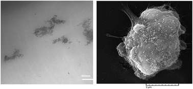
The present study demonstrates HEMA-grafted nanodiamond (ND-HEMA)/acrylate-terminated polyurethane-acrylate diluents (APUA) composites as promising materials for bone implant applications. Neat APUA and APUA composites containing ND-HEMA at different loadings up to 2 wt% were prepared by an in situ polymerization method. Morphological analysis demonstrated that ND-HEMAs were actually in the form of tightly bound aggregates which led to formation of big agglomerates at a concentration of 2 wt%. It was also suggested that ND-HEMAs were preferentially localized in the continuous soft domain of APUA; however it interacted by both soft and hard domains. Moreover, ND-HEMAs caused considerable phase separation between soft and hard domains as well as increased crystallinity. Maximum improvement in tensile properties of APUA was observed at 1 wt% loading of ND-HEAMs, namely 175% improvement in modulus and 40% increase in strength. The hydrophilic nature of ND-HEMA enhanced water absorption of composites resulted in higher hydrolysis degradation of APUA. In vitro biocompatibility evaluation via culturing human osteosarcoma cells (MG-63 osteoblast-like cell line), demonstrated no adverse effect on the cell viability of samples. Furthermore, the composites showed favorable cell adhesion and growth, and ND-HEMAs did not cause any negative effect on proliferation, ALP production and osteoblast attachment by MG-63 cells compared with neat APUA.
Incorporation of appropriate functional or reinforcing particles is versatile route to improve the performance of PUs and its derivatives.7 Reinforcement of PUs in hard tissue applications has been attempted through incorporating rigid particles such as β-tricalcium phosphate (β-TCP),1 mineralized allograft bone particles (MBPs)2 and hydroxyapatite nanoparticles (nHA)8 in the recent decade. However, current functional nanoparticles have been shown to be much influential on the property enhancement of polymer matrices compared to micron sized particles. Amongst many nanoparticles investigated in the field of polymer nanocomposites, nanodiamond (ND) is increasingly receiving much attention in research community due to its superior mechanical properties (Young's modulus 1220 GPa),9 high thermal conductivity,10 low friction coefficient,11 unique electrical resistivity and optical properties.9 These unique characteristics along with the rich surface chemistry with tunable surface,4 biocompatibility and low toxicity have made ND favorable candidate for biomedical applications.12 Additionally, mass production of ND with reasonable price is promising because of availability of economically viable production techniques such as detonation method.10
Recent studies have shown that ND is able to improve significantly the mechanical, tribological and thermal conductivity of polymers at both low, e.g. ∼1 wt% 13 and high, i.e. above 20 wt%,14 ND loadings. However, the studies on PU/ND nanocomposites dealing with microstructure, mechanical properties and functional performance have been rarely reported in literature. Recent investigation on nanostructure, dynamics and elastic properties of hybrid nanocomposites containing semi-interpenetrating polymer network of PU and poly(2-hydroxyethyl methacrylate) with 3-D diamond exhibited maximal effects at low contents, as low as 0.25 wt%.10 It was also shown that incorporation of a few percent of ND, e.g. 0.5 wt%, led to a remarkable improvement in tribological properties of elastomeric PU, while its elastic behavior and tensile strength remained almost unchanged.15
Enhancement of the mechanical properties in the polymer matrix containing ND is rather complicated due to the formation of ND agglomeration,4 difficulties in obtaining a homogeneous dispersion of nanoparticles8 and challenges in generation of sufficient interfacial adhesion.7 The poor interfacial interactions often lead to weak load transferring between hosting polymer and the reinforcements. Therefore, it is necessary to tailor the surface chemistry of NDs according to intrinsic chemical nature of polymer matrix. Consequently, appropriate surface functionalization of ND along with a suitable mixing method such as melt mixing and in situ polymerization could be effective in preventing the formation of large agglomerates in polymer/ND composites.16
Literature shows that the polymer/ND composites targeted for biomedical applications has also been investigated more or less by researchers. Zhang et al.4 reported that ND filled poly(L-lactic acid) (PLLA) composite exhibited significantly enhanced mechanical properties and increased biomineralization with no toxicity on murine osteoblast cell (7F2) proliferation which was suggested to be used as bone scaffolds and regenerative medicine. The attempt conducted by Protopapa et al.17 to increase overall performance of poly methyl methacrylate (PMMA) by ND for fixed interim prostheses was found to be successful.
Recently, we synthesized biodegradable APUs based on low molecular weight poly(ε-caprolactone)diol (PCL), aliphatic 1,6-hexamethylene diisocyanate (HDI) and hydroxyethyl methacrylate (HEMA) as potential thermosetting polymers for tissue applications.18 It was shown that these polymers exhibited complex multiphase microstructure containing soft and hard domains with either amorphous and/or crystalline morphologies which were chemically cross-linked. The objective of the present work was to examine the potential aptitude of ND on the improvement of mechanical properties of biodegradable APU. The attempts were made to explore the structure–properties relationship in such complicated multiphase polymeric system. As the composite was targeted for biomedical applications, its cytotoxicity and in vitro degradation properties were also investigated thoroughly.
The average diameter and size distribution of a very dilute (0.0003 wt%) ND dispersion in chloroform was determined via dynamic light scattering (DLS) on a Malvern Zetasizer Nano S instrument (Red badge), ZEN1600 (Malvern Instruments Ltd, UK) equipped with a He–Ne-laser 4 mW (633 nm).
The morphology and dispersion state of ND-HEMA filled APUA were investigated using a field emission scanning electron microscope (FE-SEM) (JEOL, JSM-6700F) at an accelerating voltage of 10 kV and transmission electron microscopy (TEM, Philips, cm30) at 200 kV. The cured composites were sectioned by a diamond knife with an Ultramicrotome Leica Reichert, OMU-3, Austria. X-ray diffraction analysis (XRD) was used to evaluate the crystallinity of APUA film with a STADI P diffractometer (STOE, Germany). The X-ray source was Cu/Kα radiation at a voltage of 40 kV, 30 mA current with a radiation wavelength of 1.542 Å. Spectra were recorded in the range of Bragg's angle 2θ = 10° to 120° at a scanning rate of 0.2° min−1.
Dynamic mechanical analyses (DMA) were carried out with a Tritec 2000 DMA (Triton, Tritec Technology, England). The scans were performed in a single cantilever bending mode operating under a dry nitrogen purge. The temperature-dependent characterization (storage modulus, loss factor) of each sample was evaluated with a temperature sweep from −100 °C to 150 °C, at a frequency of 1 Hz, with a heating rate of 4 °C min−1 and at 0.02% strain. The rectangular specimens with dimensions of 10 mm × 5 mm × 1 mm were used. The density of the specimens was determined based on Archimedean principle in water according to ASTM D792 with Mettler Toledo density meter XS104. Mechanical performance of the samples was evaluated by tensile testing on dumbbell shaped specimens at a cross-head speed of 2 mm min−1 according to ASTM-D638 using HIWA 2126 universal machine (HIWA Engineering Co., Iran). The tensile testing machine was equipped with a 50 kgf (490.3 N) load cell with incremental extensometer to measure the strain precisely. Three to five tensile tests were carried out for each sample to obtain reliable repeatability.
The contact angle measurements were performed using OCA15 plus (data physics instruments, Germany) equipped with a video measuring system for the evaluation of surface hydrophilicity of APUA and its composites with ND-HEMA. The solvent droplet was put on the air-side surface of a film at room temperature. The average of four measurements was reported as water contact angle of each sample. In vitro degradation experiments were performed according to ASTM F 1635 (samples were sterilized using UV treatment and then sterilized media were used). Each sample was placed in an individual 50 mL glass vial containing 0.1 M PBS (phosphate buffered saline solution) with a pH = 7.4 ± 0.2. Polymers were kept in a 50 rpm shaking incubator at 37 °C for up to 180 days. At each time interval, samples were removed from the PBS, placed in distilled water for 5 h to remove buffer salts then dried under vacuum for 2 days at 37 °C, and weighed to determine dry mass. Mass loss of the samples was obtained as follows:
![]() | (1) |
As shown in Fig. 1, FTIR spectrum of ND-HEMA shows characteristic peaks at 1712 cm−1 (CO) and 1635 cm−1 (C
C) which are relevant to HEMA attached on ND surfaces.16 Presence of HEMA on ND-HEMA was also examined by TGA. As shown in Fig. 2a, ND-COOH is thermally stable up to almost 500 °C, because it does not show sensible mass losses up to that temperature. However, ND-HEMA exhibits a step mass loss of almost 5 wt% between 200 and 400 °C suggesting presence of chemically attached HEMA on the surface of ND which is consistent with literature for HEMA grafted carbon nanotube.16
![]() | ||
Fig. 2 (a) TGA and DTG thermograms of functionalized ND particles, (b) particle size distribution of as-received ND and functionalized NDs. |
The size distribution of all three NDs obtained via DLS is shown in Fig. 2b. The average hydrodynamic diameter of as-received ND is found to be 66 nm which is higher than the single primary ND particles with average diameter of 5 nm. This suggests that the smallest entity of as-received ND is indeed an aggregate, consisting of tightly bound ND primary particles, which is consistent with previously reported size for ND.24 Functionalized NDs show slightly larger hydrodynamic diameter and little wider size distribution compared to as-received ND. This is consistent with literature reporting on the average size of functionalized NDs;4,24 which could be attributed to the coverage of outer surface of NDs by the organic groups. Dispersibility of all NDs in acetone and chloroform is compared in Fig. 3. It was revealed that the suspension stability of the as-received and oxidized NDs in both solvents were poor while ND-HEMA showed slightly better dispersion stability.21
![]() | ||
Fig. 4 Micrographs of APUA/ND-HEMA (a) FESEM of 1 wt% ND-HEMA, and (b) FESEM of 2 wt% ND-HEMA, (c) TEM of 1 wt% ND-HEMA, and (d) TEM of 2 wt% ND-HEMA. |
XRD diffraction pattern of pristine polymer and the composites are displayed in Fig. 5. APUA exhibits one broad diffraction peak at 2θ of 19° which is attributed to the presence of small size crystallite originating from its soft segment, i.e. PCL.18 In APUA/ND-HEMA composites the position of this peak remains unchanged while its intensity increases proportionally with the amount of ND-HEMA content in the samples. This could be indicative of increased crystallinity of composites compared to neat APUA film.
A similar increase in crystallinity of the polymer matrix with ND nanoparticle was described in literature as well4,26 which was often explained by the nucleating role of ND originated by its affinity with matrix. The crystallinity (XC) of samples was calculated using the method described by Young and Lovell.1,27 Crystallinity of neat APUA was found to be 38% which was completely consistent with DSC data reported in our previous publication.18 The crystallinity of ND-HEMA filled APUA increased compared to neat APUA as 42.83%, 43.4% and 41.96% for 0.5 wt%, 1 wt% and 2 wt% loadings, respectively, showing a maximum at 1 wt% loading. The relative loss in crystallinity at 2 wt% loading of ND-HEMA may be due to the spatial hindrance caused by closer particle distance or promotion of agglomeration which constrained further growth of crystallite. The small and new peak observed at 2θ of 43.6° for composites was attributed to the (111) plane of diamond as expected for nanocrystalline diamond.28 With the increase of ND-HMEA concentration the intensity of this peak becomes more prominent as well.
Basically, partial phase separation between hard (isocyanate constituent in polymer) and soft (polydiol) segments of PUs is a unique characteristic resulting in a polymer with two-phase morphology in which hard domain of 5–20 nm average diameter29 is distributed uniformly in the soft domain. The extent of phase separation of hard and soft domains is often described by the degree of phase separation (DPS) which is mostly determined using the FTIR spectrum by measuring the hydrogen bonding index (R),30,31 i.e. area ratio between the peak intensity of free-of-hydrogen bonding carbonyl (1730 cm−1) to hydrogen-bonded carbonyl (1702 cm−1), as DPS = R/R + 1.30,31
FTIR spectrum of APUA and APUA composites was first determined (Fig. 6a) and then the corresponding peak for carbonyl groups was deconvoluted to five Gaussian peaks using Origin software (Fig. 6b). As given in Table 1, DPS of pristine APUA is increased by incorporation of ND-HEMA demonstrating its capability in formation of further hard domains. Consistent with crystallinity, there is a maximum DPS at 1 wt% loading of ND-HEMA suggesting that the agglomeration prevents the formation of further separated hard domains possibly due to the spatial hindrance.
![]() | ||
Fig. 6 (a) FTIR spectra of APUA nanocomposites, (b) deconvoluted FTIR spectra in the carbonyl stretching region of APUA/ND-HEMA (1 wt%). |
Sample | R | DPS | ρ (g cm−3) | Tg,S (°C) | Tg,H (°C) |
---|---|---|---|---|---|
APUA | 0.97 | 0.49 | 1.161 | −58.5 | 39.8 |
APUA/ND-HEMA (0.5 wt%) | 1.41 | 0.58 | 1.171 | −58 | 46.9 |
APUA/ND-HEMA (1 wt%) | 1.63 | 0.62 | 1.174 | −56.4 | 48 |
APUA/ND-HEMA (2 wt%) | 1.36 | 0.57 | 1.171 | −55.5 | 46.5 |
![]() | ||
Fig. 7 DMA curves of APUA and APUA/ND-HEMA composites at frequency of 1 Hz (a) loss factor (tan![]() |
As shown in Fig. 7b, a stepwise loss in storage modulus of neat APUA is observed at temperatures corresponding with prominent glass–rubber transition peak in tanδ curve. However, there is no distinct storage modulus step loss for the minor relaxation peak, as mentioned by tan
δ curve, probably due to overlapping with that of prominent transition tan
δ peak observed below −10 °C. Accordingly, glass–rubber transition temperature of soft domain (referred to as Tg,S) was obtained using the storage modulus–temperature curve by intersecting two tangents; one below transition loss and the other near inflection point of the modulus transition loss according to ASTM-E1640. As a result, Tg,S of neat APUA was found to be −58.5 °C (see Table 1). According to Fig. 7b, as storage modulus of APUA exhibits a significant stepwise drop around Tg,S (see Fig. 7b), it can be deduced that the soft domain forms the continuous phase in which the hard domains are distributed uniformly as dispersed phase.32
As reported in Table 1, incorporation of ND-HEMA into APUA increases Tg,S slightly, however, it influences greatly the tanδ peak of neat APUA at 10 °C. Accordingly, the minor relaxation peak of neat APUA becomes larger and shifted to higher temperatures in presence of ND-HEMA (see Fig. 7a). This behavior could be attributed to the increment of DPS and crystallinity in presence of nanodiamonds, as well as presence of nanodiamonds itself in soft domain inducing more heterogeneity in relaxation behavior of amorphous region.
As deduced in Fig. 7, the small tanδ peak around 45 °C which is correspondent with a step loss in storage modulus of APUA, is indeed related to the glass transition of hard phase (crosslinked acrylate/isocyanate containing domain), while a small sharp peak observed around 60 °C indicates the melting point (Tm) of crystalline phase, as discussed by Alishiri et al.18 Glass transition temperature of hard domain (denoted by Tg,H) was obtained using the storage modulus data as well. As reported in Table 1, addition of ND-HEMA to APUA results in the enhancement of Tg,H which could be due to the interaction of hard domain with the ND-HEMA probably via the chemical bonding through the HEMA functional groups involved in the crosslinking reaction.
DMA data suggest that ND-HEMA is interacted by both hard and soft domains of APUA. According to literature,29,32,33 average diameter of dispersed hard domain ranges between 5 and 20 nm with a hard inter-domain distances within 5–25 nm which are much smaller than the ND-HEMA aggregate size obtained from DLS and TEM. Such morphological feature led us to assume that ND-HEMA is preferentially localized at soft continuous domain, however, there should be possibility for dispersed hard domains (due to its nano-scale size) to attach on the outer surface of ND-HEMA aggregates periodically (not like a uniform layer at outer surface). Somewhat similar morphological model was suggested by Fernández-d'Arlas et al. for thermoplastic PU/carbon nanotube composites.34 Presence of HEMA functional group on outer surface of ND-HEMA and in APUA structure provides a tendency for attachment of hard domain on the outer surface of ND-HEMA by involving in crosslinking reaction.
Storage modulus of APUA was observed to enhance with incorporation of ND-HEMA particles, indicating reinforcement effect of ND-HEMA on APUA.17 According to Fig. 7b, the extent of improvement in storage modulus is much higher below Tg,S, where the soft domain stays in its glassy state, suggesting ND-HEMAs interact mainly with APUA soft domain.35,36 Higher storage modulus of ND-HEMA filled APUA is attributed to restricted mobility of polymer chains due to interfacial bonding between nanodiamonds and APUA matrix,37 restricting composite ability to deform by strengthening APUA molecules.38 The extent of enhancement in storage modulus is found to be dependent on the ND-HEMA content. APUA composite with 1 wt% ND-HEMA particles exhibits the highest improvement in storage modulus, while at 2 wt% loading a relative decrease is observed which is more likely due to the agglomeration of ND-HEMA in APUA.39
As shown in Fig. 8a, incorporation of ND-HEMA enhances the tensile properties of APUA significantly, e.g. 175% in elastic modulus and 40% in tensile strength at 1 wt% loading. Such significant improvements in tensile properties of bone implantable PU are much higher than other reinforcements reported in literature like β-TCP (maximum improvement 15% for compressive modulus and 5% for compressive strength at 10 wt% β-TCP1) and nHA (maximum 17 and 20% improvements in the young modulus and tensile strength, respectively, at 10 wt% loading40).
![]() | ||
Fig. 8 Tensile properties of the samples, (a) modulus and tensile properties and (b) elongation at break and fracture energy. |
The effectiveness of ND-HEMA in tensile properties of APUA can be attributed to the strong interfacial interaction between polymer–filler and uniform dispersion of ND-HEMA within the matrix.41 Surface functional group of ND-HEMA is effective in fine dispersion in the matrix and achievement of good interfacial interaction in the composite. Furthermore, enhancement in mechanical properties can be attributed partly to the increase of crystallinity of APUA matrix in presence of ND-HEMA13,42 and increment in DPS.30,31 Crystalline structure has stronger mechanical properties36 and can contribute significantly on overall mechanical properties of semi-crystalline polymers. It is noteworthy that localization of ND-HEMA in soft domain, as discussed above, provides unique opportunity to enhance the mechanical properties of mechanically weaker phase of APUA, because, the hard domain is rigid and fully crosslinked phase18 with higher mechanical properties acting as rigid filler itself in APUA matrix. The relative decrease in mechanical properties of 2 wt% loading ND-HEMA is completely in line with crystallinity and DPS data as well which is more likely related to the loss in degree of dispersion of ND-HEMA at this concentration. Fracture energy was also calculated from the area under the stress–strain curves, see Fig. 8b. Addition of ND-HEMA causes a decrease in fracture energy of APUA, as expected, however APUA/ND-HEMA (1 wt%) shows the largest fracture energy amongst the composites, probably due to its unique morphological feature, i.e. higher DPS and crystallinity with almost fine ND dispersion.
To investigate the role of ND-HEMA on hydrolytically degradation rate, in vitro degradation test method used in surgical implants was utilized. Fig. 9b shows the mass remaining profile of neat APUA and APUA/ND-HEMA composites versus incubation time in PBS over 6 months. It was expected that the sample would undergo hydrolysis degradation through the ester bonds of PCL.49 It is observed that all samples shows mass loss with time indicating their biodegradation. Interestingly, ND-HEMA has increased the degradability of APUA so the difference between mass loss of neat APUA and APUA/ND-HEMA becomes much prominent at longer times suggesting the positive role of ND-HEMA on acceleration of hydrolytic degradation of APUA. Such behavior can be ascribed to hydrophilic nature of ND-HEMA enhancing the water absorption that would improve the rate of hydrolysis of PCL ester bonds.50 Consistent with mechanical properties and water contact angle data, the biodegradation is dependent on ND-HEMA content so a relative loss in biodegradation is observed for 2 wt% loading possibly due to its poor dispersion.
The degradation property is suitable in tissue engineering for the bone repair.51 It is necessary that the degradation rate of composites should tailor the healing rate of bone. In this way the initial weight loss of composites during the early days should be low as bone is defected,52 and in the course of regeneration of bone tissue, the bone substitute should be degraded in line with the bone reconstruction rate.53 It is to be noted that many biodegradable bone substitutes reported in literature1,49 showed low degradation rate, therefore any acceleration is indeed desirable.
The potential toxicity of carbon nanoparticles reported in literature has shown a great deal of controversy and confusion. Several authors have reported favorable cellular interactions with carbon nanoparticles,55,56 while some others have reported decreased cell viability.57,58 The controversy may be originated from a number of variables used in their studies including cell type, nano particle type (MWNT, SSWNT, ND, CNF), shapes and size, surface chemistry, level of impurities as well as local concentration and surface functionalization. Schrand et al.59 reported that ND is less cytotoxic than other carbon materials. Moreover, several in vitro experiments with different cell lines generally showed no or very little toxicity of all tested NDs.60 However, there may be the situations in which NDs can exhibit certain cytotoxic effects as reported by Burleson.60 Nevertheless, according to open literature,4,61 the ND powder itself up to the concentration of 1000 μg mL−1 did not produce significant changes in the viability of neuroblastoma, kidney, 7F2 osteoblasts and epithelial cells. Therefore, it is imperative to investigate the toxicity and biocompatibility of ND-based biomaterials before they can be considered for widespread acceptance and use. As APUA/ND-HEMA composite is targeted to be introduced as potential material for bone applications, comprehensive understanding about toxicity and biocompatibility of this composite is crucial requirement.
The cells viability next to APUA and three different fabricated composites (0.5, 1 and 2 wt% loadings) on MG-63 cell line were quantified by MTT assay as shown in Fig. 10a. It is to be noted that the carcinoma cell line is normally used in biocompatibility studies because of its rapid rate of growth and proliferation along with easy separation through the viability test compared to normal cell line. According to the MTT assay, exposure of the cells to neat APUA and APUA/ND-HEMA composites did not influence cell viability in a significant manner compared to the control group (TPS). According to Fig. 10a, viable cells for all samples are more than 80% that stands within acceptable biocompatibility range, because a material is considered to be cytotoxic when its viable cells becomes less than 70%.62 Nevertheless, the attached cells were well spread on APUA and APUA/ND-HEMA composites suggesting their unaffected cell functions. The results indicate that in presence of NDs, the cells still survive and grow well which is in line with literature data.4 Actually, Burleson et al.60 reported that the effect of NDs on carcinoma cell line was more pronounced than on normal human cells.
It is found that viability of MG-63 cells for APUA/ND-HEMA is slightly lower than that of neat APUA, but the difference is not considerable. Also consistent with literature data,44 the result suggested that the hydrophilic surface of the lower content of ND-HEMA in composites was more favorable for the cell spreading and growth than the hydrophobic surface of the higher content films. It could be observed that cytotoxicity of all samples shows a slight reduction at 5th day but at longer incubation time of 10th day inverse result is obtained (with p > 0.05 which is statistically insignificant). The obtained results demonstrated that there was not any significant difference in the cell viability and the overall cell morphology of the samples during the incubation period.
The number of cells adhered to the samples is shown in Fig. 10b. No clear differences are observed in MG-63 cell attachment of APUA/ND-HEMA composites with increasing ND content in the first day. The lack of clearly distinguishable effect may be because of negligible difference in their wettability (water contact angle) which is believed to cause significant changes in cell adsorption during the 24 h cell culture incubation period. Furthermore, the carcinogenic cell-type used (MG-63) may have been less sensitive to wetting conditions than primary cells.6 Similar to some studies63 no correlations between chemical composition and cell attachment were observed. An increase in the total osteoblast number was found with increasing culture incubation period (from 5th to 10th day) in all samples (Fig. 10b).
The effect of ND-HEMA on MG-63 osteoblast differentiation of APUA was further analyzed by ALP activity, since ALP is regarded to be an important phenotype of bone-forming cells.4 The ALP gene's expression levels in MG-63 cells cultured at three different times (1st, 5th and 10th days) on neat APUA and its composite with ND-HEMA were evaluated and the results were compared to those in MG-63 cells cultured on TPS for similar periods. According to Fig. 10c, no considerable ALP is produced by seeding the MG-63 cells on all films in comparison with TPS. The incorporation of ND-HEMA into the films did not cause any significant differences in ALP production by MG-63 cells in 1st and 10th days compared with neat APUA polymer. ALP activity was found in all samples to be time-dependent. When comparing the data at 5th and 10th days to those at 1st day, we found a significant decrease in ALP gene expression (p < 0.05). Other osteogenic cell lines, such as murine osteoblast (7F2) cells on PLLA/ND nanocomposites also exhibited a similar inverse relationship between incubation time and ALP gene expression in vitro.4
Results of osteoblast developmental stages indicated that production of ALP decreased as the proliferation was speeded up. Therefore, an inverse relationship between growth and ALP production were obtained. Similar results were reported in previous in vitro investigations for many cell types, e.g. osteoblast-like cell lines (MC3T3-E1).64
Overall, the data from ALP assay as well as MTT assay suggest that APUA composites containing up to 2 wt% ND-HEMA are non-cytotoxic, support MG-63 cell proliferation and osteogenic differentiation, and therefore can be used for biomedical applications. Furthermore the results indicated that the incorporation of ND-HEMA in the samples had no adverse effect on the proliferation, ALP production and osteoblast attachment in comparison with neat APUA.
In order to assess the morphology and cytoskeletal architecture of MG-63 cells on APUA and APUA/ND-HEMA composites, the cells were fixed after 4 days post-seeding. SEM micrographs (Fig. 10d) show proliferation and attachment of cells on to the films. SEM microphotographs of cells grown on samples suggested that the proliferation of cells could maintain their normal morphology while adhered on the surface of films. Therefore SEM results confirmed that MG-63 cells have become fully confluent on all samples. Moreover, the in vitro appearance of the cells was consistent with that reported in literature.
This journal is © The Royal Society of Chemistry 2016 |