DOI:
10.1039/C5RA19109B
(Review Article)
RSC Adv., 2016,
6, 2835-2853
The metamorphosis of vascular stents: passive structures to smart devices
Received
16th September 2015
, Accepted 17th December 2015
First published on 22nd December 2015
Abstract
Drug-eluting stents (DES) represent a major milestone in stent technology, saving the lives of millions worldwide afflicted with cardiovascular disease. Though a wide range of therapeutic approaches has been employed for treatment of coronary artery diseases, stent deployment has become the most extensively practiced method to open blocked arteries in comparison with surgical revascularisation. In recent years, the bare metal stent has evolved into more effective drug-eluting systems and the encouraging results from initial clinical trials have instilled hope among cardiologists to reduce the number of fatalities caused by cardiovascular diseases through the deployment of drug-eluting stents. Over the years, the stents have evolved from a passive framework that maintained blood flow through the blood vessels into multi-functional devices that could also serve to mitigate conditions leading to the blockage of the blood vessels while promoting re-endothelialization of the blood vessel. This review traces the evolution of the stents from the first generation structures to the modern versions that overcome their limitations. A glimpse into the exciting future of stent technology is also provided.
1. Introduction
Coronary artery disease is a major cardiovascular disorder that develops due to escalation of lipid oxidation. The oxidized products get deposited in the sub-endothelial space of coronary arteries leading to atherosclerosis.1 This results in a complex synergy between blood components leading to disruption in blood flow, artery wall irregularities, and other pathological conditions such as inflammation with monocyte accumulation, proliferation of smooth muscle cells and thrombosis upon platelet activation and fibrin formation. These manifestations cause severe obstruction of arteries, paving way for a spectrum of clinical events ranging from angina, myocardial infarction to sudden cardiac arrest.2 Among the leading causes of death around the globe, coronary artery diseases top the list accounting for 35% fatality.3 Treatment of cardiovascular disorders is highly invasive and the odds of survival are also minimal.4 Traditionally, balloon angioplasty has been used to treat obstructed arteries, but this strategy is limited due to tearing of the vessel wall, causing acute vessel closure. It results from the disruption of the plaques by the expanding balloon, weakening the artery and also induce damage to the inner layers of the artery leading to deep intimal arterial injury contributing to restenosis at a later date.5 To avoid such occurrences, a permanent metal framework known as stent is inserted through a blocked artery, which gives support to the artery as well as preserves the integrity of the vessels.6
Bare metal stent (BMS) are considered to be advancement over conventionally employed balloon angioplasty for the treatment of coronary artery diseases. Bare metal stents were the first to be approved by the Food & Drug Administration (FDA) and were considered highly efficient for reopening blocked arteries.2 The BMS is fabricated as a typical mesh-like metal thin tube called as struts.7 Stents are used as a frame to support the artery walls and confine the plaques to the wall.6 Metals like stainless steel, cobalt-chromium, platinum and titanium possess sufficient strength to undergo plastic deformation thereby preventing the collapse of the artery due to the force imposed during stent implantation apart from restoring the original dimensions of the atherosclerotic artery.6 BMS prevents restenosis through suppression of early arterial recoil and reduction of contraction as well as reduce tearing of vascular space through sealing the dissection.8 However, stents being non-native to the body, are recognized as foreign objects and the immune system mounts a cascade of immune and inflammatory reactions at the time of implantation rendering the stent thrombogenic and non-biocompatible.9 It has been observed that about 10–20% patients every year suffer from restenosis due to excess proliferation of the neointima.10 Neointima is the inflammatory and proliferative reaction of the vessel wall to the injury created by angioplasty and stenting. The proliferative response is a normal healing process which includes smooth muscle proliferation and endothelial coverage of stent struts making the stent non-thrombogenic. But the quantum of neointimal proliferation becomes excessive in many individuals, causing narrowing of the opened artery, which results in-stent restenosis. None of the newer alloys could mitigate the problem of restenosis. These limitations have resulted in the emergence of drug eluting stents (DES) that act as a localized drug delivery system to mitigate the adverse effects of a bare metallic stent.
Drug eluting stents (DES) use a wide variety of pharmacological agents such as anti-proliferative and anti-inflammatory drugs namely sirolimus, paclitaxel, resveratrol and curcumin are coated on the metal stent using a polymer coating. The polymers commonly employed include the biostable polymers such as poly(ethylene-co-butyl methacrylate), poly(styrene-isobutylene-styrene) and phosphorylcholine as well as biodegradable polymers like polylactic acid, poly(L-lactide-co-glycolic acid)11 as a carrier. DES has shown promising results in retarding the rate of in-stent restenosis as well as inflammatory response.12 DES has made major headway in the field of cardiovascular intervention by reducing post-stenting complications such as restenosis or neointimal hyperplasia as well as the need for subsequent surgical interventions. It has enabled site-specific controlled drug release, which inhibits intimal hyperplasia and provides positive remodeling to the stenosed artery.13 However, since the appearance of first-generation drug eluting stents, a new complication namely thrombosis, has emerged due to the excess accumulation of platelets and enhanced fibrolytic processes at the stent site. This has raised concerns on the safety and efficacy of DES.
Diverse strategies involving modification of the coating material, stent material, stent design, drugs employed, etc., have been attempted to overcome several drawbacks of the DES in order to develop more robust delivery systems. Several novel approaches in DES are also under different phases of pre-clinical and clinical trials. The present review attempts to explore the various advances in the field of stent technology including the different types of stents, their merits and demerits, on going research and development in drug eluting stents and emerging generation of ‘smart stents’ that will be discussed in the following sections.
2. Evolution of stent materials
Metals are the most commonly employed material for stents due to their strength and stability. Mechanical and physical characteristics of metals such as high toughness and fatigue strength apart from possessing corrosion- and abrasion-resistance have contributed to their use for various load-bearing implants. Stents are a type of vascular implants that require a durable scaffold that can reinforce the weak vessel wall, resist continuous pressure and in addition be biocompatible in the vascular space. Conventionally metals are the main choice for a stent material.2 Based on the their characteristic features, safety and responsive nature, different metals and their alloys have been employed for the fabrication of a stent.2 The earliest and most common stent material has been the alloy, stainless steel.2 Other materials that have been used for fabricating stents include alloys like cobalt-chromium, platinum-iridium, and nitinol. Pure metals like gold, magnesium and titanium has also been used for making stents.2 Other alternative non-metallic materials that have been employed as stent materials are biodegradable polymers.2
2.1 Stainless steel coronary stent
316L stainless steel has been the traditional choice of material for fabrication of bare metal stents.14 Steel could potentially react with the biological system and undergo corrosion in the physiological conditions but stainless steel, an alloy of steel combines high strength, formability and corrosion resistance with a broad range of mechanical properties thereby making it an excellent choice of material for stents.15 It also has a high radial strength and its ductility enables fabrication of stents with thinner strut dimensions. Stainless steel stents generally possess ultimate tensile strengths between 600 and 1000 MPa depending on their composition.16 The tensile strengths of many iron-based stents that also possess degradation properties range from 200–500 MPa. Several iron-based stents alloyed with platinum possess tensile strengths as high as 1500 MPa.17 Since stainless steel is alloyed to various metals such as nickel, chromium and molybdenum, their release can trigger a local immune response.2 In case of stent, struts design play an important role for endothelialization but due to poor strength of stainless steel it restricts the dimensions of the struts that could be fabricated using this alloy leads to restenosis.18 In addition, their ferromagnetic nature makes them non-MRI (magnetic resonance imaging) compatible.19
2.2 Cobalt-chromium stent
Cobalt-chromium is an advanced alloy that is stronger and denser than stainless steel. The tensile strength of cobalt-chromium stents generally lies between 800 and 1200 MPa.16 Due to their inherent strength, stents with thinner struts can be fabricated without compromising their radiopacity.2 Thinner struts enable superior tissue coverage due to better endothelialization.18 Such design is not feasible with stainless steel owing to its poorer strength. Cobalt-chromium alloy also possesses better fatigue-resistance, which leads to excellent radial strength and is more corrosion-resistant than stainless steel.19 In addition, they possess high non-ferromagnetic property and hence the cobalt-chromium alloys are more MRI compatible than stainless steel.20 However, fatigue fracture is one of the common disadvantages of such alloy-based stents.19
2.3 Platinum-iridium stent
Apart from corrosion resistance and stability, platinum metal has also been found to be biocompatible and radiopaque that makes it an excellent candidate for biomedical applications.21 But the prohibitive cost of platinum limits its widespread use. Therefore various platinum alloys have been developed such as platinum-iridium, rhodium, palladium and ruthenium. Among these noble metal alloys. Platinum-iridium shows qualities for a stent material such good radiopacity, corrosion resistance, low inflammatory response hemocompatibility and biocompatibility.22 However, lack of strength limits its application as a stent material as its recoiling percentage is higher as compared to stainless steel stent.2
2.4 Titanium stent
Titanium has been used extensively for biomedical applications such as in artificial hip and knee joints, cardiac valve prostheses, screws used for fracture fixation, bone plates, pacemakers, etc.2 Biocompatibility, low electronic conductivity, stability at physiological pH and corrosion resistance are the main characteristics of titanium, which makes it a promising candidate for coronary stents.23 However, titanium has low tensile strength and low ductility that may increase the chance of stents to fail under stress test during deployment. By alloying with other metals, the mechanical properties of titanium can be improved.24 Stents made from various titanium alloys have tensile strengths ranging from 300–1000 MPa.16
2.5 Nitinol stent
Nitinol is a nickel-titanium alloy with shape memory property and has been used to develop self-expanding stents.25 Nitinol stents have been found to be super-elastic; crush recoverable and more physiologically compatible than balloon expandable bare metal stents.26 The shape setting property of nitinol lowers the risk of damage to the stent both during the deployment into the body and at the time of vessel recoil.2 The major drawbacks of this material are its high cost and increased metal fatigue, which leads to its failure due to bending or twisting.27 Another issue with nitinol stents is its bulky delivery system, which makes it difficult to track through tortuous vessels. Currently nitinol is used in the manufacture of the self expanding stents used in peripheral arteries and aorta. Fig. 1 summarizes the major merits and demerits of the commonly employed metallic stents.
 |
| Fig. 1 Outline the different types of stent material that have emerged with time along with their shortcomings. | |
2.6 Other stent materials
Tantalum is a promising material for many biomedical applications especially as scaffolds for orthopaedic and dental applications.28 Tantalum causes low inflammatory response and has negligible dissolution properties.28 It possesses excellent strength, ductile properties and biocompatibility that have been useful for designing scaffolds for load-bearing tissues like bone.16 Tantalum stents have been used as a substitute for platinum stents because of their high corrosion resistance and chemical inertness due to the highly stable oxide layer.29 In addition, they are more cost-effective than platinum.30 Tantalum alloys are widely used for medical implants due to their biocompatibility and non-irritating nature. Non-irritating stents cause no inflammatory responses thereby avoiding adverse effects such as vessel spasms.31 However, their poor mechanical properties limit their application, as there exists an increased possibility of material fracture during stent deployment.2 Also, stent recoil has been observed in tantalum-based stents when compared to stainless steel stents.2 Gold is one of the least reactive metal found in nature and it is biocompatible as well as a blood compatible material with higher radiopacity.32 These properties make it well suited as a stent material. However, improper gold coating during galvanization leads to surface defects during the stent manufacture because of which no significant difference in the performance of these stents were observed in clinical trials when compared to other stents.33 Kastrati et al., have demonstrated that gold stents increase the risk of restenosis and target vessel revascularization over the first year of stent placement.34 Hence, these stents are rarely used in the present era. However in recent years, many approaches have been made to improve the performance of bare metal stents. Nanotechnology-enabled techniques like nanotexturing and nanopatterning have emerged, which could enhance the drug release efficiency of the stent surface. In addition, novel metal alloys such as platinum-chromium have been explored for stenting applications due to their better mechanical properties and radiopacity.35
2.7 Covered stents
Covered stent is a unique, flexible, biocompatible metal stent with an extra layer of membrane covering the stent completely or partially.36 It is a multipurpose stent design that can be used for cardiovascular, neurovascular and peripheral interventions.37,38 Covered stents represent an improved version of stent-grafts. They reduce fibrosis that is commonly encountered in bare metal stents.39 The covered stent acts as a physical barrier that reduces the blockages occurring due to tissue in-growth and overcomes several limitations of other categories of stents.40 These membranes can be effective against restenosis by preventing direct contact of the metal with the biological tissues.41 Mechanical properties of the membrane material also are key factors that determine the performance of a covered stent. Biostable polymers such as poly(styrene-b-isobutylene-b-styrene) (SIBS),42 expanded poly(tetrafluoroethylene) (ePTFE) and silicon membranes43 have been the most successful materials for covered stents.40 Natural and modified polymers have also been explored extensively for use in covered stents.41,44 Chitosan-based polymers (polyethylene oxide CH-PEO), PVA (polyvinyl alcohol) cryogel membranes, polyethylene terephthalate (PET) or segmented polyether polyurethane (SPP) (SPU), bio-inspired salmon collagen fibrillar gel, heparin-impregnated collagen stent-graft, etc., have been used as coatings on stents.45–47
Covered stents face many clinical challenges when it comes to a small vascular treatment and aneurysms. They possess the risk of promoting thrombosis, hyperplasia and have been reported to prolong the healing process.48 Hence, a novel technique employing a drug-loaded membrane coating on metal stent was introduced to overcome the limitations of covered stents. The drug releases gradually from the polymer membrane thereby achieving localized delivery at the target site that serves to mitigate any possible toxic effects arising due to the introduction of the covered stent. Table 1 describes the different types of covered stent systems that have been reported for the treatment of various conditions. Although the covered stent is superior to other stents it displays high risk of stent migration, perforation, tissue in-growth or hyperplasia.49 To overcome the problem of stent migration, partially covered stents have been developed with broad uncovered ends as dumbbells to grip on to the tissue to impart better migration-resistance to the stents40 (Fig. 2).
Table 1 Different types of covered stents used in cardiovascular complications
Stents |
Application |
Covered stent graft |
Treatment of de novo or traumatic arterial aneurysms and pseudo-aneurysms46 |
Membrane-covered Express Monorail stents |
Treatment of aneurysms, therapy for complications arising due to sealing coronary perforations47 |
Paclitaxel-loaded poly(ε-caprolactone) electrospun fibrous membrane covered stent |
Treatment of benign cardiac strictures to avoid inflammation and scar formation48 |
Heparin loaded poly(L-lactide-co-caprolactone) nanofibre covered stent |
Treatment of aneurysm50 |
 |
| Fig. 2 Schematic representation of a partially covered stent. | |
Fabric stents are a type of covered stents and are endovascular stent grafts. They are commonly used for the treatment of aneurysms and they provide support to the collapsing vessels. It is a permanent graft and consists of a metal stent (nitinol, stainless steel or cobalt-chromium) fitted with a fabric made up of woven polyester (PET, Dacron), expanded polytetrafluorethylene (ePTFE) and polyether type polyurethane which serves as a membrane to support the inner wall of vessels and prevent it from rupture and maintains pulsatile blood pressure.51,52
3. Evolution of drug eluting stents from bare metal stents
Drug eluting stents (DES) represent the first breakthrough advancement in stent technology, as they were primarily designed to prevent high rates of restenosis encountered in bare metal stents. Delivering drugs directly to the target site reduces further stent-related complications associated with coronary artery disease.53 Typically, DES consists of a bare metal backbone with thin polymer coating containing a drug formulation. The superiority of drug eluting stents in reducing in-stent restenosis in comparison with bare metal stents has been established beyond doubt through animal experiments and large clinical trials. Regardless of the fact that DES has transformed the field of interventional cardiology, certain stent-related complications mar its successes. The anti-proliferative drug in the DES restricts the intimal growth and migration of smooth muscle cells thus preventing restenosis, but it also inhibits endothelialization of the stent surface.54 The resulting exposed stent surface increases the risk of thrombosis at the implant site. Clinical data reveals that the first drug eluting stents loaded with sirolimus and paclitaxel as anti-proliferative drugs though successful in limiting the growth of vascular smooth muscle cells, also impaired the endothelial cell proliferation leading to incomplete neointimal coverage and eventually stent thrombosis.55 The polymer coating also may activate the immune response that in turn cause inflammation and in-stent restenosis as well as stent thrombosis at the site of deployment.56 Therefore, an ideal drug for DES should promote rapid development of a normal endothelial lining of the stented vessel segment and prevent the proliferation of vascular smooth muscle cells (VSMCs) without hindering the healing process of endothelial cells. Many drugs and drug combinations are under active investigation to achieve desired repair and action. Many new classes of drug eluting stents have emerged based on the drugs employed and these represent the next generation drug eluting stents.57
4. Progress in choice of drugs for DES
Drugs that possess anti-proliferative, anti-inflammatory, anti-thrombotic or immunosuppressive properties have been employed as pharmacological agents in drug eluting stents. These drugs are loaded in to polymers that are coated on the stents and are released by either degradation of the polymer in the case of biodegradable coatings or through diffusion through the polymer chains in the case of a bio-stable coating. These drugs have diverse molecular targets to exert their anti-proliferative or anti-inflammatory property. These targets include mTOR pathway, NF-κB, calcineurin, cyclooxygenases, p27, P-selectin etc. (Fig. 3).
 |
| Fig. 3 Depicts the major sites of drugs action used in stents. COX: cyclooxygenase, LOX: lipoxygenase, NF-κB: nuclear factor-κB, VSMC; vascular smooth muscle cells, mTOR: mammalian target of rapamycin. | |
4.1 Anti-proliferative drugs used in DES
4.1.1. Sirolimus. Sirolimus (rapamycin) belongs to the class of natural macrocyclic lactones and has been found to be a potent immunosuppressive and anti-proliferative agent that binds to an intracellular receptor protein and eventually induces cell-cycle arrest.12 It also retards vascular smooth cell migration, proliferation and growth through inhibition of mammalian target of rapamycin (mTOR).58,59Extensive studies and numerous clinical trials have demonstrated that sirolimus can retard the proliferation of smooth muscle cells of both rat and human origin in vitro. It also effectively decreased the thickness of the intima in animal models of vascular injury.12 Commercially available sirolimus eluting stents CYPHER (Company-Cordis, Platform Bx VELOCITY™), have been subjected to clinical trials (RAVEL and European SIRIUS) that revealed their high anti-proliferation activity and ability to decrease major adverse cardiac events (MACE). Significant reduction in neointimal hyperplasia was also observed due to the deployment of sirolimus eluting stent.60 But their slow release profile and inability to promote re-endothelialization results in stent thrombosis, which could negate their clinical significance of reducing restenosis as compared to bare metal stent.61
4.1.2. Sirolimus analogues. The initial success with sirolimus had triggered a search of sirolimus analogues that possess similar anti-proliferative effect but lesser adverse effects. Among these analogues, everolimus (a new macrocylic triene derivative), biolimus A9, immunosuppressive agents such as cyclosporine, mycophenolic acid and tacrolimus, which induce G1 arrest leading to reduced proliferation and immune response, have been incorporated in stents.10 Everolimus is a macrolide antibiotic that possesses the ability to suppress the immune response and cell proliferation leading to prevention of restenosis similar to sirolimus.12 It possesses greater polarity than sirolimus and hence has better bioavailability and is rapidly absorbed on the arterial wall and attains peak concentration within few hours thereby offering better efficacy.12 Experimental data have shown that orally administered everolimus inhibits in-stent neointimal formation and significantly promotes neointimal healing.62 Evorolimus has been found to act at a later stage than the calcineurin inhibitor cyclosporin and tacrolimus.63Biolimus A9 also known as umirolimus is a novel sirolimus derivative, which is specifically designed for stent applications. It exhibits immunosuppressive properties similar to sirolimus, but due to its lipophilic nature it is absorbed rapidly about 10 times faster than sirolimus or everolimus.64 It can easily cross the cell membrane and achieve therapeutic effects in its target tissue when compared to sirolimus and hence leads to lesser systemic exposure.65 In terms of potency, biolimus readily enters the membrane layer of smooth muscle cells and induces cell-cycle arrest at G0 phase.66 Tacrolimus (FK506) is another 23-membered macrolide lactone, a calcineurin inhibitor employed as an immunosuppressive drug during organ transplantation to avoid rejection.67 Unlike sirolimus it exerts its pharmacologic effect by inhibiting calcineurin, a calcium-dependent serine–threonine phosphatase that activates the T cells.68 Novolimus, an active metabolite of sirolimus, is an mTOR inhibitor macrocyclic lactone, which has anti-proliferative and anti-inflammatory properties and has received a great deal of attention.69 It has been basically used for drug eluting stent applications and has a well documented safety and efficacy profile.70 Zotarolimus is a semi-synthetic rapamycin derivative, which has been especially developed for drug eluting stents using phosphorylcholine (PC) as carrier.71 It is a distinct cytostatic drug, which has an inherent growth factor inhibitory effect as well as immunosuppressive nature.72 Its exceptional lipophilic nature has an advantage over other sirolimus analogues, making it a better choice for drug eluting stents.72 Abbott clinical trials PREFER, ENDEAVOUR and ZoMaxx carried out with zotarolimus eluting stent (ZES) coated with PC has proved its safety and efficacy by reducing thrombus formation and restenosis incidence up to 2%.72 The major drawback of ZES is its higher rate of late stent thrombosis.73 Myolimus is a versatile rapamycin analogue with excellent anti-proliferative property, site-specific drug delivery and wide therapeutic index.74 The Elixir clinical study using DESolve – a myolimus eluting bioresorbable coronary stent consisting of myolimus-loaded bioresorbable polymer coating, indicated significant success with good efficacy profile, lower incidence of restenosis and high radial strength.75,76
4.1.3. Paclitaxel. Paclitaxel is a type of taxane and a potent anti-proliferative agent used to treat breast and ovarian cancer.77 It alters micro-tubular dynamics and stabilizes the microtubule assembly during the mitotic cycle. This interferes with vital cell processes like cell motility, mitosis, stimuli-responsive activation, secretory functions and signal transduction.59 Being a lipophilic agent, the systemic levels of paclitaxel effectively inhibit the migration and proliferation of vascular smooth muscle cells.78 High dose of paclitaxel has been shown to cause inflammatory cell loss, medial thinning and increased risk of stent thrombosis. Moderate doses of paclitaxel have been reported to elicit biocompatible responses, promote re-endothelialization and remodeling.79 Low doses of paclitaxel completely inhibits growth, proliferation and migration of smooth muscle cells for an extended duration due to the structural alterations caused in the cytoskeleton.78 Paclitaxel eluting stents and sirolimus eluting stents are considered to be the first generation of drug eluting stents.80 Clinical trials TAXOL, DELIVER-I, ELUTES and ASPECT conducted with paclitaxel-eluting stent exhibited dose-dependent reduction in restenosis and target-lesion revascularization but were also found to increase the sub-acute thrombosis.59
4.2 Anti-inflammatory drugs used in DES
4.2.1. Curcumin. Curcumin, a major polyphenol present in turmeric, has been used as a medicine in traditional Indian medicinal system due to its wound healing, anti-inflammatory as well as anti-oxidant properties.81 Due to its wide range of actions, curcumin can play an inhibitory role on the growth of smooth muscle cells, reducing in-stent restenosis and enhancing endothelialization. Curcumin was shown to suppress the expression of leukocyte adhesion molecules such as intracellular adhesion molecule-1, endothelial leukocyte adhesion molecule-1, vascular cell adhesion molecule-1, and E-selectin on endothelial cells induced by TNF-α. This has been postulated to occur through its ability to inhibit TNF-α-induced NF-κB activation.82 Curcumin also inhibits collagen and adrenaline-induced aggregation of platelets.83 Platelet aggregation triggered by collagen has been associated with an increase in the thromboxane level.84 It is, therefore, likely that curcumin may have anti-thrombotic activity. Additionally, curcumin has been also been reported to block the action of arachidonic acid, platelet-activating factor and collagen thereby suppressing the aggregation of platelets triggered by epinephrine, ADP and collagen.85Various studies have reported that curcumin-eluting stents are highly thrombo-resistant and display dose dependent reduction in restenosis.82,86,87 However, extensive clinical trials are required to establish the potential of curcumin-eluting stents in preventing stent-associated complications. Fig. 4 depicts the various molecular targets of curcumin in blood that are beneficial for its use in DES.
 |
| Fig. 4 Shows main targets of curcumin in vascular space. CRP: C-reactive protein, IL-8: interleukin-8, MPC-1: monocyte chemoattractant protein-1, TNF-α: tumor necrotic factor alpha, NO: nitric oxide. | |
4.2.2. Resveratrol and quercetin. Red wine contains numerous beneficial polyphenols with resveratrol and quercetin being the major components. Both these molecules have been recognized for their anti-inflammatory and anti-allergic response.88 It has been established that platelet activation is the main cause of thrombosis in drug eluting stents. The potential mechanism behind platelet activation is through adenosine dinucleotide phosphate (ADP) as well as protein kinase C (PKC).89 Resveratrol-loaded stents exhibited a tendency to inhibit platelet aggregation as well as P-selectin-positive platelets in a dose-dependent manner and subsequently reduced thrombus formation.90 Additionally, both flavonoids have demonstrated anti-migratory and anti-proliferative actions on vascular smooth muscle cells (VSMCs).91 They have been found to inhibit activation of inflammatory cells and aid endothelial cell function.92 Quercetin's anti-inflammatory effects may be partly due to its ability to inhibit inflammation-inducing enzymes cyclooxygenase (COX) and lipo-oxygenase (LOX), which in turn decreases the inflammatory mediators such as prostaglandins and leukotrienes.93 Kleinedler et al., demonstrated that polyphenols coated stents show reduction in neointimal hyperplasia, accelerate re-endothelization and also show a dose-dependent activation of platelets and macrophages.94,95 Table 2 lists other therapeutic molecules that are under active investigation for addressing stent-related complications.
Table 2 Other therapeutic molecules that have been employed in stent coatings for the treatment of cardiovascular diseasesa
Therapeutic agent |
Salient properties |
SMC: smooth muscle cells, MI: myocardial infraction, HMG-CoA: 3-hydroxy-3-methylglutaryl-coenzyme A. |
Dexamethasone |
Anti-inflammatory, immunosuppressive glucocorticoid, which inhibit cytokines, chemokines and other cell adhering molecules during fibrosis. It is also an anti-proliferative agent that inhibits vascular smooth muscle cell proliferation to lower the risk of restenosis96,97 |
Phytoncide |
A volatile plant extract with anti-oxidative, anti-inflammatory property. Studies also indicate its anti-thrombogenic nature98 |
Emodin |
A naturally existing anthraquinone derivative that inhibits TNF-α-induced proliferation, migration, and expression of matrix metalloproteins (MMP-2 and MMP-9) as well as inflammatory responses by vascular smooth muscle cells99 |
Epigallocatechin gallate |
A catechin derivative that plays an important role in inhibiting protein tyrosine kinase activity and reduces the proliferative response of vascular smooth muscle cells. Galloyl group has hypolipidemic and anti-oxidant effect reduce the serum cholesterol level by inhibiting low-density lipoprotein (LDL) oxidation following the degradation of the oxidized-LDL by human monocyte-derived macrophages100 |
Echinomycin |
Anti-cancer agent, possesses anti-thrombotic and anti-inflammatory properties and an effective response to vessel injury101 |
Tamibarotene |
Anti-thrombotic and endothelialization properties102 |
Cytochalasin |
Reversibly blocks actin polymerization and inhibits SMC contraction and geometric remodeling resulting in inhibition of restenosis103 |
Leflunomide |
Immunomodulatory molecule that suppress systemic inflammation with regard to cardiovascular disease along with significant reduction in the rate of MI. It inhibits the NFκB signaling pathway involved in both inflammation and atherogenesis. It also interfaces leukocyte-endothelial cell adhesion104 |
Statins |
Used to decrease cholesterol levels by inhibition of the enzyme HMG-CoA reductase. Demonstrates anti-coagulating activity by stimulating the protein C pathway105 |
Though different drugs have been investigated in DES, each drug has several drawbacks that have resulted in additional therapy-related complications in patients. The use of anti-proliferative agents to suppress SMC proliferation also retards endothelialization thereby enhancing the risk of thrombosis. Hence additional medication in the form of anti-thrombotic agents has to be administered routinely to the patient to reduce the risk of thrombosis. The prolonged use of such drugs leads to several side effects. Therefore, the possibility of shifting from single drug eluting stents to multiple drug eluting systems is being actively explored. However, the best combination of drugs that will not interfere with each other's action or cause undesirable adverse effects, while enhancing endothelialization and suppressing smooth muscle cell proliferation is yet to be established in a clinical setting.
5. Polymer choices for coating stents
Polymers have wide-ranging applications in the biomedical field, especially for fabrication of DES.106 Polymer coatings have been used for entrapping a broad spectrum of therapeutic agents including drug molecules, proteins, peptides and nucleic acids107 and enable their sustained release over an extended period of time. Polymer coatings for drug-eluting stents present a significant improvement in the overall clinical outcome and reduce the problems associated with bare metal stents.108 The incidence of thrombosis and restenosis has reduced up to 50% with the drug eluting polymer coatings on the stent surface.109 Both non-biodegradable and biodegradable polymers have been studied extensively for drug eluting stents. Non-biodegradable polymers are found to be non-reactive, bio-stable and give permanent support to lumen.110 However, long exposure times associated with such coatings give rise to concerns over chronic foreign body inflammation that consequently results in intense proliferation of smooth muscle cells leading to in-stent restenosis.111 Due to this shortcoming, biodegradable or bioabsorbable polymers have been explored as an alternate option for coating. Table 3 described some of the biodegradable polymers used for stent coating. Their tuneable degradation and sustained drug release properties make them as a preferred coating material for stent applications.112 As these coatings degrade into non-toxic products with time, they possess the advantage of not eliciting any long-term foreign body response in arteries.113 These features of biodegradable polymer are mainly dependent on their physicochemical properties such as molecular weight, crystallinity, molar ratio, hydrophobicity and glass transition temperature.114 Lower molecular weight polymers tend to degrade faster than higher molecular weight polymers, whereas the molar ratio of the monomers in a copolymer can affect its crystallinity and mechanical properties.115,116 The other significant advantage of biodegradable polymer coatings is that they can serve as controlled drug delivery systems in order to attain sustained release of the drug.2 In addition, a bioabsorbable polymer-coated stent will restore blood flow in the blocked artery and support the vessel similar to conventional stents. After a specific time interval, the coating gradually gets resorbed thereby reducing the possibility of long-term inflammatory response.
Table 3 List of biodegradable polymers used for stent application
Polymers |
Characteristic features |
Poly(L-lactide-co-glycolic acid) (PLGA) |
Biocompatible, tunable mechanical properties with controlled drug release.117 Degradation can be modified to avoid long term polymer exposure.118 Drawback: its poor flexibility can cause defects |
Poly D,L-lactic acid (PDLLA) and poly L-lactic acid (PLLA) |
PLLA has high tensile strength and toughness when compared to PDLLA.119,120 Drawback: PDLLA degrades faster than PLLA |
Polycaprolactone (PCL) |
Highly viscoelastic and superior rheological properties.121 Drawback: slow degradation rate and lower strength modulus |
Poly(L-lactide-co-caprolactone) (PLCL) |
Copolymer integrates the properties of poly-L-lactide such as biocompatibility and higher degradation rate, and the characteristic mechanical properties of poly(caprolactone) such as flexibility.122,123 Tuneable elasticity, biodegradability, non-toxic nature of poly(lactide-co-caprolactone) make it promising for use in drug eluting stents124 |
5.1 Hydrogels as stent coating materials
Hydrogels are emerging as a unique coating option for drug eluting stents due to their biocompatibility and biomimicking nature.125 Natural polymers such as collagen, gelatin and polysaccharides as well as synthetic polymers like poly(ethylene oxide) and poly(vinyl alcohol) are the most common hydrogels investigated in the field of tissue engineering and drug delivery systems.126,127 High water retention, anti-thrombogenic nature, tunable structure, stimuli sensitive features and drug diffusion properties are typical characteristics of hydrogels that have resulted in their widespread use as a biomaterial.128 Hydrogels have been considered as good delivery systems for a diverse range of molecules such as therapeutic molecules, proteins, cells and growth factors, but their hydrophilic nature limits their ability to deliver lipophilic molecules.129 Therefore, hydrogel composites have been designed to load nano and microcarriers in their 3D matrix for a higher encapsulation and controlled drug release.130 Various studies indicate that chitosan, heparin-collagen and hyaluronic acid are the most promising hydrogel matrices for DES coating. These hydrogels are charged at physiological pH and hence can be easily assembled through layer-by-layer assembly on the stent to form multilayered hydrogel film encapsulating drug-loaded nanoparticles.129,131 Coating methods such as dip- and spray coating followed by freeze drying or lyophilisation have been employed to bind hydrogels through hydrogen bond associations or non-covalent bonds to the stent surface.132 But, the hydrogel attachment is not stable for long-term and hence new methods such as photo-crosslinking and free radical Fenton reactions have been explored to increase the stability of hydrogel coating.133 However, the poor mechanical strength and fast degradation rate of hydrogel coatings limit their extensive application in DES.128
Surprisingly, a scan of literature reveals that the range of polymers investigated for stent coatings have been very limited and additional biodegradable systems that are biocompatible, hemocompatible with adequate mechanical strength and elasticity needs to be explored.
6. Smart alternatives to conventional polymer coating
6.1 Bioresorbable stents
Metal stents remain in the coronary artery for the life time of the patient. Even in the absence of restenosis, a metal stent has the potential to prevent normal dilatation of coronary arteries in response to physiological conditions like exercise. The effect may be negligible when a short stent is used in a single vessel, but when multivessel long stents are used, the coronary reserve can be affected. Long stents in major arteries like left anterior descending (LAD) and posterior descending artery (PDA) can prevent the attachment of surgical grafts in the event of restenosis. This challenge has promoted the development of special polymers that can elute drugs to inhibit neointimal proliferation, provide radial strength to prevent acute vessel closure and disappear gradually leaving a normal vessel wall.
Poly-L-lactic acid (PLLA) scaffold is the world's first biodegradable stent and is made of high molecular weight PLLA. PLLA is most frequently used in current generation of bioabsorbable stents because of its key mechanical properties. It is a semi-crystalline polymer, which degrades in vivo predominantly through hydrolysis.134 It has high elastic modulus, can withstand deformation and reduces the amount of recoil in the vessel.135 After deployment of the polymeric absorbable stent, the polymer starts absorbing water from the surrounding tissue. With time, the stent loses mass as the polymer starts fragmenting into smaller lower molecular weight oligomers.136 Due to its semi-crystalline nature, PLLA initially provides mechanical support while the amorphous segments facilitate a uniform dispersion of drug and serves as a controlled drug delivery system. A study performed using a porcine model has shown that stents coated with high molecular weight PLLA was well tolerated and effective.137 The first absorbable stent that was implanted in humans is the poly L-lactic acid (PLL)-based Igaki-Tamai stent (Igaki Medical Planning Company, Kyoto, Japan). This stent underwent clinical trial in coronary artery disease, but further development and marketing was not carried out possibly due to vascular injuries report during trials.138 Other PLLA based BRS (bioresorbable scaffold) which are under clinical trials is Abbott Vascular and DeSolve BRS. The BRS (bioresorbable scaffold) everolimus-eluting stent manufactured by Abbott Vascular, Santa Clara, California, is the first in the category of bioabsorbable stents that was found to possess similarities with the metallic drug eluting stent in its clinical outcome (ABSORB Cohart A and ABSORB Cohart B study) two years post-implantation. In addition, this stent exhibited more beneficial outcomes due to full-stent absorption. The BVS eluting stent comprises a coating of everolimus-entrapped poly(D,L-lactide) over a bioabsorbable PLLA backbone. BVS has reached the market and thousands of stents have been deployed in Europe and Asia and is awaiting FDA approval in USA.138 Another revolutionary bio-absorbable scaffold is the Elixir Medical Novolimus-eluting bioresorbable coronary stent scaffold system. It involves a PLLA based scaffold coated with polymer drug matrix that takes about a year to degrade.139 The degradation time was chosen based on earlier studies where it was found that it takes approximately a year to restore the patient's coronary vessel eventually to its normal de novo state. The stent scaffolds have the capability to retain the radial strength, support the blood vessel during the vessel-healing period apart from possessing significant expandability and degradability. In cases of ineffective contact between the stent strut and the arterial wall, the stent scaffolds have been found to self appose to the vessel wall of nominal sizes.138,140
Bioabsorbable magnesium stents: the modern trend in stenting technology is the development of short-term implants composed of biocompatible and fully degradable materials, which reinforce the vessel at the time of recoil followed by their complete degradation.141 Such degradable implants offer better healing and reformation of local vasculature with positive remodeling.142 Bioabsorbable magnesium stents are the first generation bioabsorbable metal alloys that were developed to overcome the limitations imposed by bare metal stents such as late in-stent restenosis, inflammation, late stent thrombosis, prolonged antiplatelet therapy and low radiopacity.143 Magnesium stent exhibits a controlled biodegradation and is generally absorbed within the body in two months.144 In addition, magnesium stents are also magnetic resonance compatible and are radiolucent.135
Magnesium alloys were previously used in biodegradable orthopedic implants.145 Magnesium stents have low elastic recoil when compared to stainless steel stents.145 Their ultimate tensile strength is around 250 MPa.16 They are hypo-thrombogenic at the time of degradation thus making them a promising candidate for coronary stent applications.146 The stent is constructed with a magnesium alloy (WE43) and also contains a small percentage of zirconium, yttrium and rare earth metals.2 The stent is fabricated from a single tube of an absorbable magnesium alloy using a laser beam.147 These bioresorbable metallic stents display controlled erosion and possess additional benefits of anti-thrombotic, anti-arrhythmic and anti-proliferative properties.148 Since magnesium is the fourth most important micronutrient in the biological system, local toxicity of the degradation products of these stents is unlikely.149 Surprisingly, this stent system failed in the clinical trials with unacceptable rates of restenosis (45%) and poor radio opacity and radial strength.150
6.2 Polymer-free drug eluting stents
Due to concerns arising on the safety of the polymer coating used for an extended duration, development of a theoretically safer “no-polymer” strategy for drug delivery has gained momentum in recent years.53,54 It has been suggested that the degradation products of biodegradable polymers and the presence of unreacted monomers in the polymer may lead to the induction of the inflammatory cascade.151 In an effort to avoid polymer coating, the surface of the drug eluting stents have been designed to be porous to serve as reservoirs for the sustained release of the anti-restenotic agents. This strategy enables accomplishment of “programmable” drug elution without the use of polymers.53,54 A typical polymer-free DES comprises a microporous or nanoporous surface finishing on a 316L stainless steel stent. This design enables direct drug adhesion to the stent surface and facilitates polymer-free drug-delivery.152 In another method, multiple micro-reservoirs loaded with anti-proliferative drugs were incorporated into the abluminal side of the stent and this design ensures drug delivery to the abluminal-side only.152 To incorporate thrombo-resistance, a high density ultrathin pyrolytic carbon film coating of high density was made on the stent surface. This coating has been suggested to be more biocompatible.152
7. Evolution of endothelialization strategies
The major drawback of using anti-proliferative drugs is delayed re-endothelialization, which results in increased risk of thrombosis and disturbs the vascular space. Hence, innovative attempts to promote endothelialization have been made in the recent years. Though many of these strategies are currently in the laboratory and pre-clinical stages only, it is expected that some of them may progress to clinical trials soon.
7.1 Endothelial progenitor cells (EPC) capturing therapy
Endothelial progenitor cells and nitric oxide-based strategies aim to address the re-endothelialisation related complications and also expand the therapeutic options for treatment of coronary artery disease. This stent coating enables a cell-based therapy by capturing the circulating endothelial progenitor cells and/or employs biopharmaceuticals such as proteins, peptides, monoclonal antibodies and nucleic acids to modulate nitric oxide synthesis thereby promoting endothelialisation on the stent surface.153
Antibody therapy is a unique and effective way to repair the vascular injury by targeting circulating endothelial progenitor cell to the stent surface. Endothelial progenitor cells (EPC) have specific surface markers such as CD34, CD31, CD105, CD133 and CD144, which play a key role in endothelium lining.154 The key concept in this therapy is to covalently immobilize monoclonal antibodies of endothelial cell markers on the stent surface through polymeric carriers such as hyaluronan–chitosan, poly L-lactic acid (PLLA), which can capture the circulating endothelial progenitor cells and facilitate re-endothelialisation on the stented area155 (Fig. 5). The most extensively used antibodies are anti-CD34, anti-CD133, CD144 and anti-CD105. Purified mouse anti-CD34 monoclonal antibody has been found to display specific interaction with the human CD34 antigen present in hematopoietic precursor cells and endothelial cells.156 Experiments carried out with mouse anti-CD34 monoclonal antibody revealed that they do not react with normal peripheral lymphocytes, monocytes, erythrocytes, granulocytes, or platelets.156 Human vascular endothelial (VE)-cadherin also known as CD144, is a typical endothelial cells marker found in veins, arteries, capillary and large vessels and is found localized at the cell–cell junctions. They are reported to play a key role in angiogenesis and in endothelial cell biology by regulating the cohesion and also the organization of the intercellular junctions.157 Antibodies against specific types of cadherins have also been attempted to recruit endothelial cells at the stent surface for re-endothelialization.158 A recent attempt reports the development of a ‘dual therapy stent’ also known as Combo stent (OrbusNeich), which essentially comprises of a combination of abluminal sirolimus eluting polymer coated-stent along with an antibody that attracts endothelial progenitor cells to address the challenges of delayed coronary artery healing associated with drug eluting stents.159 Antibodies against CD34 were employed to demonstrate the superiority of this stent over conventional sirolimus eluting stents.
 |
| Fig. 5 Depicts the schematic representation of antibody therapy. | |
Stem cell therapy is a promising strategy to re-endothelialize stents with rapid endothelial cell (EC) adhesion and proliferation by using exogenous stem cells.160,161 This therapy uses various isolated cells such as cardiac stem cells, mesenchymal stem cells, umbilical cord blood cells and endothelial progenitor cells to coat stent.162 The EPCs coated stent can either differentiate into endothelial cells or stimulate the migration of neighboring endothelial cells (ECs) or enable direct capture of the circulating endothelial cells (CEC) from the blood stream, which stimulate re-endothelialization of the inner lining of the stented blood vessel.163 However, ethical issues involving stem cells have limited progress in the development of stem cell based stenting techniques.
In situ endothelialization was attempted through specific binding of endothelial cells on the stent surface using the EC-specific peptide sequence Arg–Glu–Asp–Val (REDV) immobilized on the polymer layer of the stent.159 However, clinical trials revealed that this strategy did not effectively prevent in-stent restenosis (ISR). This setback highlights the fact that in situ endothelialization on the stent surface involves more complex factors than endothelial seeding alone and additional parameters to curtail restenosis need to be introduced.
Other biomolecules investigated to improve endothelialization include the metabolic intermediate sphingosine-1-phosphate,164 organoselenium compounds such as 3,3′-dipropionicdiselenide (SeDPA) and selenocystamine.165 These molecules induce the production of nitric oxide through the catalytic activity of endogenous S-nitrosothiols present in the blood166 as well as by stimulating growth factors like VEGF (vascular endothelial growth factor) and bFGF167 (basic fibroblast growth factor).
Most of the reports on antibody and stem cell therapies have employed stainless steel stents for coatings.
7.2 Gene therapy
An emerging paradigm in stent technology is the evolution of gene eluting stents (GES) to overcome the shortcomings of DES and stent-related complications. Gene therapy is an efficient therapeutic approach to address the disease processes leading to the vascular injury by focusing both on the structural and biological basis of restenosis and other molecular level complications.168 It employs therapeutic genes for growth factors and cytokines that are specific to their molecular targets, which could be used to treat in-stent restenosis and late-stent thrombosis.169 Fig. 6 summarizes the different classes of GES based on the type of vectors, coating, immobilization methods and therapeutic genes employed.
 |
| Fig. 6 Pictorial presentations of classes of GES (gene eluting stent). VEGFs: vascular endothelial growth factors, eNOS: endothelial nitric oxide synthase, iNOS: inducible nitric oxide synthase, MCP-1: monocyte chemo attractant protein, PPARγ: peroxisome proliferative activity gamma, COX: cyclooxygenase. | |
A typical gene eluting stent consists of a permanent stent metal platform, a properly sequenced therapeutic gene product specific to a specific molecular target, a safe and potent gene delivery system and a coating to immobilize the vector on the stent.170 For delivery, both viral and non-viral gene delivery systems have been explored in the development of effective gene eluting stents (Fig. 7). Cationic non-viral delivery systems formulated from cationic polymers and cationic lipids have been complex with plasmid DNA or small interference RNA (si-RNA).171 Decoy oligonucleotides, which are short 10 base pair oligonucleotides, act as a regulator for the expression of target gene and have been reported to protect the therapeutic gene from the degradative enzymes.172 The therapeutic efficacy of the non-viral gene delivery systems are not very high and hence attempts to employ viral vectors for the release of the therapeutic genes have also been documented in literature. Retroviruses such as Semliki forest viruses,173 Sendai viruses,174 baculoviruses,175 hemagglutinating viruses,171 lentiviruses,176 and adeno-associated viruses,161,176 have been employed for the delivery of therapeutic genes from the stent. A range of polymers had been used in gene eluting stents for immobilizing the therapeutic genes. These include the biodegradable poly(L-lactide-co-glycolide),171 gelatin,177 phosphatidyl choline,178 docecylated chitosan,179 poly(allyl aminebisphosphonate)180 and liposomal vesicles.181 The immobilization of the therapeutic genes and their vectors with the stent have been accomplished through affinity binding techniques182 or through stent tethering182 or using bulk immobilization strategies.182
 |
| Fig. 7 Illustrates the detail of the two types of delivery systems, non-viral [A] and viral vector [B] stents for therapeutic gene delivery. | |
Different types of therapeutic genes have been employed in stents. These include genes encoding for vascular endothelial growth factor (VEGF),183 endothelial nitric oxide synthase (eNOS),168 inducible nitric oxide synthase (iNOS),180 monocyte chemotactic protein (MCP-1),184 peroxisome proliferative activated receptor (PPAR-γ),185 cyclooxygenase (Cox),186 thrombomodulin,187 p53,188 p21, p27 and p16,189 and thymidine phosphorylase.190 Genes encoding for regulatory proteins such as thymidine kinase and cytosine deaminase, which impair cell cycle during the S phase of the cell cycle, have been employed in GES.191 GES with cytosine deaminase gene have exhibited higher apoptosis levels of vascular smooth muscle cells and enhanced inhibition of neointimal formation.192 Recent strategies have employed vascular gene therapy that simultaneously targeted multiple pathways resulting in pleiotropic effects that accelerated re-endothelialization apart from decreasing intimal hyperplasia.193 Several groups have investigated the therapeutic potential of vascular endothelial growth factors (VEGFs) for enhanced anatomical and functional recovery of the endothelium post-injury. Some of the studies have employed a naked plasmid DNA encoding for a 165-amino acid isoform of VEGF (phVEGF165).194 Nitric oxide (NO) is another pleiotropic agent that is produced in the blood vessels by nitric oxide synthases (NOS). NO is considered to be an important molecule as it performs diverse functions that include inhibition of platelet activation apart from proliferation and migration of vascular smooth muscle cells (VSMC), prevention of leukocyte chemotaxis, triggering VSMC apoptosis, vasodilation and re-endothelialization.195 These therapeutic genes are always accompanied with the co-delivery of a reporter gene that serves as a selectable marker to the vascular tissue.170 The use of GES embodies an attractive mechanism for delivery of therapeutic gene to the site of the atherosclerotic blood vessel with the following advantages: (i) enables targeted therapy at the pathophysiological site; (ii) enhances local concentration of the therapeutic gene at the target site due to immobilization of the vector to the stent struts resulting in reduced risk of systemic toxicity and enhanced transfection potential at the targeted site; (iii) combines revascularization with gene delivery in a single standard procedure; (iv) stents can facilitate extended vector release and (v) stent-conjugated vectors are able to withstand the shearing force due to blood flow and hence exhibit better therapeutic outcomes.170
However, using stent as a platform for gene delivery poses several challenges. These include delivery issues of the therapeutic gene to the target site that still remains to be optimized, stent efficiency, biocompatibility of coatings, surface modification for viral vectors to reduce immunogenicity, complex vector immobilization techniques and inadequate delivery systems for feasible vector to achieve optimized gene expression and potency.196 Nevertheless, this concept holds much promise for therapeutic interventions in the near future.
8. Recent advances in stent technology
Stent technology has incorporated frequently evolving concepts in the field of interventional cardiology that have served to advance the treatment for cardiovascular diseases. The bare metallic stent has gradually transformed into drug eluting stents that have also undergone a radical change from first generation to third generation drug-eluting stents. However, the present types of drug-eluting stents still suffer from complications arising from re-narrowing of the stented blood vessels in high-risk patients and occurrence of late in-stent thrombosis due to premature discontinuation of dual anti-platelet therapy that is prescribed to stented patients and other clinical consequences.197 Therefore the need of the hour is integration of ideal stent design, perfect choice of materials and drug molecules based on the underlying pathology of disease complications, to obtain a more efficacious and safer drug eluting stent. Development of newer stents with very low rates of restenosis and stent thrombosis are further limited by high cost and invasiveness. In the following sections, we have described some of the latest stratagems that have emerged in drug eluting stents with special emphasis on the unique advancements achieved through the incorporation of nano-dimensional features on stents.
8.1 Nanotechnology-enabled stenting strategies
Nanotechnology is a promising strategy that has improved the quality of both conventional therapy and diagnosis. The use of nano-dimensional carriers loaded with drugs has been found to improve the drug solubility, stability, bioavailability, reduce immune recognition, facilitate regulated drug release and most importantly ensure site-specific delivery of the drug.198 Numerous examples of nano-dimensional drug delivery systems are available in literature. These include nanocrystals, vesicles, micelles, nanoparticle–drug conjugates, magnetic nanoparticles, nanogels and biodegradable nanoparticles.198 Nanocarriers have also enabled use of uncommon routes of drug administration for better therapeutic outcomes.199 Nanoparticles can be surface modified to avoid immune recognition and can be used to bind to specific receptors on the cell surface thereby enabling targeting of specific tissues, cells, organelles, receptors and hence has been extensively explored for drug, gene and vaccine delivery applications.171 Nanoporous membranes have also been reported for controlled-delivery drug devices.199 Eliciting specific responses from cells through manipulation of the scaffold topography through incorporation of nano-dimensional features on the surface or through peptide sequences that bind to specific cell surface receptors has also been extensively investigated.200 Integration of such nano-interventions in stents have also been attempted to address complications arising due to conventional stents. The following section highlights some of the nanotechnology-mediated stent concepts.
8.1.1. Nanofiber-coated stents. During stent deployment, the polymer coating on the drug eluting stent tends to crack and delaminate due to poor mechanical properties and weak adherence on the metal. To avoid such defects, a semi-permeable membrane with minimal imperfections and good physico-mechanical properties can be a good substitute for therapeutic carriers.201 Nanofibers are an appropriate choice to form a therapeutic membrane on stents with flexible surface, high loading capacity as a result of its large surface area-to-volume ratio and can provide an additional reinforcement for the vessel wall.202 A nanofibrous membrane consists of uniform nano-dimensional fibers, which are aligned directly on the abluminal side of stent. These nanofibers can be easily fabricated by electrospinning.203 As a delivery system, it can be used to encapsulate drugs, peptides, growth factors or stem cell by using loading techniques like co-axial electrospinning, surface coating, blending and core–shell electrospinning.204 Such systems can minimize frequent loss of therapeutic agent in bloodstream as well as act as a barrier between the stent and vessel wall.205 The delivery mechanisms operational in nanofibrous membranes can be diffusion or polymer degradation-based depending on the choice of the polymer used for fabricating the nanofibrous membrane.206 The high surface area to volume ratio of the nanofibers and their microporous structure favours endothelial cell proliferation and migration, which are highly desirable in drug eluting stents.207 The nanofiber-coated stents also provide uniform laminar flow throughout the blood vessel, which is a principal requirement of a drug eluting stent.208 It has been demonstrated that coating of hormone-loaded nanofibers of PLGA and PLA on the stent surface offered superior coating stability and a high endothelial proliferation rate suggesting that such strategies may soon replace existing concepts in stent design. Concerted efforts to identify the appropriate material from which the nanofibers will be fabricated are required to transform this concept from bench to bedside.208 Studies have demonstrated the successful use of nanofiber-coated non-vascular stent for tracheal regeneration, treatment of benign cardiac stricture and in an esophageal stent for treatment of cancer.209,210
8.1.2. Nano-textured stents. Nanotexturing is a nanotechnology-based approach to improve the surface property of stent, which can enhance its cell interaction. It also enables efficient drug delivery and reduces the polymer content.211 It is a cost effective process leading to the formation of a uniform nano-porous structure on the abluminal surface of the stent with the help of pulsed laser.212 As a result, the surface to volume ratio of the drug eluting stent will significantly increase their drug-loading capacity213 and also such surface modification promote cell adhesion via contact guidance by the nano-patterns.214 Laser based nanofabrication is the most common and successful method for fabricating nanotextured drug delivery system on the stent surface due to their controlled and flexible micro- and nano-patterning capability.212 Other methods include photolithography, X-ray and electron beam lithography, colloidal and electrostatic assembly, self-assembly, sol–gel process and electrochemical modification such as anodic oxidation.213,215 Femtosecond laser patterning and plasmonics are other techniques that have been reported for nanotexturing vascular stents. These are simple and inexpensive methods causing less thermal damage.212,216,217 Ultra short laser pulses upon interaction with the stent surface generate a non-equilibrium state of matter to cause ablation. By varying the amplitude and other experimental parameters, different sizes and shapes can be patterned on the surface.212,218 Various surface topographies like dimples, cones, pores, rods, spikes and spheres can be created through nanofabrication.218 In vitro release studies have confirmed that the increased surface area of the nanotextured stent enables three times greater retention of drug molecules when compared to its untextured counterpart.212
8.1.3. Nanotube coating. Among the various nanotexturing structures, nanotubes proved to be most efficacious drug delivery systems due to their immense drug loading capability and large surface area to entrap multiple molecules for a sustained drug release and controlled degradation.219 A typical nanotube-coating consists of hollow cylindrical nanostructures with uniform dimension, made up of both organic and inorganic materials such as polymer, lipid, carbon, silica, and metal oxides.219 Carbon nanotubes, alumina and titanium dioxide nanotubes are most widely studied as drug eluting stent coating when compared to other materials. This is chiefly due to their easy synthesis, nano-porous nature, prolonged drug release properties and biocompatibility.220 Electrochemical anodization and capillary driven assembly of nanotubes are the main methods that have been employed to fabricate nanotubes on stent substrates.221,222 Furthermore, tailoring the dimensions such as thickness, length and diameter of the nanotubes enables controlled release of the drugs.223 Various studies have been performed with nanotube coated stents. These include nanoporous anodic aluminium oxide (AAO), single walled carbon nanotube and anodized titanium oxide nanotube array. These coatings have been proved to be biocompatible and can effectively serve as a controlled drug delivery system.224–227 However, such coatings have also been reported to induce vascular inflammation and toxicity apart from concerns on their robustness and stability of the coating.228,229
8.1.4. Nanoparticle coating. Nanoparticulate delivery system is an emerging paradigm in stent technology for coating the stent surface.171,230 Nanoparticles are nano-dimensional spherical carriers with high loading capacity, uniform distribution and possess fast permeability in vascular space.231 As a local delivery system they are the most studied systems due to their permeability in biological systems.232 Materials that can be used as nanoparticulate carrier systems include metallic nanoparticles (gold and silver), mesoporous silica and polymeric nanoparticles such as dendrimers, micelles (liposomes and block copolymer).233 For stent coatings, polymeric nanoparticles have been extensively explored for therapeutic delivery due to their biocompatibility, sustained release properties and easy delivery of hydrophilic and hydrophobic molecules.170 Nanoparticles encapsulating therapeutic agents can be immobilized on the stent surface using spray, dip coating or by other methods such as cationic electrodeposition.234,170 Studies have shown that the nanoparticle layer is regularly distributed on the stent surface without any voids giving a uniform surface and thickness, which can be regulated by controlling the deposition parameters.235 Besides enabling targeted drug delivery at the diseased site, these nanoparticle-coated stents possess a therapeutic potential for the treatment of coronary artery disease by ensuring negligible focal inflammation.171,230 However, this concept is still in its infancy and further in-depth studies are required to establish this as an effective stratagem.236
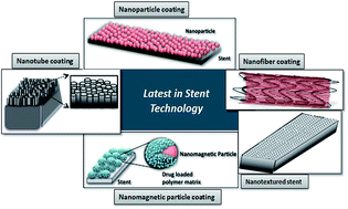
8.1.5. Magnetic nanoparticle-based coatings. Magnetic nanoparticles have emerged as an “on-demand approach” for highly controlled drug delivery.237 These nanoparticles consist of magnetic elements which exhibit superparamagnetism such as iron, cobalt and nickel and their derivatives.238 Under an external magnetic field gradient, they release the therapeutic agents for site-specific and stimuli-responsive therapy.239 Such systems have also been explored in stent technology in recent years. An in vitro study conducted by B. Polyak and M. Chorny clearly indicated the biocompatibility of magnetic nanoparticle coating on the stent surface under a homogenous magnetic field.240,241 Biophan technology has developed novel magnetic nanoparticle-based stenting technologies that provide a higher degree of controlled and targeted drug delivery.242 Nanomagnetic drug eluting stents represent a radical shift from the current passive approach in drug eluting stents to an active drug delivery system that releases specified amounts of the drug in response to an external electromagnetic stimulus.241 This gives the opportunity to control the release of the anti-proliferative drug, constant release of which in the later stages is a potential reason of late-stent thrombosis. Magnetic mesoporous nanocomposite has also been recently explored as stent coatings due to their high surface area, porous nature, biocompatibility and better endothelialzation.228 Yet another exciting application for this technology is that nanoparticles can be tuned to respond selectively to different wavelengths, thus enabling multiple drugs to be delivered at different times, controlled by unique electromagnetic signals. This multiple drug coating can be used to treat different complications simultaneously with specific electromagnetic signals for each drug.242 However, this concept is yet to be validated through experiments. Fig. 8 depicts the various nanotechnology-enable concepts in stents that have evolved in recent years.
 |
| Fig. 8 Emerging nanotechnology enabled-stent strategies. | |
It is evident from the above discussion that stents have evolved from being mere structural supports to functional materials that can aid better healing of the diseased region. However, though newer strategies have been developed for stent-based interventions, these have also given rise to new complications that need to be addressed. It is heartening to note that the novel stenting options have resulted in better survival and clinical outcomes than their predecessors. But further efforts are required to mitigate some of the complications associated with these modern stents. Fig. 9 depicts the evolution of stenting technologies over the decades along with their associated complications.
 |
| Fig. 9 Pictorial presentation of stent evolution. DES: drug eluting stent, EPC: endothelial progenitor cells, PEVA/PMBA: polyethylene-co-vinyl acetate/poly-n-butyl methacrylate, PC: phosphorylcholine, SIBS: poly(styrene-b-isobutylene-b-styrene), PLA: poly lactic acid, PLGA: poly(lactic-co-glycolic acid), PCL: polycaprolactone, PLLA: poly L-lactide acid. | |
9. Conclusions
The evolution of stent technology was marred by the occurrence of restenosis in bare metal stents. Drug eluting stents solved the problem of restenosis, but at the cost of increased risk of stent thrombosis. The dual issues of stenosis due to intimal proliferation and stent thrombosis due to delayed endothelialisation, remains the Achilles heel of this form of interventional therapy. Stents have evolved from being mere supporting frameworks to multi-functional devices that also attenuate the complications arising due to stent deployment. Novel strategies in metallurgy, instrumentation, drug molecules and polymer technology has made stents thinner, stronger and polymer free, to enhance endothelialization while inhibiting smooth cell proliferation. Exciting new stents like immunostents and gene eluting stents are undergoing preclinical testing with promising results. A wider repertoire of stent materials, polymers and therapeutic molecules has catalyzed the development of the next generation stents that promise longevity, better therapeutic efficacy and lesser post-implantation complications. Nanotechnology-enabled interventions promise to further improve the stent performances though more in-depth studies and clinical trials are required in this direction to evaluate their efficacy. Coating techniques employed to modify the stent surface with polymer and drugs also can influence the stent performance. Thus identifying the ideal combination of material, therapeutic molecules, stent design and fabrication method is the need of the hour to establish the supremacy of stents for treatment of cardiovascular disorders.
Acknowledgements
The authors wish to acknowledge funding from the Department of Science & Technology under the FIST programme (SR/FST/LSI-453/2010) and infrastructural support from SASTRA University. PR gratefully acknowledges the financial assistance from SASTRA University under the TA fund.
References
- E. Falk, P. K. Shah and V. Fuster, Circulation, 1995, 92, 657–671 CrossRef CAS PubMed
. - G. Mani, M. D. Feldman, D. Patel and C. M. Agrawal, Biomaterials, 2007, 28, 1689–1710 CrossRef CAS PubMed
. - V. L. Roger, A. S. Go, D. M. Lloyd-Jones, E. J. Benjamin, J. D. Berry, W. B. Borden, D. M. Bravata, S. Dai, E. S. Ford, C. S. Fox, H. J. Fullerton, C. Gillespie, S. M. Hailpern, J. A. Heit, V. J. Howard, B. M. Kissela, S. J. Kittner, D. T. Lackland, J. H. Lichtman, L. D. Lisabeth, D. M. Makuc, G. M. Marcus, A. Marelli, D. B. Matchar and C. S. Moy, Circulation, 2012, 125, 2–220 CrossRef
. - B. Lagerqvis, K. Säfström, E. Ståhle, L. Wallentin and E. Swahn, J. Am. Coll. Cardiol., 2001, 38, 41–48 CrossRef
. - R. L. Mueller and T. A. Sanborn, Am. Heart J., 1995, 129, 146–172 CrossRef CAS PubMed
. - L. A. Alicea, J. I. Aviles, I. A. Lopez, L. E. Mulero and L. A. Sanchez, Applications of Engineering Mechanics in Medicine, GED–University of Puerto Rico, Mayaguez, 2004, p. F1 Search PubMed
. - F. Amin, M. N. Ali and M. A. Minhas, Int. J. Biomed. Adv. Res., 2013, 4, 764–775 CrossRef
. - D. L. Fischman, M. B. Leon, D. S. Baim, R. A. Schatz, M. P. Savage, I. Penn, K. Detre, L. Veltri, D. Ricci, M. Nobuyoshi, M. Cleman, R. Heuser, D. Almond, P. S. Teirstein, R. D. Fish, A. Colombo, J. Brinker, J. Moses, A. Shaknovich, J. Hirshfeld, S. Bailey, S. Ellis, R. Rake and S. Goldberg, N. Engl. J. Med., 1994, 331, 496–501 CrossRef CAS PubMed
. - L. Denes, L. Entz and V. Jancsik, Int. J. Vasc. Med., 2012, 2012, 9 Search PubMed
. - R. Waksman, Cardiovasc. Radiat. Med., 2002, 3, 226–241 CrossRef PubMed
. - Textbook of Interventional Cardiovascular Pharmacology, ed. N. Kipshidze, J. Fareed, P. W. Serruys and J. Mose, 2007, p. 291, informa healthcare Search PubMed
. - A. M. Salam, J. A. Suwaidi and D. R. Holmes, Curr. Probl. Cardiol., 2006, 31, 8–119 CrossRef PubMed
. - G. S. Mintz, Circulation, 2007, 115, 2379–2381 CrossRef PubMed
. - Heart stents, Appropedia 2012, http://www.appropedia.org/Heart_stents.
- B. A. L. Mangour, R. Mongrain and S. Yue, Mater. Sci. Appl., 2013, 4, 606–621 Search PubMed
. - P. Poncin and J. Proft, Material and processes for medical devices conference, 8–10 September, 2003 Search PubMed
. - M. Moravej and D. Mantovani, Int. J. Mol. Sci., 2011, 12, 4250–4270 CrossRef CAS PubMed
. - K. Milewski, A. Zurakowski, J. Pajak, E. Pajak-Zielinska, L. Liszka and P. P. Buszman, et al., Med. Sci. Monit., 2010, 16(1), BR40–BR44 Search PubMed
. - O. F. Bertrand, R. Sipehia, R. Mongrain, J. Rodes, J. C. Tardif and L. Bilodeau, et al., J. Am. Coll. Cardiol., 1998, 32, 562–571 CrossRef CAS PubMed
. - A. Klocke, J. Kemper, D. Schulze, G. Adam and B. Kahl-Nieke, J. Orofac. Orthop., 2005, 66(4), 279–287 CrossRef PubMed
. - N. Gilbert, Platinum in Biomedical Applications. AZO Materials, July 5th 2015 Search PubMed
. - B. Bhargava, I. De Scheerder, Q. B. Ping, H. Yanming, R. Chan and H. Soo Kim, et al., Cathet. Cardiovasc. Interv., 2000, 51(3), 364–368 CrossRef CAS PubMed
. - H. Hermawan, D. Ramdan and J. R. P. Djuansjah, Metals for Biomedical Applications (book) Biomedical Engineering–From Theory to Applications chapter 17, Intech, 2011 Search PubMed
. - T. Hanawa, J. Artif. Organs, 2009, 12(2), 73–79, DOI:10.1007/s10047-008-0456-x
, Epub 2009 Jun 18. - D. Stoeckel, Minimally Invasive Therapy & Allied Technologies, 2000, 9(2), 81–88 Search PubMed
. - M. Azaouzi, N. Lebaal, A. Makradi and S. Belouettar, Mater. Des., 2013, 50, 917–928 CrossRef CAS
. - Nitinol: The shape of things to come, http://www.medicaldevicenetwork.com/features/feature116/1March2006TomDuerigMedicinaldevices-network.com.
- C. G. Paganias, G. A. Tsakotos, S. D. Koutsostathis and G. A. Macheras, Indian J. Orthop., 2012, 46, 505–513 CrossRef PubMed
. - V. Burt, Worth Their Weight: Precious metals have ideal properties for medical devices micro manufacturing, 2011, p. 4, http://www.micromanufacturing.com/content/worth-their-weight-precious-metals-have-ideal-properties-medical-devices Search PubMed
. - Gold-Applications and Developments in the Electronics, Biomaterials and Catalysis, AZO Materials, 2015, http://www.azom.com/article.aspx?ArticleID=1899.
- R. J. Harling, Biocompatibility of Tantalum, 2002, http://www.x-medics.com/tantalum_biocompatibility.htm Search PubMed
. - H. Zitter and H. Plenk, J. Biomed. Mater. Res., 1987, 21, 881–896 CrossRef CAS PubMed
. - E. R. Edelman, P. Seifert, A. Groothuis, A. Morss, D. Bornstein and C. Rogers, Circulation, 2001, 103, 429–434 CrossRef CAS PubMed
. - A. Kastrati, A. Schömig, J. Dirschinger, J. Mehilli, N. V. Welser, J. Pache, H. Schühlen, T. Schilling, C. Schmitt and F. J. Neumann, Circulation, 2000, 101, 2478–2483 CrossRef CAS PubMed
. - C. Jorge and C. Dubois, Med. Devices: Evidence Res., 2015, 8, 359–367 Search PubMed
. - B. Peters, P. Ewert and F. Berger, Ann. Pediatr. Cardiol., 2009, 2, 3–23 CrossRef PubMed
. - Y. Farhatnia, A. Tan, A. Motiwala, B. G. Cousins and A. M. Seifalian, Biotechnol. Adv., 2013, 31, 524–542 CrossRef PubMed
. - Y. Zhu, C. Hu, B. Li, H. Yang, Y. Cheng and W. Cui, Acta Biomater., 2013, 9, 8328–8336 CrossRef CAS PubMed
. - T. H. Baron, Curr. Opin. Gastroenterol., 2011, 27, 262–267 CrossRef CAS PubMed
. - ASGE Technology Committee, S. Varadarajulu, S. Banerjee, B. Barth, D. Desilets, V. Kaul, S. Kethu, M. Pedrosa, P. Pfau, J. Tokar, A. Wang, L. M. W. K. Song and S. Rodriguez, Gastrointest. Endosc., 2011, 74, 455 CrossRef PubMed
. - I. C. T. Santos, A. Rodrigues, L. Figueiredo, L. A. Rocha and J. M. R. S. Tavares, Proc. Inst. Mech. Eng., Part L, 2012, 226, 330–341 CAS
. - L. Pinchuk, G. J. Wilson, J. J. Barry, R. T. Schoephoerster, J. M. Parel and J. P. Kennedy, Biomaterials, 2008, 29(4), 448–460 CrossRef CAS PubMed
. - Natural and Synthetic Biomedical Polymers, ed. S. Kumbar, C. Laurencin and M. Deng, Elsevier, 2014, p. 302 Search PubMed
. - S. Venkatraman, F. Boey and L. L. Lao, Prog. Polym. Sci., 2008, 33, 853–874 CrossRef CAS
. - M. C. Chen, F. L. Mi, Z. X. Liao and H. W. Sung, Adv. Polym. Sci., 2011, 243, 185–230 CrossRef CAS
. - N. Nagai, R. Kubota, R. Okahashi and M. Munekata, Mechanical and Biological Properties of Bio-Inspired Nano-Fibrous Elastic Materials from Collagen, Intech, 2011, ch. 13 Search PubMed
. - M. Pavlides and D. A. Gorard, Stents in Gastrointestinal Endoscopy, Intech, 2011, ch. 8 Search PubMed
. - P. Hindy, J. Hong, Y. L. Tsai and F. Gress, Gastroenterol. Hepatol., 2012, 8, 526–534 Search PubMed
. - American Society for Gastrointestinal Endoscopy (ASGE), Pancreatic and Biliary stents: Technology status evaluation report, Gastrointest. Endosc., 2013, 77, 319–327 CrossRef PubMed
. - D. Maras, C. Lioupis, G. Magoufis, N. Tsamopoulos, K. Moulakakis and V. Andrikopoulos, Cardiovasc. Intervent. Radiol., 2006, 29, 958–968 CrossRef PubMed
. - C. K. Zarins, F. R. Arko, T. Crabtree, D. A. Bloch, K. Ouriel, R. C. Allen and R. A. White, J. Vasc. Surg., 2004, 40, 1–11 CrossRef PubMed
. - J. N. Albertini, M. A. DeMasi, J. Macierewicz, R. E. I. Idrissi, B. R. Hopkinson and C. Clément, Vascular, 2005, 13, 321–326 CrossRef PubMed
. - A. Abizaid and J. R. Costa, Circ.: Cardiovasc. Interventions, 2010, 3, 384–393 CrossRef CAS PubMed
. - J. F. Granada, M. J. Price, P. A. French, S. R. Steinhubl, D. E. Cutlip, R. C. Becker, S. S. Smyth and H. L. Dauerman, Circ.: Cardiovasc. Interventions, 2011, 4, 629–637 CrossRef CAS PubMed
. - M. Joner, A. V. Finn, A. Farb, E. K. Mont, F. D. Kolodgie, E. Ladich, R. Kutys, K. Skorija, H. K. Gold and R. Virmani, J. Am. Coll. Cardiol., 2006, 48, 203–205 CrossRef PubMed
. - T. F. Lüscher, J. Steffel and F. R. Eberli, Circulation, 2007, 115, 1051–1058 CrossRef PubMed
. - M. Cassagnol and M. Saad, Drug-eluting Stents, US Pharm., 2012, p. 37 Search PubMed
. - P. Wu and D. W. Grainger, Biomaterials, 2006, 27, 2450–2467 CrossRef CAS PubMed
. - B. L. van der Hoeven, N. M. M. Pires, H. M. Warda, P. V. Oemrawsingh and B. J. M. van Vlijmen, Int. J. Cardiol., 2005, 99, 9–17 CrossRef PubMed
. - A. Colombo, G. Stankovic and J. W. Moses, J. Am. Coll. Cardiol., 2002, 40, 1021–1033 CrossRef PubMed
. - K. McKeage, D. Murdoch and K. L. Goa, Am. J. Cardiovasc. Drugs, 2003, 3, 211–230 CrossRef PubMed
. - G. I. Kirchner, I. Meier-Wiedenbach and M. P. Manns, Clin. Pharmacokinet., 2004, 43, 83–95 CrossRef CAS PubMed
. - M. Zeier and M. van Der Giet, Transpl. Int., 2011, 24, 30–42 CrossRef CAS PubMed
. - E. O. de Abreu-Silva, R. A. Costa, A. Abizaid, A. Ramondo, P. Brenot, H. Benamer, A. Desideri, J. Berland, B. O. Almeida, F. Digne, M. A. Perin, J. P. de Castro, J. R. Costa, R. Staico, L. F. Tanajura, A. Abizaid and PAX-B study Investigators, Rev. Bras. Cardiol. Invasiva, 2012, 20, 146–154 CrossRef
. - N. Tada, R. Virmani, G. Grant, L. Bartlett, A. Black, C. Clavijo, U. Christians, R. Betts, D. Savage, S. H. Su, J. Shulze and S. Kar, Circ.: Cardiovasc. Interventions, 2010, 3, 174–183 CrossRef CAS PubMed
. - J. Matthew, Coronary Stenting: A Companion to Topol's Textbook of Interventional Cardiology, Elsevier Saunders, 2013, ch. 8 Search PubMed
. - K. Tanabe, Drugs, 2003, 63, 1535–1548 CrossRef CAS PubMed
. - J. M. Venstrom and J. W. Young, Essential Clinical Immunology, ed. J. B. Zabriskie, The Rockefeller University, Cambridge University Press, 2009, ch. 18 Search PubMed
. - Elixir Medical Corporation, Drug Catalog, http://elixirmedical.com/index.php?page=ous-drug-catalog.
- Elixir Medical Corporation, Novolimus, http://elixirmedical.com/index.php?page=ous-novolimus.
- J. Y. Cho, Y. Ahn and M. H. Jeong, Korean Circ. J., 2013, 43, 645–654 CrossRef PubMed
. - S. E. Burke, R. E. Kuntz and L. B. Schwartz, Adv. Drug Delivery Rev., 2006, 58, 437–446 CrossRef CAS PubMed
. - C. E. Cox, Resolute Zotarolimus-Eluting Stent Noninferior to Xience V, 2010, The Source for interventional cardiovascular news and education, http://stage.tctmd.com/txshow.aspx?tid=1000742%<?pdb_no 26id?>26id<?pdb END?>=90184.
- W. Rutsch, B. Witzenbichler and H. Schuhlen, Multicentre first-in-man study with the lowest known limus dose on the Elixir medical myolimus™ eluting coronary stent system with a durable polymer: 12-month clinical and six month angiographic and IVuS follow-up. Proceedings of Euro PCR, 2009, http://elixirmedical.com/index.php?page=ouos-myolimus Search PubMed
. - S. Verheye, M. Webster, J. Stewart, A. Abizaid, R. Costa, J. R. Costa, J. Yan, V. Bhat, S. Toyloy, L. Morrison and J. Ormiston, JACC: Cardiovasc. Interv., 2013, 6, S1–S62 Search PubMed
. - EuroIntervention 2013, Acute scaffold recoil with the novel myolimus-eluting bioresorbable vascular scaffold in the treatment of de novo, non-complex coronary lesions: a subanalysis of the DESolve first-in-man trial, http://www.pcronline.com/eurointervention/Abstracts2013_online_issue/39/.
- T. Meštrović, What is Paclitaxel? News medical 2014, http://www.news-medical.net/health/What-is-Paclitaxel.aspx.
- D. I. Axel, W. Kunert, C. Göggelmann, M. Oberhoff, C. Herdeg, A. Küttner, D. H. Wild, B. R. Brehm, R. Riessen, G. Köveker and K. R. Karsch, Circulation, 1997, 96, 636–645 CrossRef CAS PubMed
. - L. K. Pendyala, J. Li, T. Shinke, S. Geva, X. Yin, J. P. Chen, S. B. King, K. A. Robinson, N. A. Chronos and D. Hou, JACC: Cardiovasc. Interv., 2009, 2, 253–262 CrossRef PubMed
. - G. Nakazawa, A. V. Finn, M. Vorpahl, E. R. Ladich, F. D. Kolodgie and R. Virmani, J. Am. Coll. Cardiol., 2011, 57, 390–398 CrossRef CAS PubMed
. - S. Mishra and K. Palanivelu, Ann. Indian Acad. Neurol., 2008, 11, 13–19 CrossRef PubMed
. - K. T. Nguyen, N. Shaikh, K. P. Shukla, S. H. Su, R. C. Eberhart and L. Tang, Biomaterials, 2004, 25, 5333–5346 CrossRef CAS PubMed
. - R. Srivastava, V. Puri, R. C. Srimal and B. N. Dhawan, Arzneim. Forsch., 1986, 36, 715–717 CAS
. - H. Emms and G. P. Lewis, Br. J. Pharmacol., 1986, 87, 109–115 CrossRef CAS PubMed
. - B. H. Shah, Z. Nawaz, S. A. Pertani, A. Roomi, H. Mahmood, S. A. Saeed and A. H. Gilani, Biochem. Pharmacol., 1999, 58, 1167–1172 CrossRef CAS PubMed
. - H. S. Jang, H. Y. Nam, J. M. Kim, D. H. Hahm, S. H. Nam, K. L. Kim, J. R. Joo, W. Suh, J. S. Park, D. K. Kim and H. C. Gwon, Cathet. Cardiovasc. Interv., 2009, 74, 881–888 CrossRef PubMed
. - C. J. Pan, J. J. Tang, Y. J. Weng, J. Wang and N. Huang, J. Controlled Release, 2006, 116, 42–49 CrossRef CAS PubMed
. - H. Shapiro, P. Singer, Z. Halpern and R. Bruck, Gut, 2007, 56, 426–435 CrossRef CAS PubMed
. - D. E. Golan, A. H. Tashjian and E. J. Armstrong, Principles of Pharmacology: The Pathophysiologic Basis of Drug Therapy, Lippincott Williams and Wilkins, Philadelphia, 3rd edn, 2011, ch. 1 Search PubMed
. - Y. M. Yang, X. X. Wang, J. Z. Chen, S. J. Wang, H. Hu and H. Q. Wang, Am. J. Chin. Med., 2008, 36, 603–613 CrossRef CAS PubMed
. - B. Poussier, A. C. Cordova, J. P. Becquemin and B. E. Sumpio, J. Vasc. Surg., 2005, 42, 1190–1197 CrossRef PubMed
. - A. R. Khandelwal, V. Y. Hebert, J. J. Kleinedler, L. K. Rogers, S. L. Ullevig, R. Asmis, R. Shi and T. R. Dugas, J. Nutr., 2012, 142, 1487–1494 CrossRef CAS PubMed
. - A. Garcia-Lafuente, E. Guillamón, A. Villares, M. A. Rostagno and J. A. Martínez, Inflammation Res., 2009, 58, 537–552 CrossRef CAS PubMed
. - J. J. Kleinedler, J. D. Foley, E. A. Orchard and T. R. Dugas, J. Controlled Release, 2012, 159, 27–33 CrossRef CAS PubMed
. - J. Goepp, Life Extension Magazine April 2009 Report. Quercetin Enhanced Antioxidant Protection Against Heart Disease, Cancer, Allergies, and More. http://www.lifeextension.com/magazine/2009/4/quercetin/page-02.
- J. Zhang, Y. Liu, R. Luo, S. Chen, X. Li, S. Yuan, J. Wang and N. Huang, Appl. Surf. Sci., 2015, 328, 154–162 CrossRef CAS
. - M. Hamalainen, R. Nieminen, I. Uurto, J. P. Salenius, M. Kellomaki, J. Mikkonen, A. Kotsar, T. Isotalo, T. Tammela LJ, M. Talja and E. Moilanen, Basic Clin. Pharmacol. Toxicol., 2013, 112, 296–301 CrossRef CAS PubMed
. - S. N. Kang, S. E. Kim, J. Choi, K. Park, J. H. Goo, D. S. Sim, Y. J. Hong, J. H. Kim, Y. K. Joung, J. Lee, M. H. Jeong and D. K. Han, Biomaterials, 2015, 44, 1–10 CrossRef CAS PubMed
. - L. Meng, D. Yan, W. Xu, J. Ma, B. Chen and H. Feng, Int. J. Mol. Med., 2012, 29, 999–1006 CAS
. - S. Jin, JSM Biotechnology & Biomedical Engineering, 2013, 1(1002), 1/7–7/7 Search PubMed
. - Y. K. Lee, J. H. Park, H. T. Moon, D. Y. Lee, J. H. Yun and Y. Byun, Biomaterials, 2007, 28, 1523–1530 CrossRef CAS PubMed
. - M. Inoue, M. Sasaki, Y. Katada, K. Fujiu, I. Manabe, R. Nagai and T. Taguchi, J. Biomed. Mater. Res., Part A, 2013, 101, 2049–2057 CrossRef PubMed
. - L. L. Kunz, L. M. Tatalick, P. G. Anderson, R. W. Schroff and G. S. Roubin, J. Am. Coll. Cardiol., 1995, 25, 302A CrossRef
(abstract). - T. Deuse, R. G. Erben, F. Ikeno, B. Behnisch, R. Boeger, A. J. Connolly, H. Reichenspurner, C. Bergow, M. P. Pelletier, R. C. Robbins and S. Schrepfer, Atherosclerosis, 2008, 200, 126–134 CrossRef CAS PubMed
. - F. Violi, C. Calvieri, D. Ferro and P. Pignatelli, Circulation, 2013, 127, 251–257 CrossRef PubMed
. - S. Venkatraman, F. Boey and L. L. Lao, Prog. Polym. Sci., 2008, 33, 853–874 CrossRef CAS
. - P. Agrawal, J. Pharmacovigilance, 2014, 3, e127 Search PubMed
. - G. G. Stefanini, R. A. Byrne, P. W. Serruys, A. de Waha, B. Meier, S. Massberg, P. Jüni, A. Schömig, S. Windecker and A. Kastrati, Eur. Heart J., 2012, 33, 1214–1222 CrossRef CAS PubMed
. - B. Doyle and D. R. Holmes, Med. Devices: Evidence Res., 2009, 2, 47–55 CAS
. - Y. Fu and W. J. Kao, Expert Opin. Drug Delivery, 2010, 7, 429–444 CrossRef CAS PubMed
. - A. V. Finn, G. Nakazawa, M. Joner, F. D. Kolodgie, E. K. Mont, H. K. Gold and R. Virmani, Arterioscler., Thromb., Vasc. Biol., 2007, 27, 1500–1510 CrossRef CAS PubMed
. - J. Lv, Y. Wu, X. Zhang, T. Jing, L. Zhang, S. Tong, Z. Song, M. Wang, G. Wang and L. Chi, Eur. J. Med. Res., 2015, 20, 21 CrossRef PubMed
. - M. Vorpahl, A. Finn, M. Nakano and R. Virmani, EuroIntervention, 2009, 5, F28–F35 CrossRef PubMed
. - S. H. Chaudhary, V. Arora, K. Kholi and V. Kumar, Polym. Rev., 2013, 53, 546–567 CrossRef
. - Polymeric Biomaterials, Revised and Expanded, ed. S. Dumitriu, 2nd edn, 2002, p. 95 Search PubMed
. - L. Barghi, D. Asgari, J. Barar and H. Valizadeh, Adv. Pharm. Bull., 2015, 5(1), 51–56 Search PubMed
. - Z. Pan and J. Ding, Interface Focus, 2012, 2, 366–377 CrossRef PubMed
. - R. Jabara, N. Chronos, D. Conway, W. Molema and K. Robinson, JACC: Cardiovasc. Interv., 2008, 1, 81–87 CrossRef PubMed
. - Biomaterials Science: An Introduction to Materials in Medicine, ed. B. D. Ratner, 2nd edn, 2004 Search PubMed
. - M. Zilberman and R. C. Eberhart, Annu. Rev. Biomed. Eng., 2006, 8, 153–180 CrossRef CAS PubMed
. - M. A. Woodruff and D. W. Hutmacher, Prog. Polym. Sci., 2010, 35, 1217–1256 CrossRef CAS
. - J. Fernández, A. Etxeberria, J. M. Ugartemendia, S. Petisco and J. R. Sarasua, J. Mech. Behav. Biomed. Mater., 2012, 12, 29–38 CrossRef PubMed
. - S. I. Jeong, B. S. Kim, S. W. Kang, J. H. Kwon, Y. M. Lee, S. H. Kim and Y. H. Kim, Biomaterials, 2004, 25, 5939–5946 CrossRef CAS PubMed
. - J. Fernández, A. Etxeberria and J. R. Sarasua, J. Mech. Behav. Biomed. Mater., 2012, 9, 100–112 CrossRef PubMed
. - H. Geckil, F. Xu, X. Zhang, S. J. Moon and U. Demirci, Nanomedicine, 2010, 5, 469–484 CrossRef CAS PubMed
. - I. Gibas and H. Janik, Chem. Chem. Technol., 2010, 4 Search PubMed
. - B. Semmling, S. Nagel, K. Sternberg, W. Weitschies and A. Seidlitz, J. Pharm. Technol. Drug Res., 2013, 2 Search PubMed
. - T. R. Hoare and D. S. Kohane, Polymer, 2008, 49, 1993–2007 CrossRef CAS
. - M. C. Chen, H. W. Tsai, C. T. Liu, S. F. Peng, W. Y. Lai, S. J. Chen, Y. Chang and H. W. Sung, Biomaterials, 2009, 30, 2102–2111 CrossRef CAS PubMed
. - W. W. Yang and E. Pierstorff, J. Lab. Autom., 2012, 17, 50–58 CrossRef CAS PubMed
. - S. Park, S. H. Bhang, W. G. La, J. Seo, B. S. Kim and K. Char, Biomaterials, 2012, 33, 5468–5477 CrossRef CAS PubMed
. - L. Indolfi, F. Causa, C. Giovino, F. Ungaro, F. Quaglia and P. A. Netti, J. Biomed. Mater. Res., Part A, 2011, 97(2), 201–211 CrossRef PubMed
. - B. Butruk, M. Trzaskowski and T. Ciach, Mater. Sci. Eng., C, 2012, 32, 1601–1609 CrossRef CAS PubMed
. - A. Atala, R. Lanza and R. Nerem and J. A. Thomson, Principles of Regenerative Medicine, 2008, pp. 594–603 Search PubMed
. - R. Waksman, J. Invasive Cardiol., 2006, 18, 70–74 Search PubMed
. - J. Ferdous, V. B. Kolachalama and T. Shazly, Acta Biomater., 2013, 9, 6052–6061 CrossRef CAS PubMed
. - Y. Onuma and P. W. Serruys, Circulation, 2011, 123, 779 CrossRef PubMed
. - C. V. Bourantas, Y. Zhang, V. Farooq, H. M. Garcia-Garcia, Y. Onuma and P. W. Serruys, Curr. Cardiol. Rep., 2012, 14, 626–634 CrossRef PubMed
. - Elixir Medical Corporation, January 15, 2014, Elixir Medical Announces First Commercial Implant Of The DESolve® Novolimus Eluting Coronary Scaffold System in Europe, http://elixirmedical.com/index.php?mact=News,cntnt01,detail,0%26cntnt01articleid=25%26cntnt01origid=101%26cntnt01returnid=110.
- http://elixirmedical.com/index.php%3Fpage=ous-desolve.
- R. C. Eberhart, S. H. Su, K. T. Nguyen, M. Zilberman, L. Tang, K. D. Nelson and P. Frenkel, J. Biomater. Sci., Polym. Ed., 2003, 14, 299–312 CrossRef CAS PubMed
. - J. A. Ormiston and P. W. Serruys, Circ.: Cardiovasc. Interventions, 2009, 2, 255–260 CrossRef CAS PubMed
. - C. M. Campos, T. Muramatsu, J. Iqbal, Y. J. Zhang, Y. Onuma, H. M. G. Garcia, M. Haude, P. A. Lemos, B. Warnack and P. W. Serruys, Int. J. Mol. Sci., 2013, 14, 24492–24500 CrossRef PubMed
. - E. J. Topol and P. S. Teirstein, Textbook of Interventional Cardiology, Elsevier PA, 6th edn, 2012, pp. 171–196 Search PubMed
. - J. Reifenrath, D. Bormann and A. M. Lindenberg, Magnesium Alloys as Promising Degradable Implant Materials in Orthopaedic. Chapter 6 Magnesium Alloys-Corrosion and Surface Treatments, 2011 Search PubMed
. - B. Heublein, R. Rohde, V. Kaese, M. Niemeyer, W. Hartung and A. Haverich, Heart, 2003, 89, 651–656 CrossRef CAS PubMed
. - M. Moravej and D. Mantovani, Int. J. Mol. Sci., 2011, 12, 4250–4270 CrossRef CAS PubMed
. - J. Daemen and P. W. Serruys, Circulation, 2007, 116, 316–328 CrossRef PubMed
. - C. D. Mario, H. Griffiths, O. Goktekin, N. Peeters, J. Verbist, M. Bosiers, K. Deloose, B. Heublein, R. Rohde, V. Kasese, C. Ilsley and R. Erbel, J. Intervent. Cardiol., 2004, 17, 391–395 CrossRef PubMed
. - R. Erbel, C. Di Mario, J. Bartunek, J. Bonnier, B. de Bruyne, F. R. Eberli, P. Erne, M. Haude, B. Heublein, M. Horrigan, C. Ilsley, D. Böse, J. Koolen, T. F. Lüscher, N. Weissman and R. Waksman, Lancet, 2007, 369, 1869–1875 CrossRef CAS
. - E. Marin, M. I. Briceño and C. C. George, Int. J. Nanomed., 2013, 8, 3071–3091 Search PubMed
. - D. M. Martin and F. J. Boyle, Med. Eng. Phys., 2011, 33, 148–163 CrossRef PubMed
. - H. Takahashi, D. Letourneur and D. W. Grainger, Biomacromolecules, 2007, 8, 3281–3293 CrossRef CAS PubMed
. - G. J. Padfield, D. E. Newby and N. L. Mills, J. Am. Coll. Cardiol., 2010, 55, 1553–1565 CrossRef PubMed
. - J. Aoki, P. W. Serruys, H. van Beusekom, A. T. L. Ong, E. P. McFadden, G. Sianos, W. J. van der Giessen, E. Regar, P. J. de Feyter, H. R. Davis, S. Rowland and M. J. B. Kutryk, J. Am. Coll. Cardiol., 2005, 45, 1574–1579 CrossRef CAS PubMed
. - F. Timmermans, J. Plum, M. C. Yöder, D. A. Ingram, B. Vandekerckhove and J. Case, J. Cell. Mol. Med., 2009, 13, 87–102 CrossRef PubMed
. - E. Dejana, M. G. Lampugnani, O. Martinez-Estrada and G. Bazzoni, Int. J. Dev. Biol., 2000, 44, 743–748 CAS
. - C. K. Kang, W. H. Lim, S. Kyeong, W. S. Choe, H. S. Kim, B. H. Jun and Y. S. Lee, Colloids Surf., B, 2013, 102, 744–751 CrossRef CAS
. - O. K. Lin, Y. Hou, K. F. Ren and J. Ji, Thin Solid Films, 2012, 520, 4971–4978 CrossRef
. - Q. Lin, X. Ding, F. Qiu, X. Song, G. Fu and J. Ji, Biomaterials, 2010, 31, 4017–4025 CrossRef CAS PubMed
. - J. Aoki, P. W. Serruys, H. V. Beusekom, A. T. L. Ong, E. P. McFadden, G. Sianos, W. J. van der Giessen, E. Regar, P. J. de Feyter, H. R. Davis, S. Rowland and M. J. Kutryk, J. Am. Coll. Cardiol., 2005, 45, 1574–1579 CrossRef CAS PubMed
. - A. J. Boyle, S. P. Schulman and J. M. Hare, Circulation, 2006, 114, 353–358 CrossRef PubMed
. - J. M. Lee, W. S. Choe, W. W. Seo, W. H. Lim, C. K. Kang, S. Kyeong, H. J. Cho, B. K. Kim, Y. C. Kim, J. Hur, H. M. Yang, K. W. Park, H. J. Cho, Y. S. Lee and H. S. Kim, Abstract 9971, Circulation, 2012, 126 Search PubMed
. - X. D. Jing, X. M. Wei, S. B. Deng, J. L. Du, Y. J. Liu and Q. She, Clin. Chim. Acta, 2015, 446, 248–252 CrossRef CAS PubMed
. - Z. Yang, Y. Yang, K. Xiong, X. Li, P. Qi, Q. Tu, F. Jing, Y. Weng, J. Wang and N. Huang, Biomaterials, 2015, 63, 80e92 Search PubMed
. - Y. Weng, J. Chen, Q. Tu, Q. Li, M. F. Maitz and N. Huang, Interface Focus, 2012, 2, 356–365 CrossRef PubMed
. - H. Xu, K. T. Nguyen, E. S. Brilakis, J. Yang, E. Fuh and S. Banerjee, Journal of Cardiovascular Translational Research, 2012, 5, 519–527 CrossRef PubMed
. - F. Sharif, S. O. Hynes, R. Cooney, L. Howard, J. McMahon, K. Daly, J. Crowley, F. Barry and T. O'Brien, Mol. Ther., 2008, 16, 1674–1680 CrossRef CAS PubMed
. - F. Sharif, K. Daly, J. Crowley and T. O'Brien, Cardiovasc. Res., 2004, 64, 208–216 CrossRef CAS PubMed
. - D. Goh, A. Tan, Y. Farhatnia, J. Rajadas, M. S. Alavijeh and A. M. Seifalian, Mol. Pharmaceutics, 2013, 10, 1279–1298 CrossRef CAS PubMed
. - R. X. Yin, D. Z. Yang and J. Z. Wu, Theranostics, 2014, 4, 175–178 CrossRef CAS PubMed
. - M. J. Mann and V. J. Dzau, J. Clin. Invest., 2000, 106, 1071–1075 CrossRef CAS PubMed
. - A. J. Roks, R. H. Henning, H. Buikema, Y. M. Pinto, M. J. Kraak, R. A. Tio, D. de Zeeuw, H. J. Haisma, J. Wilschut and W. H. van Gilst, Gene Ther., 2002, 9, 95–101 CrossRef CAS PubMed
. - I. Masaki, Y. Yonemitsu, K. Komori, H. Ueno, Y. Nakashima, K. Nakagawa, M. Fukumura, A. Kato, M. K. Hasan, Y. Nagai, K. Sugimachi, M. Hasegawa and K. Sueishi, FASEB J., 2001, 15, 1294–1296 CAS
. - A. Paul, C. B. Elias, D. Shum-Tim and S. Prakash, Sci. Rep., 2013, 3, 2366 Search PubMed
. - P. D. Williams, P. Ranjzad, S. J. Kakar and P. A. Kingston, Viruses, 2010, 2, 334–371 CrossRef CAS PubMed
. - Y. Takemoto, H. Kawata, T. Soeda, K. Imagawa, S. Somekawa, Y. Takeda, S. Uemura, M. Matsumoto, Y. Fujimura, J. Jo, Y. Kimura, Y. Tabata and Y. Saito, Arterioscler., Thromb., Vasc. Biol., 2009, 29, 857–862 CrossRef CAS PubMed
. - S. Ganly, S. O. Hynes, F. Sharif, A. Aied, V. Barron, K. McCullagh, J. McMahon, P. McHugh, J. Crowley, W. Wang, T. O'Brien and U. Greiser, J. Controlled Release, 2013, 167, 109–119 CrossRef CAS PubMed
. - D. Zhu, X. Jin, X. Leng, H. Wang, J. Bao, W. Liu, K. Yao and C. Song, Int. J. Nanomed., 2010, 5, 1095–1102 CrossRef CAS PubMed
. - I. Fishbein, I. S. Alferiev, O. Nyanguile, R. Gaster, J. M. Vohs, G. S. Wong, H. Felderman, I. W. Chen, H. Choi, R. L. Wilensky and R. J. Levy, Proc. Natl. Acad. Sci. U. S. A., 2006, 103, 159–164 CrossRef CAS PubMed
. - T. S. Levchenko, W. C. Hartner and V. P. Torchilin, Cardiovasc. J., 2012, 8, 36–41 Search PubMed
. - J. Li, D. Li, F. Gong, S. Jiang, H. Yu and Y. An, BioMed Res. Int., 2014, 9 Search PubMed
. - D. H. Walter, M. Cejna, L. Diaz-Sandoval, S. Willis, L. Kirkwood, P. W. Stratford, A. B. Tietz, R. Kirchmair, M. Silver, C. Curry, A. Wecker, Y. S. Yoon, R. Heidenreich, A. Hanley, M. Kearney, F. O. Tio, P. Kuenzler, J. M. Isner and D. W. Losordo, Circulation, 2004, 110, 36–45 CrossRef CAS PubMed
. - K. Egashira, K. Nakano, K. Ohtani, K. Funakoshi, G. Zhao, Y. Ihara, J. Koga, S. Kimura, R. Tominaga and K. Sunagawa, Arterioscler., Thromb., Vasc. Biol., 2007, 27, 2563–2568 CrossRef CAS PubMed
. - W. Koch, V. Jung, N. von Beckerath, A. Schomig and A. Kastrati, Diabetologia, 2004, 47, 1126–1127 CrossRef CAS PubMed
. - Q. Liu, Z. Q. Chen, G. C. Bobustuc, J. M. McNatt, H. Segall, S. Pan, J. T. Willerson and P. Zoldhelyi, Circulation, 2005, 111, 1833–1840 CrossRef CAS PubMed
. - J. M. Waugh, E. Yuksel, J. Li, M. D. Kuo, M. Kattash, R. Saxena, R. Geske, S. N. Thung, S. M. Shenaq and S. L. Woo, Circ. Res., 1999, 84, 84–92 CrossRef CAS PubMed
. - Y. Yonemitsu, Y. Kaneda, S. Tanaka, Y. Nakashima, K. Komori, K. Sugimachi and K. Sueishi, Circ. Res., 1998, 82, 147–156 CrossRef CAS PubMed
. - F. C. Tanner, M. Boehm, L. M. Akyürek, H. San, Z. Y. Yang, J. Tashiro, G. J. Nabel and E. G. Nabel, Circulation, 2000, 101, 2022–2025 CrossRef CAS PubMed
. - T. T. Rissanen and S. Ylä-Herttuala, Mol. Ther., 2007, 15, 1233–1247 CrossRef CAS PubMed
. - S. R. Opie and N. Dib, J. Endovasc. Ther., 2004, 151 CrossRef
. - R. L. Harrell, S. Rajanayagam, A. M. Doanes, R. J. Guzman, E. A. Hirschowitz, R. G. Crystal, S. E. Epstein and T. Finkel, Circulation, 1997, 96, 621–627 CrossRef CAS PubMed
. - L. Brito and M. Amiji, Int. J. Nanomed., 2007, 2, 143–161 CrossRef CAS PubMed
. - E. van Belle, F. O. Tio, D. Chen, L. Maillard, D. Chen, M. Kearney and J. M. Isner, J. Am. Coll. Cardiol., 1997, 29, 1371–1379 CrossRef CAS PubMed
. - J. E. Barbato and E. Tzeng, J. Vasc. Surg., 2004, 40, 187–193 CrossRef
. - K. Ohtani, K. Egashira, K. Nakano, G. Zhao, K. Funakoshi, Y. Ihara, S. Kimura, R. Tominaga, R. Morishita and K. Sunagawa, Circulation, 2006, 114, 2773–2779 CrossRef CAS PubMed
. - M. Egred, A. Alahmar and R. A. Perry, Br. J. Cardiol., 2008, 15, 326–328 Search PubMed
. - J. Safari and Z. Zarnegar, J. Saudi Chem. Soc., 2014, 18, 85–99 CrossRef CAS
. - S. Ahmad, Internet J. Nanotechnol., 2006, 2, 1–5 Search PubMed
. - M. Nune, P. Kumaraswamy, U. M. Krishnan and S. Sethuraman, Curr. Protein Pept. Sci., 2013, 14, 70–84 CrossRef CAS PubMed
. - C. H. Park, M. Y. Chung, A. R. Unnithan and C. S. Kim, Mater. Sci. Eng., C, 2015, 50, 133–140 CrossRef CAS PubMed
. - F. H. M. Hojjati, M. Okamoto and R. E. Gorga, J. Compos. Mater., 2006, 40, 1511–1565 CrossRef
. - ElectrospinTech, Introduction to Nanofiber Covered Stent http://electrospintech.com/coveredstent.html#.VizW3K4rKDX, 2014 Search PubMed
. - N. Goonoo, A. Bhaw-Luximon and D. Jhurry, J. Biomed. Nanotechnol., 2014, 10, 2173–2199 CrossRef CAS PubMed
. - C. W. Hwang, P. V. Johnston, G. Gerstenblith, R. G. Weiss, G. F. Tomaselli, V. E. Bogdan, A. Panigrahi, A. Leszczynska and Z. Xia, Biomaterials, 2015, 52, 318e326 CrossRef PubMed
. - A. J. Meinel, O. Germershaus, T. Luhmann, H. P. Merkle and L. Meinel, Eur. J. Pharm. Biopharm., 2012, 81, 1–13 CrossRef CAS
. - R. Vasita and D. S. Katti, Int. J. Nanomed., 2006, 1, 15–30 CrossRef CAS PubMed
. - B. Oh and C. H. Lee, Mol. Pharmaceutics, 2013, 10, 4432–4442 CrossRef CAS PubMed
. - Y. Zhu, C. Hu, B. Li, H. Yang, Y. Cheng and W. Cui, Acta Biomater., 2013, 9, 8328–8336 CrossRef CAS PubMed
. - C. G. Park, M. H. Kim, M. Park, J. E. Lee, S. H. Lee, J. H. Park, K. H. Yoon and Y. B. Choy, Macromol. Res., 2011, 19, 1210–1216 CrossRef CAS
. - J. M. Caves and E. L. Chaikof, J. Vasc. Surg., 2006, 44, 1363–1368 CrossRef PubMed
. - R. Nair, V. Molian and P. Molian, J. Manuf. Sci. Eng., 2012, 134 Search PubMed
. - A. W. Martinez and E. L. Chaikof, Wiley Interdiscip. Rev.: Nanomed. Nanobiotechnol., 2011, 3, 256–268 CrossRef CAS PubMed
. - G. Mendonça, D. B. Mendonça, F. J. Aragão and L. F. Cooper, Biomaterials, 2008, 29, 3822–3835 CrossRef PubMed
. - F. Variola, J. B. Brunski, G. Orsini, P. T. de Oliveira, R. Wazen and A. Nanci, Nanoscale, 2011, 3, 335–353 RSC
. - K. Kurselis, R. Kiyan and B. N. Chichkov, Appl. Surf. Sci., 2012, 258, 8845–8852 CrossRef CAS
. - P. A. Molian, Journal of Micro and Nano-Manufacturing, 2015, 3, 10 Search PubMed
. - I. Etsion, J. Tribol., 2005, 127, 248–253 CrossRef
. - G. Chen, Pharm. Anal. Acta, 2012, 3, e136 Search PubMed
. - E. Gultepe, D. Nagesha, S. Sridhar and M. Amiji, Adv. Drug Delivery Rev., 2010, 62, 305–315 CrossRef CAS PubMed
. - D. Losic and S. Simovic, Expert Opin. Drug Delivery, 2009, 6, 1363–1381 CrossRef CAS PubMed
. - N. Chakrapani, B. Wei, A. Carrillo, P. M. Ajayan and R. S. Kane, Proc. Natl. Acad. Sci. U. S. A., 2004, 101, 4009–4012 CrossRef CAS PubMed
. - K. C. Popat, M. Eltgroth, T. J. LaTempa, C. A. Grimes and T. A. Desai, Small, 2007, 3, 1878–1881 CrossRef CAS PubMed
. - A. M. Md Jani, D. Losic and N. H. Voelcker, Prog. Mater. Sci., 2013, 58, 636–704 CrossRef CAS
. - A. Paul, W. Shao, D. Shum-Tim and S. Prakash, Biomaterials, 2012, 33, 7655e7664 CrossRef PubMed
. - M. S. Aw, K. Gulati and D. Losic, J. Biomater. Nanobiotechnol., 2011, 2, 477–484 CrossRef CAS
. - T. Shokuhfar, S. Sinha-Ray, C. Sukotjo and A. L. Yarin, RSC Adv., 2013, 3, 17380–17386 RSC
. - Y. Wang, W. Zhang, J. Zhang, W. Sun, R. Zhang and H. Gu, ACS Appl. Mater. Interfaces, 2013, 5, 10337–10345 CAS
. - T. L. Moore, J. E. Pitzer, R. Podila, X. Wang, R. L. Lewis, S. W. Grimes, J. R. Wilson, E. Skjervold, J. M. Brown, A. Rao and F. Alexis, Part. Part. Syst. Charact., 2013, 30, 365–373 CrossRef CAS
. - K. Nakano, K. Egashira, S. Masuda, K. Funakoshi, G. Zhao, S. Kimura, T. Matoba, K. Sueishi, Y. Endo, Y. Kawashima, K. Hara, H. Tsujimoto, R. Tominaga and K. Sunagawa, JACC: Cardiovasc. Interv., 2009, 2, 277–283 CrossRef PubMed
. - V. Labhasetwar, C. Song and R. J. Levy, Adv. Drug Delivery Rev., 1997, 24, 63–85 CrossRef CAS
. - U. Westedt, L. Barbu-Tudoran, A. K. Schaper, M. Kalinowski, H. Alfke and T. Kissel, Eur. J. Pharm. Biopharm., 2004, 58, 161–168 CrossRef CAS PubMed
. - J. Safari and Z. Zarnegar, J. Saudi Chem. Soc., 2014, 18, 85–99 CrossRef CAS
. - V. Serbezov, S. Sotirov and S. Serbezov, 17th International School on Quantum Electronics: Laser Physics and Applications, Conference Volume 8770, Nessebar, Bulgaria, 2013 Search PubMed
. - Y. K. Joung, B. N. Jang, J. H. Kang and D. K. Han, Biomaterials and Biomedical Engineering, 2014, 1, 131–150 CrossRef
. - P. A. Lemos, V. Farooq, C. K. Takimura, P. S. Gutierrez, R. Virmani, F. Kolodgie, U. Christians, A. Kharlamov, M. Doshi, P. Sojitra, H. M. van Beusekom and P. W. Serruys, EuroIntervention, 2013, 9, 148–156 CrossRef PubMed
. - J. Zhong, W. Liu, L. Kong and P. C. Morais, Sci. Rep., 2014, 4, 6338 CrossRef CAS PubMed
. - http://en.wikipedia.org/wiki/Magnetic_nanoparticles.
- R. Sensenig, Y. Sapir, C. MacDonald, S. Cohen and B. Polyak, Nanomedicine, 2012, 7(9), 1425–1442 CrossRef CAS PubMed
. - B. Polyak, I. Fishbein, M. Chorny, I. Alferiev, D. Williams, B. Yellen, G. Friedman and R. J. Levy, Proc. Natl. Acad. Sci. U. S. A., 2008, 105, 698–703 CrossRef CAS PubMed
. - M. Chorny, I. Fishbein, S. Forbes and I. Alferiev, IUBMB Life, 2011, 63, 613–620 CrossRef CAS PubMed
. - Biophan Technologies, Nanomagnetic Drug Delivery: Eluting Devices, http://www.biophan.com/index.php?option=com_content%26task=view%26id=262%26Itemid=426.
|
This journal is © The Royal Society of Chemistry 2016 |
Click here to see how this site uses Cookies. View our privacy policy here.