In vitro and in vivo study of a colon-targeting resin microcapsule loading a novel prodrug, 3,4,5-tributyryl shikimic acid

Kai Dong a, Aiguo Zenga, Maoling Wangb, Yalin Dongc, Ke Wanga, Chenning Guoa, Yan Yana, Lu Zhanga, Xianpeng Shia and Jianfeng Xing*a
aSchool of Pharmacy, Xi'an Jiaotong University, 76 Yanta West Road, Xi'an 710061, Shaanxi, China. E-mail: xajdxjf@mail.xjtu.edu.cn; Fax: +86-29-82655139; Tel: +86-29-82655139
bQilu Hospital of Shandong University, Qingdao, Shandong, China
cDepartment of Pharmacy, The First Affiliated Hospital of Medical College, Xi'an Jiaotong University, Xi'an, Shaanxi, China

Received 22nd August 2015 , Accepted 16th January 2016

First published on 20th January 2016


Abstract

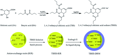
Prodrugs synthesized by different drugs not only overcome the defects of the original drugs, but also significantly enhance their treatment effects. In this study, a novel prodrug, 3,4,5-tributyryl shikimic acid (TBS), for the treatment of ulcerative colitis (UC) was synthesized by shikimic acid (SA) and butyric acid (BA) through the esterification reaction. Furthermore, the anion exchange resin, Amberlite 717 was employed to load the sodium salt of TBS through a batch process. Then the drug-loaded exchange resin (TBSS-IER) was encapsulated in the coating material, Eudragit S100, to prepare the colon-targeting drug resin microcapsule (TBSS-DRM) through an in-liquid drying method. The morphology and structure of TBSS-IER and TBSS-DRM were characterized by scanning electron microscopy (SEM). The in vitro release study demonstrated the good colon-targeting of TBSS-DRM. In the in vivo study, the TBSS-DRM exhibited a good therapeutic effect on the experimental colitis mouse induced by 2,4,6-trinitrobenzenesulfonic acid (TNBS). All results indicated that the prodrug was effective for colitis and the resin microcapsule system had good colon-targeting and could be used for the development of colon-targeting preparations.
1. Introduction

Ulcerative colitis (UC) is a representative type of inflammatory bowel disease and its pathogenesis still remains unknown.1–3 UC has a long course of disease and is prone to recur, thus seriously affecting the quality of patients' life. Currently, medication is still the main therapeutic regimen of UC. Generally, the drugs used for the treatment of UC include corticosteroids and aminosalicyclic acids. However, some of them have often induced incidences of serious side-effects.4,5 Nowadays, many researchers are pursuing potent novel drugs for UC management and many papers have reported that some components of vegetables and herbs can inhibit the inflammatory response and show new potential therapeutics for UC.6–8

Shikimic acid (3,4,5-trihydroxy-1-cyclohexene-1-carboxylic acid; SA) which is extracted from the fruits of Chinese star anise (Illicium verum Hook. fil.) can be used to improve the symptoms of UC because of its good anti-inflammation and anti-coagulation effects.9,10 Butyric acid (BA) is a short chain fatty acid and plays an important role in the human body.11–13 Many experiments have confirmed that BA had good anti-inflammatory and therapeutic effects on UC.14,15 But its relatively short half-life of oral administration limits its application. In this study, we synthesized a novel prodrug, 3,4,5-tributyryl shikimic acid (TBS), through the formation of an ester bond between SA and BA for the treatment of UC. However, from the in vivo experiment on mice, we found that the prodrug was easily hydrolyzed in the upper gastrointestinal tract. So it was necessary to design a colon-targeting preparation to transport the prodrug to the colon to prevent its hydrolysis in the upper gastrointestinal tract and play a better treatment role.

Ion-exchange resin (IER) is a kind of water insoluble inert polymer material which has been widely used in several scientific investigations due to its ion-exchange reaction with external-ions through their own functional groups.16–18 In recent years, IER has been employed as a drug carrier in a variety of drug delivery systems for the aims of improving effectiveness and safety of drugs, site-specific releasing, taste-masking and prolonging the duration of drug action.19–26 In this study, the anion-exchange resin, Amberlite 717 was employed as the carrier to load TBS through the ion-exchange reaction. Simultaneously, in order to prevent the drug-loaded resin from releasing drug at the site of high ionic strength (e.g., the stomach), the enteric coating material, Eudragit S100, was introduced to encapsulate the drug-loaded resin to form the drug-loaded resin microcapsule. The microcapsule hardly released drug in the upper gastrointestinal tract. However, in the lower gastrointestinal tract, it could release drug in the colon environment of low ionic strength through the degradation of Eudragit S100, thus achieving the aim of colon-targeting.

2. Materials and methods

2.1 Materials and reagents

Shikimic acid (SA, purity > 98%) was purchased from Shaanxi Sciphar Biotechnology, China. Butyric acid (BA) was purchased from Shanghai Aladdin Chemistry, China. 717 anion-exchange resin (Amberlite 717) was obtained from Xi'an LanXiao Technology, China. Eudragit S100 was purchased from Rohm, German. Liquid paraffin, Span 80, PEG 400 and PEG 4000 were purchased from Tianjin Kermel Chemical, China. All other reagents were analytical grade and obtained from commercially available sources.

2,4,6-Trinitrobenzenesulfonic acid (TNBS) was purchased from Sigma, USA. O-Dianisidine dihydrochloride (ODD) and ethylenediaminetetraacetic acid (EDTA) were purchased from Shanghai Aladdin Chemistry, China. Hexadecyl trimethyl ammonium bromide (HTAB) and 30% H2O2 solution were purchased from Tianjin Kermel Chemical, China. All other chemicals were analytical grade, commercially available products.

2.2 Animals

Male BALB/c mice weighing 20 ± 2 g were purchased from the Laboratory Animal Center of Xi'an Jiaotong University, and housed under controlled temperature and relative humidity conditions of 20–25 °C, and 50–60%, respectively, and under a 12/12 h light/dark cycle. All animals had ad libitum access to water and food. And they were quarantined for 1 week prior to treatment. All animal care and experimental protocols complied with the guidelines of animal ethics committee at Xi'an Jiaotong University.

2.3 Synthesis of TBS

In this study, a simple but efficient esterification reaction was used to synthesize TBS. Specifically, 50 g SA and 200 ml BA were added to a 1000 ml round-bottomed flask and stirred until evenly. 3 drops of 98% concentrated sulfuric acid as catalyst was added to the mixture and stirred for 10 min followed by adding 500 ml ice water to the flask. After being extracted by dichloromethane, the product was loaded on a silica gel open column and eluted with petroleum ether/ethyl acetate (5/1). The 3,4,5-tributyryl shikimic acid was obtained after removing solvents through flash evaporation (102.64 g, yield: 93.0%).

The products were characterized by 1H NMR, infrared spectroscopy (IR) and mass spectrometry (MS). 1H NMR spectra (DMSO-d6) were recorded on a 300 MHz 1H NMR spectrometer (Bruker, Germany). Chemical shifts (δ) were reported in ppm downfield from the internal standard tetramethylsilane (TMS). The IR and MS spectra were recorded with FTIR-8400S IR spectrometer (Shimadzu, Japan) and GCMS-QP2010 Mass Spectrometer (Shimadzu, Japan), respectively.

2.4 Preparation of TBSS-resin microcapsule

2.4.1 Pretreatment of ion-exchange resin (IER). In this work, Amberlite 717 which was mainly composed of styrene–divinylbenzene copolymer was used to encapsulate TBS. It has quaternary ammonium cationic groups and the active chloride ions. Through replacing the chloride ion which was connected with quaternary ammonium groups, the drug ions could be loaded in the resin. Specifically, a certain quantity of Amberlite 717 was immersed and washed in 50 °C deionized water to remove water-soluble impurities, then transferred them into 95% ethanol with stirring to remove the organic impurities. After washed with deionized water until no residual ethanol existed, the IER was dried under vacuum at 50 °C. The pre-dried IER was immersed in 0.1 mol l−1 hydrochloric acid solution with constant stirring for 24 h, then washed with deionized water until neutral and dried to obtain the anion (Cl) exchange resin.
2.4.2 Preparation of TBSS-IER complex. Since IER mainly encapsulates drugs through the ion-exchange reaction, drugs which can be loaded into IER must be ionized. In this study, TBS first reacted with sodium hydroxide (NaOH) to obtain its sodium salt. Specifically, 21.0 g TBS was added to 200 ml NaOH solution (0.275 mol l−1) with constant stirring, the 3,4,5-tributyryl shikimic sodium (TBSS) was obtained after extraction, filtration and drying of the water-soluble product. The 3,4,5-tributyryl shikimic sodium-loaded resin (TBSS-IER) complex was prepared through a batch process. The purified IER (0.50 g) was suspended in a 6.0 g l−1 TBSS aqueous solution under magnetic stirring at 30 °C. Samples were collected from TBSS aqueous solution at each predetermined time internal and determined by high-performance liquid chromatography (HPLC) (methanol[thin space (1/6-em)]:[thin space (1/6-em)]water[thin space (1/6-em)]:[thin space (1/6-em)]acetic acid 75[thin space (1/6-em)]:[thin space (1/6-em)]25[thin space (1/6-em)]:[thin space (1/6-em)]0.5, v/v, C18 BDS Hypersil, column temperature 30 °C, flow rate 1 ml min−1, detection wavelength 254 nm, LOQ: 0.15 μg ml−1). The drug-loading capacity (Q) of IER was determined by the measurement of the residual TBSS in solution, which was calculated according to the following equation:
Qt = V/WR(C0Ct)
where Qt was the drug-loading capacity of resin at time t. C0 was the initial drug concentration and Ct was the drug concentration at time t. V was the volume of drug solution and WR was the quality of resin.
2.4.3 Preparation of TBSS-resin microcapsule (TBSS-DRM). Eudragit S100 is a kind of methyl acrylic acid–methyl methacrylate copolymer. It is stable in the environment below pH 7.0 and degrades at pH > 7.0, thus making it a suitable colon-targeting preparation.27,28 In this study, in order to enhance the colon-targeting of TBSS-IER, Eudragit S100 was used to encapsulate TBSS-IER and the in-liquid drying process was employed to prepare the TBSS-DRM.29 Specifically, 20 ml liquid paraffin and 2.5 ml Span 80 were mixed and stirred evenly to form the continuous phase. 0.1 g Eudragit S100 and 0.01 g PEG 400 were dissolved in 7.5 ml acetone to form the dispersed phase, 0.5 g TBSS-IER (containing about 0.5 g prodrug) was firstly immersed in 50 ml 20% (w/w) PEG 4000 solution and then added in the dispersed phase with stirring. The prepared dispersed phase was dropped into the continuous phase to form emulsion with constant stirring for 6 h at 40 °C to remove acetone. The obtained TBSS-DRM was filtered and washed by petroleum ether to remove liquid paraffin and dried at 40 °C.

The morphology of the TBSS-IER and TBSS-DRM was characterized by scanning electron microscopy (SEM). The drug-loaded IER and DRM were frozen in liquid nitrogen, and then lyophilized for 72 h. The lyophilized IER and DRM were then sputtered with gold, and their morphology and microstructure were observed by a scanning electron microscope (TM-1000, HITACHI, Japan).

2.5 In vitro drug release study

In this part, two in vitro release experiments were investigated according to the paddle method for dissolution test in Chinese Pharmacopoeia (2010 edition), respectively. The first experiment was to investigate the impact of the ionic concentration on the ion-exchange ability of TBSS-IER. Specifically, 0.1 g TBSS-IER was added in three sodium chloride (NaCl) solutions of different concentrations (0.05 mol l−1, 0.15 mol l−1 and 0.80 mol l−1), respectively. The operation was carried under the condition of 37 ± 0.5 °C and 50 rpm. 5 ml samples were collected and replaced with the same volume of release medium at predetermined time internals. The HPLC method was used to determine the TBSS content in the release medium. The second experiment was to investigate the release behavior of TBSS-DRM and to simulate the process of the gastrointestinal transit in vivo. Specifically, 0.5 g TBSS-DRM was successively added into artificial gastric juice (pH = 1.2) for 2 h, artificial small intestinal juice (pH = 6.8) for 4 h and artificial colon juice (pH = 7.4) for 6 h. And the operation was carried under the condition of 37 ± 0.5 °C and 50 rpm. 5 ml samples were collected and replaced with the same volume of release medium at predetermined time internal (1 h). In the in vitro study, the volumes of all dissolution media were 900 ml, and after filtering through 0.45 μm filter membrane, samples of the two experiments were determined by HPLC according to 2.4.2 section, and calculated the percentage of cumulative release (%).

2.6 Therapy of TBSS-DRM on the experimental colitis induced by TNSB in mice

2.6.1 Induction of colitis and experimental protocols. Colitis was induced according to the procedure described by Wang et al.30 Specifically, after 12 h of fasting, mice were anesthetized with ether before induction of colitis. 0.1 ml 50% (v/v) ethanol which contained 2.5% (v/v) TNBS was instilled into the colon 3.5–4.0 cm from the anus by a gavage needle. Mice were kept in a head-down position for 30 s to prevent the leakage of the intracolonic instillation. Mice in the control group received physiological saline instead of TNBS solution. From the results of our pre-experiments, TBS was undetectable in colon due to the rapid hydrolysis of the ester bond in the upper gastrointestinal tract. Therefore, dexamethasone sodium phosphate (DXSP), which was also used in the treatment of UC, was selected to replace TBSS as the positive control. The mice were randomly divided in to 4 groups: (1) control-no colitis induced (p.o., 0.5% CMC-Na, n = 15), (2) TNBS (p.o., 0.5% CMC-Na, n = 15), (3) TNBS + DXSP (p.o., 2 mg kg−1 DXSP, n = 15), (4) TNBS + TBSS (p.o., 200 mg kg−1 TBSS, n = 15). The treatment was given after the induction of colitis for 12 h. During the experiment, the mice body weight changes of each group were recorded and mice feces were collected to investigate their characteristics and determine the fecal occult blood daily. At the end of the experiment, the number of the ultimately surviving mice was recorded and the final survival rate was calculated. Then all of the mice in the respective groups were killed, and the entire colon was excised and cleaned of adherent adipose tissue, opened longitudinally, and rinsed with cold physiological saline to remove fecal. The intestinal segment from each mouse was stored at −70 °C for subsequent measurement.
2.6.2 Measurement of disease activity index (DAI). Disease activity index (DAI) which reflects the therapeutic effect of different drugs on colitis induced by TNBS is the sum of scores given for body weight loss (scored as: 0, none; 1, 1–5%; 2, 5–10%; 3, 10–15%; 4, over 15%), stool consistency (scored as: 0, well-formed pellets; 2, loose stools; 4, diarrhea) and fecal occult blood (scored as: 0, normal; 1, occult blood+; 2, occult blood++; 3, occult blood+++; 4, visible blood in the stool).31 The test of mouse fecal occult blood was performed using o-tolidine. Specifically, a small amount of mouse feces was smeared on a white plate. 2–3 drops of 10 g l−1 of o-tolidine in glacial acetic acid solution was dripped into the feces and blended evenly. Then 2–3 drops of 3% H2O2 solution was added in the mixture, which was followed by timing as well as observing the results immediately. The evaluation criteria of fecal occult blood level were shown in Table 1. In this study, the DAI of each group were recorded daily and the figure of DAI varying trend was drawn. Moreover, the differences between groups were evaluated simultaneously.
Table 1 The assay criteria of o-benzidine to determine the fecal occult blood of mice
Results Phenomenon
Normal No color occurred after adding reagents for 2 min
Occult blood+ Light blue occurred at first, then turn blue after adding reagents for 10 s
Occult blood++ Light blue occurred at first, then turn brown after adding reagents
Occult blood+++ Brown occurred immediately after adding reagents


2.6.3 Measurement of MPO activity. The measurement of myeloperoxidase (MPO) is to determine the activity of myeloperoxidase which is used to measure the accumulation of neutrophils.32 Specifically, the weighted tissue samples were homogenized in ten volumes of ice-cold phosphate buffer (50 mM K2HPO4, pH 6.0) containing 0.5% (w/v) HTAB. The homogenate was centrifuged at 10[thin space (1/6-em)]000 rpm for 10 min at 4 °C, and the supernatant was discarded. Then the remaining precipitate was homogenized with an equivalent volume of 50 mM K2HPO4 containing 0.5% (w/v) HTAB and 10 mM EDTA. MPO activity was assessed by measuring the hydrogen-peroxide-dependent oxidation of o-dianisidine dihydrochloride (ODD). One enzyme unit was defined as the amount of enzyme producing one absorbance change per minute at 460 nm and 37 °C. Enzyme activity was calculated as U g−1 tissue.33

2.7 Statistical analysis

Statistical analyses were performed with SPSS version 13.0 for Windows. All results were expressed as mean ± SD. Data between groups were compared using Wilcoxon rank sum test. And data among multiple groups were compared using Kruskal–Wallis test. p < 0.05 was considered to be statistically significant.

3. Results

3.1 Synthesis and characterization of TBS

In this study, the prodrug, 3,4,5-tributyryl shikimic acid was obtained through the formation of ester bond between BA and SA (Scheme 1). The structure of TBS was determined by 1H NMR in CDCl3, and the spectrum was shown in Fig. 1. Specifically, the peaks at 1.0, 1.7 and 2.2 ppm belonged to the methyl group (CH3), the β and the γ methylene group (CH2), respectively. While the peaks at 2.3, 2.8 and 5.8 ppm were attributed to the methylidynes (CH) of the hexatomic ring which directly connected with the BA groups. The peaks at 5.3 and 6.9 belonged to the methylene group (CH2) and the double bond of the hexatomic ring. The 7.3 ppm corresponded to the protons of the carboxyl group (–COOH).
image file: c5ra16971b-s1.tif
Scheme 1 Synthesis scheme of 3,4,5-tributyryl shikimic acid (TBS).

image file: c5ra16971b-f1.tif
Fig. 1 1H NMR spectrum of TBS (in CDCl3).

The FTIR spectra of 5-BA was shown in Fig. 2. In general, the absorption bands at 3256 cm−1 and 1743 cm−1 were attributed to the carboxyl group (–COOH). The peak at 1697 cm−1, 1250, 1165, 1103 cm−1 belonged to the ester (νC[double bond, length as m-dash]O and νC–O–C) formed between SA and BA. Furthermore, the molecular ion peak of TBS was MS m/z 384 (MH+). These results indicated that TBS was successfully synthesized through the formation of the ester band between BA and SA.


image file: c5ra16971b-f2.tif
Fig. 2 FTIR spectrum of TBS.

3.2 Preparation of TBSS-IER and TBSS-DRM

In this work, the batch process was used to encapsulate the prodrug.34 In order to be encapsulated into the IER, the sodium salt of TBS (TBSS) was synthesized through a simple reaction with NaOH. During the drug-loading process, samples collected at each time internal were determined by HPLC. And the drug-loading capacity (Q) was the difference between the total dosage and the residual TBSS in solution. Fig. 3 showed the drug-loading capacity of resin changing over time. As shown in Fig. 3, the ion-exchange reaction got equilibrium at 1 h, and the drug-loading capacity was 1.07 g/g. In the coating process, in-liquid drying method was used to prepare TBSS-DRM. Liquid paraffin which was stable and non-volatile was selected as the continuous phase. The dispersed phase was acetone which could dissolve the coating material (Eudragit S100) and be volatilized easily. Span 80 and PEG 400 were used as emulsifier and plasticizer, respectively. Moreover, after immersing in PEG 4000 solution, the swelling degree of TBSS-IER significantly reduced, thus preventing the burst release of TBSS after the film-coated layer (Eudragit S100) degrading. In this work, the TBSS-DRM was obtained through removing the volatile dispersed phase by heating and stirring, thus guaranteeing the shape of and the dispersion of TBSS-DRM.
image file: c5ra16971b-f3.tif
Fig. 3 The variation of the drug-loading capacity (Q) of resin versus time during the TBSS-IER preparation.

The typical scanning electron microphotographs of TBSS-IER and TBSS-DRM were presented in Fig. 4. Fig. 4A showed that the drug-loaded resin was a sphere, and its particle size was about 600 μm. Fig. 4B exhibited the surface morphology of the drug-loaded resin. There were numerous tiny cavities on the surface of the resin, suggesting that the TBSS could enter the resin through them. Fig. 4C showed that, after coating with Eudragit S100, the volume of the microcapsule did not change obviously. Moreover, the leakage of drug caused by ion-exchange would be reduced in acid environment due to the coverage of the coating material.


image file: c5ra16971b-f4.tif
Fig. 4 SEM images of TBSS-IER (A), the surface morphology of TBSS-IER (B) and TBSS-DRM (C).

3.3 In vitro drug release study

Fig. 5A showed the influence of the concentration of release medium on the release behavior of TBSS-IER. With the increase of the medium concentration, the percentage of cumulative release increased as well. When the concentration of NaCl was 0.15 mol l−1, the ultimate cumulative release percentage was almost twice of that of 0.05 mol l−1 NaCl. While the cumulative release percentage of 0.8 mol l−1 NaCl had no significant difference with that of 0.15 mol l−1, indicating that with the increase of NaCl concentration, the ion-exchange capacity of resin also increased. However, when the concentration reaching a certain extent (e.g., 0.15 mol l−1), the ion-exchange capacity of resin tended to saturation. Then the increase of ion concentration would not improve the drug-loading capacity of resin. Fig. 5B was the in vitro release curve of TBSS-DRM at different pH values. This experiment imitated the variation of pH values and the transit time of the whole gastrointestinal tract (including stomach, small intestine and colon). As we know, it is desired that a colon-targeting preparation hardly releases drug in the gastric environment (pH 1.2), releases parts of drug in intestinal environment (pH 6.8) and releases lots of drug in the colon environment (pH 7.4). And the results of the in vitro release experiment confirmed the good colon-targeting property of TBSS-DRM. As shown in Fig. 5B, TBSS-DRM hardly released TBSS in the acidic environment. However, its release behavior changed significantly when the pH values varied. In the intestinal environment (pH 6.8), the accumulative release was <30%. While in the colonic environment (pH 7.4), massive TBSS released from the TBSS-DRM (>80%). This release behavior facilitated drugs to be concentrated at the targeting-site and play therapeutic effects.
image file: c5ra16971b-f5.tif
Fig. 5 The in vitro release behavior of TBSS-IER in three NaCl solutions of different concentrations (0.05 mol l−1, 0.15 mol l−1 and 0.80 mol l−1) (A) and TBSS-DRM in release medium with different pH values (B). For figure (B), the experiment was successively conducted at pH 1.2 (from 0 to 2 h), pH 6.8 (from 3 to 6 h) and pH 7.4 (from 7 to 10 h).

3.4 Effect of TBSS-DRM on the mouse survival rate and body weight changes

After 5 days of treatment, no mice died in the control group. However, 8 mice died in the TNBS group. Moreover, the death number in DXSP group and TBSS-DRM group was similar (5 mice died in DXSP group and 4 mice died in TBSS-DRM group), which might be due to the relatively large individual differences of the resistance of mouse to TNBS. Fig. 6 presented the influence of TBSS-DRM on body weight changes. From Fig. 6, the weight of the control group always maintained stable growth. However, the TNBS group showed sharp decline, then the decreasing trend slowed down. Furthermore, both weights of the TBSS-DRM group and the DXSP solution group began to rise gradually from the second day after administration and showed stable growth trend. However, compared with the DXSP solution group, the TBSS-DRM group showed a more obvious growth trend, indicating a better therapeutic effect of TBSS-DRM.
image file: c5ra16971b-f6.tif
Fig. 6 The influence of TBSS-DRM and DXSP on the body weight changes of mice. Data are shown as mean ± SD, n = 15.

3.5 Effect of TBSS-DRM on DAI and MPO activity

After the experimental colitis was successfully induced, the mice appeared some conditions, such as bloody stools, diarrhea, reduced activity and weight loss after 24 h. Fig. 7A reflected the variation tendency of the DAI of each group and Fig. 7B presented the influence of TBSS-DRM on the ultimate DAI. The DAI of control group was 0. However, due to the influence of inflammation, the DAI of TNBS group was significantly higher than the TBSS-DRM group and DXSP solution group. After treatment, both DAI of TBSS-DRM group and DXSP solution group showed obvious decline. The results of Fig. 7B indicated that both DAI of TBSS-DRM group and DXSP solution group showed significant difference compared with the TNBS group (p < 0.01), and they both showed similar decreased trend. Moreover, Fig. 8 showed the influence of TBSS-DRM on the activity of MPO. As shown in Fig. 8, compared with the TNBS group, MPO activity of TBSS-DRM group and DXSP solution group both decreased significantly (p < 0.01). And the downward trend of them was similar, indicating that both of them could effectively relieve the inflammation at the colon site. Both results of the variation of DAI and MPO activity suggested the good treatment of TBSS-DRM on the TNBS induced colitis of mice.
image file: c5ra16971b-f7.tif
Fig. 7 The influence of TBSS-DRM and DXSP on the variation of DAI index of mice (A) and the influence of TBSS-DRM and DXSP on the ultimate DAI index of mice (B). #,*Mean values with different superscript symbols were significantly different. ##p < 0.01 compared to control group; **p < 0.01 compared to TNBS group. Data are shown as mean ± SD, n = 15. Control control group, TNBS TNBS group, DXSP DXSP p.o. group, TBSS-DRM TBSS-DRM p.o. group.

image file: c5ra16971b-f8.tif
Fig. 8 The influence of TBSS-DRM and DXSP on the activity of MPO in the colon tissue of mice. #,*Mean values with different superscript symbols were significantly different. ##p < 0.01 compared to control group; **p < 0.01 compared to TNBS group. Control control group, TNBS TNBS group, DXSP DXSP p.o. group, TBSS-DRM TBSS-DRM p.o. group.

4. Discussion

Prodrug can improve the in vivo pharmacokinetics of the original drug including absorption, distribution, metabolism and excretion through changing the physical or chemical properties of the original drug by chemical modification, thereby overcoming the shortcomings and enhanced the therapeutic effect of the original drug. In this work, TBS was synthesized by SA and BA, and the prodrug could release SA and BA through the hydrolysis of ester bond, thus playing a collaborative therapeutic effect on UC. However, we found that the prodrug had poor water-solubility and was easy to be hydrolyzed in the upper gastrointestinal tract after repeated experiments. So we intended to use appropriate carriers to concentrate drugs to the lesion site according to the variation of the in vivo environment (e.g., pH values, enzyme and ionic strength), thus not only improving the treatment effect but also preventing hydrolysis of the prodrug in advance. In this study, we used IER as the carrier to load TBS through ion-exchange. Then the coating material, Eudragit S100, was used to encapsulate the drug-loaded IER so that it could be targeting to the colon site depending on the variation of the in vivo pH values. After the degradation of Eudragit S100, IER could release TBS at colon through ion-exchange. Then TBS was hydrolyzed and released SA and BA, thus having therapeutic effects (Scheme 2).
image file: c5ra16971b-s2.tif
Scheme 2 Schematic preparation, administration, and in vivo release behavior of TBSS-DRM.

As a carrier, resin can load drug depending on ion-exchange and its special structure. As depicted in Fig. 4, TBSS could enter the interior of resin through these cavities on the surface, then achieving the drug-loading through ion-exchange reaction. Moreover, the stability of drug-loaded microcapsule in acid environment could be effectively improved by coated with Eudragit S100, thereby preventing the leakage of drug in the upper gastrointestinal tract. From Fig. 3, the drug-loading capacity of resin reached saturation in a relatively short period of time (1 h, 1.07 g/g). Moreover, the release behavior could be adjusted via the media concentration. In the in vitro release study, Fig. 5A showed that with the increasing of the concentration of NaCl solution, the release percentage of the drug-loaded resin increased accordingly. The release percentage of drug-loaded resin in NaCl solutions with 0.15 and 0.8 mol l−1 concentrations were much higher than the 0.05 mol l−1 solutions, indicating that the release of drug-loaded resin was a concentration-dependent manner. With the increase of the media concentration, the number of the anions which replaced TBSS in resin increased accordingly. Therefore, more TBSS can be released. However, due to the limitation of the volume and the number of active groups, the exchange capacity of resin was limited. Therefore, when the medium concentration increased form 0.15 mol l−1 to 0.80 mol l−1, the release percentage had very small growth and the release amount got the maximum after 2 h (Fig. 5A). We can suppose that because the release of the drug-loaded resin is mainly influenced by the ionic strength, once orally taken, the resin will release drug rapidly in the strong electrolyte environment (e.g., in stomach), thus failed to achieve the colon-targeting. Therefore, we employed Eudragit S100 to encapsulate the TBS-IER through the in-liquid dying method to prepare the TBSS-DRM. The results of Fig. 5B suggested that since Eudragit S100 could not degrade at pH 1.2, TBSS was hardly released at this pH value. However, at pH 6.8, Eudragit S100 began to degrade and the TBSS-loaded resin was gradually exposed and released drugs through ion-exchange between resin and solution. At pH 7.4, Eudragit S100 was completely degraded and drugs were completely released. Moreover, with the gradually degradation of Eudragit S100, no burst release occurred during the release of TBSS. The main reason was that, because of the ion-exchange between resin and solution-ions, TBSS was gradually released. And the secondary reason was the PEG 4000 which delayed the drug release. While at pH 7.4, the Eudragit S100 was completely degraded and the TBSS-loaded resin was completed exposed to the solution, the ionic strength around the resin significantly increased and the ion-exchange degree accelerate as well. Therefore, the drug release increased significantly.

In the in vivo study, the results of mice survival rate and body weight changes showed that after the treatment of TBSS-DRM, both the survival rate and weight of mice were increased, thus confirming the good therapeutic effect of TBS on the experimental colitis. Moreover, the measurement of DAI and MPO activity also indicted that TBS had good inhibitory effects on the inflammation at the colon site. And there was no significant difference between the TBSS-DRM group and DXSP solution group about in therapeutic effect on the TNBS induced colitis mice. However, long-term or improper use of corticosteroids will cause many side-effects, such as the risk of opportunistic infection, diabetes mellitus, osteoporosis and the possible development of steroid-dependent disease.35–37 The TBSS-loaded resin encapsulated in the coating film (Eudragit S100) and hardly released drug at lower pH values (e.g., at stomach). However, in the colon environment, due to the degradation of Eudragit S100, the TBSS-loaded resin was exposed to the colon site and began to release TBSS. Moreover, due to the weak electrolyte concentration and the relatively small volume of colon, the exchange process of TBSS anions with external ions was relatively slower and TBSS could be gradually released. Also, with the hydrolysis of SA and BA, the TBSS-DRM would achieve a synergistic therapeutic effect and a sustained-release action. In conclusion, this method not only avoided TBSS being hydrolyzed in the upper digestive tract, but also achieved a similar therapeutic effect as corticosteroids.

5. Conclusion

In this study, a novel prodrug, 3,4,5-tributyryl shikimic acid (TBS), was synthesized by SA and BA through esterification reaction. Then the prodrug was encapsulated in Amberlite 717 through a batch process. Furthermore, in order to improve the colon-targeting property and the therapeutic effects of TBSS-IER, the coating polymer material, Eudragit S100, was employed to encapsulate TBSS-IER to prepare the colon-targeting drug resin microcapsule (TBSS-DRM) through the in-liquid drying process. The morphology of the TBSS-IER and TBSS-DRM were characterized by SEM. The in vitro release study showed that the TBSS-IER exhibited concentration-dependent release behavior and the TBSS-DRM displayed pH-sensitive release behavior. In the in vivo study, due to the colon-targeting and ion-exchange function, TBSS-DRM showed a similar therapeutic effect as compared with dexamethasone. All results indicated that the prodrug was effective for colitis and the resin microcapsule system had good colon-targeting property and could be used for the development of colon-targeting preparations.

Conflicts of interests

None declared.

Acknowledgements

This work was financially supported by Fundamental Research Funds for the Central Universities (xj08142016 and xjj2013054), China Postdoctoral Science Foundation Funded Project (2014M562429) and Natural Science Foundation of China (No. 30973578 and No. 81473177).

References

  1. J. Meier and A. Sturm, World J. Gastroenterol., 2011, 17, 3204–3212 Search PubMed.
  2. H. J. Freeman, Ther. Clin. Risk Manage., 2013, 9, 451–456 CrossRef PubMed.
  3. W. Blonski, A. M. Buchner and G. R. Lichtenstein, Curr. Opin. Gastroenterol., 2014, 30, 84–96 CrossRef CAS PubMed.
  4. C. De Cassan, G. Fiorino and S. Danese, Dig. Dis., 2012, 30, 368–375 CrossRef PubMed.
  5. K. Motegi, K. Nagasako, H. Nogawa, T. Akiya, Y. Kon and T. Sawada, Jpn. J. Clin. Med., 1999, 57, 2466–2471 CAS.
  6. K. Sugimoto, H. Hanai, K. Tozawa, T. Aoshi, M. Uchijima, T. Nagata and Y. Koide, Gastroenterology, 2002, 123, 1912–1922 CrossRef CAS PubMed.
  7. A. P. Bai, O. Y. Qin and R. W. Hu, Dig. Dis. Sci., 2005, 50, 1426–1431 CrossRef CAS PubMed.
  8. A. R. Martin, I. Villegas, C. La Casa and C. A. de la Lastra, Biochem. Pharmacol., 2004, 67, 1399–1410 CrossRef CAS PubMed.
  9. A. A. Bertelli, C. Mannari, S. Santi, C. Filippi, M. Migliori and L. Giovannini, J. Med. Virol., 2008, 80, 741–745 CrossRef CAS PubMed.
  10. Y. Ma, Q. P. Xu, J. N. Sun, L. M. Bai, Y. J. Guo and J. Z. Niu, Zhongguo Yaoli Xuebao, 1999, 20, 701–704 CAS.
  11. A. de Conti, V. Tryndyak, I. Koturbash, R. Heidor, J. Kuroiwa-Trzmielina, T. P. Ong, F. A. Beland, F. S. Moreno and I. P. Pogribny, Carcinogenesis, 2013, 34, 1900–1906 CrossRef CAS PubMed.
  12. S. Molina, M. I. Moran-Valero, D. Martin, L. Vazquez, T. Vargas, C. F. Torres, A. Ramirez de Molina and G. Reglero, Chem. Phys. Lipids, 2013, 175–176, 50–56 CrossRef CAS PubMed.
  13. A. Zaleski, A. Banaszkiewicz and J. Walkowiak, Przegl. Gastroenterol., 2013, 8, 350–353 CAS.
  14. A. Di Sabatino, R. Morera, R. Ciccocioppo, P. Cazzola, S. Gotti, F. P. Tinozzi, S. Tinozzi and G. R. Corazza, Aliment. Pharmacol. Ther., 2005, 22, 789–794 CrossRef CAS PubMed.
  15. P. Vernia, G. Monteleone, G. Grandinetti, G. Villotti, E. Di Giulio, G. Frieri, A. Marcheggiano, F. Pallone, R. Caprilli and A. Torsoli, Dig. Dis. Sci., 2000, 45, 976–981 CrossRef CAS PubMed.
  16. Y. Raghunathan, L. Amsel, O. Hinsvark and W. Bryant, J. Pharm. Sci., 1981, 70, 379–384 CrossRef CAS PubMed.
  17. V. Anand, R. Kandarapu and S. Garg, Drug Discovery Today, 2001, 6, 905–914 CrossRef CAS PubMed.
  18. X. Guo, R. K. Chang and M. A. Hussain, J. Pharm. Sci., 2009, 98, 3886–3902 CrossRef CAS PubMed.
  19. S. Burton, N. Washington, R. J. C. Steele, R. Musson and L. Feely, J. Pharm. Pharmacol., 1995, 47, 901–906 CrossRef CAS PubMed.
  20. S. H. Jeong and K. Park, Int. J. Pharm., 2008, 353, 195–204 CrossRef CAS PubMed.
  21. S. Narisawa, M. Nagata, C. Danyoshi, H. Yoshino, K. Murata and K. Noda, Pharm. Res., 1994, 11, 111–116 CrossRef CAS.
  22. W. Samprasit, P. Akkaramongkolporn, T. Ngawhirunpat, T. Rojanarata and P. Opanasopit, AAPS PharmSciTech, 2013, 14, 1118–1128 CrossRef CAS PubMed.
  23. H. Sohi, Y. Sultana and R. K. Khar, Drug Dev. Ind. Pharm., 2004, 30, 429–448 CrossRef CAS PubMed.
  24. C. P. Yewale, M. N. Rathi, G. G. Kore, G. V. Jadhav and M. P. Wagh, Pharm. Dev. Technol., 2013, 18, 367–376 CrossRef CAS PubMed.
  25. L. Yu, S. Li, Y. Yuan, Y. Dai and H. Liu, Int. J. Pharm., 2006, 319, 107–113 CrossRef CAS PubMed.
  26. Y. Chen, M. A. Burton, J. P. Codde, S. Napoli, I. J. Martins and B. N. Gray, J. Pharm. Pharmacol., 1992, 44, 211–215 CrossRef CAS PubMed.
  27. S. Thakral, N. K. Thakral and D. K. Majumdar, Expert Opin. Drug Delivery, 2013, 10, 131–149 CrossRef CAS PubMed.
  28. N. K. Thakral, A. R. Ray and D. K. Majumdar, J. Mater. Sci.: Mater. Med., 2010, 21, 2691–2699 CrossRef CAS PubMed.
  29. M. Li, O. Rouaud and D. Poncelet, Int. J. Pharm., 2008, 363, 26–39 CrossRef CAS PubMed.
  30. W. P. Wang, X. Guo, M. W. Koo, B. C. Wong, S. K. Lam, Y. N. Ye and C. H. Cho, Am. J. Physiol.: Gastrointest. Liver Physiol., 2001, 281, G586–G594 CAS.
  31. S. H. Patel, M. A. Rachchh and P. D. Jadav, Indian J. Pharmacol., 2012, 44, 744–748 CrossRef CAS PubMed.
  32. A. J. McCabe, M. Dowhy, B. A. Holm and P. L. Glick, J. Pediatr. Surg., 2001, 36, 334–337 CrossRef CAS PubMed.
  33. A. Bozkurt, B. Cakir, F. Ercan and B. C. Yegen, Regul. Pept., 2003, 116, 109–118 CrossRef CAS PubMed.
  34. H. Ichikawa, K. Fujioka, M. C. Adeyeye and Y. Fukumori, Int. J. Pharm., 2001, 216, 67–76 CrossRef CAS PubMed.
  35. D. R. Friend, Adv. Drug Delivery Rev., 2005, 57, 247–265 CrossRef CAS PubMed.
  36. R. Dahl, Respir. Med., 2006, 100, 1307–1317 CrossRef PubMed.
  37. A. L. Buchman, J. Clin. Gastroenterol., 2001, 33, 289–294 CrossRef CAS PubMed.

Footnote

These authors contributed to the work equally and should be regarded as co-first authors.

This journal is © The Royal Society of Chemistry 2016
Click here to see how this site uses Cookies. View our privacy policy here.