DOI:
10.1039/C6NR01161F
(Paper)
Nanoscale, 2016,
8, 10632-10641
A microwave synthesized CuxS and graphene oxide nanoribbon composite as a highly efficient counter electrode for quantum dot sensitized solar cells†
Received
9th February 2016
, Accepted 20th April 2016
First published on 21st April 2016
Abstract
To boost the photoconversion efficiency (PCE) of ever promising quantum dot sensitized solar cells (QDSSCs), and to improve the design of photoanodes, the ability of the counter electrode (CE) to effectively reduce the oxidized electrolyte needs special attention. A composite of a 15 wt% graphene oxide nanoribbon (GOR), obtained by unzipping multi-walled carbon nanotubes (MWCNTs), and CuxS intersecting hexagonal nanoplates, synthesized by a low cost, facile and scalable microwave synthesis route, is reported as a fascinating CE for QDSSCs. The best performing Cu1.18S–GOR CE could notably achieve a record PCE of ∼3.55% for CdS sensitized QDSSCs, ∼5.42% for in situ deposited CdS/CdSe co-sensitized QDSSCs and ∼6.81% for CdTe/CdS/CdS dual sensitized QDSSCs, apart from increasing the PCE of previously reported QDSSCs. A systematic investigation of the CE design revealed the high electrocatalytic activity of GOR due to the presence of organic functional groups, graphitic edge sites and a quasi-one-dimensional (quasi-1D) structure, which increases the interfacial charge transfer kinetics from the CE to the polysulfide electrolyte. The highly stable Cu1.18S–GOR CE has the added advantage of a favourable energy band alignment with the redox potential of the polysulfide electrolyte, which reduces the loss of charge carriers and thus can increase the PCE of QDSSCs.
1. Introduction
The low cost third generation solar cells fabricated using semiconductor quantum dots (QDs) as light harvesters are extremely appealing because of the solution processability of QDs and their tunable band gap, high absorption coefficient and possibilities of multiple exciton generation.1,2 Although overshadowed by the rapidly increasing efficiencies of organometal halide perovskite solar cells,3 quantum dot sensitized solar cells (QDSSCs) have started to demonstrate promising PCE,4–9 whereas the current efficiency scenario for QDSSC has just reached above 9% for the liquid junction QDSSC,10 and 10.7% for the solid state device.11 To improve the overall performance, all three basic components of QDSSCs, viz. the photoanode,12–15 the electrolyte,16,17 and the CE,18–22 need to be optimized. The role of a CE is to collect electrons from the external circuit and catalytically reduce the redox electrolyte, the latter being oxidized after hole scavenging from the sensitizer. In the popularly used polysulfide electrolyte, an ideal CE should be stable without any sulfur poisoning, possess high electrocatalytic activity, pose lower charge-transfer resistance at the CE–electrolyte interface and be cost-effective.
Pt CEs are prone to surface adsorb sulphur atoms from the polysulfide electrolytes, which decreases their conductivity and electrocatalytic reducing property, thereby affecting the PCE of QDSSCs.23 A large variety of replacement materials, such as CuxS,24–27 Co–S,28,29 NiS,30 PbS,31,32 Bi2S3,33 Cu2ZnSnS4,34 and carbon–CuxS composites,18,23 have been investigated for designing next-generation CEs. CuxS and its composite with different carbon allotropes show better performance and stability. Recently a CuS/Cu1.8S CE was reported for a CdSe-sensitized solar cell to attain 6.28% PCE,35 and FeSe2, Cu1.8S, and CuSe films were reported as efficient CEs for both dye-sensitized solar cells (DSSCs) and QDSSCs.36 As composites with carbon, the highly catalytic reduced graphene oxide (RGO)–Cu2S CE with the CdS/CdSe co-sensitizer had a PCE of 4.4%;18 however, with a similar photoanode, CuS and Cu1.8S nanosheet arrays as CEs achieved a PCE of 6.53%.37 Other than a chalcogenide based CE, composites of carbon dots and Au nanoraspberries achieved ∼5.4% PCE with a ZnO nanowire/CdS/CdSe photoanode.38
In fact, the 2D carbon nanostructure GOR with superior electrical, optical, thermal, and mechanical properties is useful as a hole extracting layer as well as electron blocking layer in solar cells.39,40 GORs with high aspect ratios and quasi-1D confinement of charge carriers are narrow elongated strips of graphene oxide (GO) with large catalytic edges and prepared by unzipping CNTs.40,41 GOR has larger surface area for interaction with the electrolyte and chalcogenide nanostructures than the parent CNTs, GO or RGO, conveniently prepared by greener routes.42 Even if GOR shows unique solution processability, there has been no report offering an optimization of the metal chalcogenide and the GOR composite that can offer high electrocatalytic performance as a CE in QDSSCs. The catalytic performance of the CE in QDSSCs depends on the available active sites and how fast the electrons flow back into the electrolyte from the external circuit, creating electron pathways to complete the circuit. So the carrier mobility of a CE should be good enough to reduce the charge transfer resistance (RCT). RCT will add up to the overall series resistance (Rs), which determines the most important parameter of QDSSC, the fill factor (FF). So the catalytically efficient CE will reduce Rs and improve FF, resulting in a higher PCE. GOR offers one of the greatest intrinsic carrier mobilities at room temperature, with a perfect atomic lattice, a promising mechanical strength, and chemical and thermal stability with additional functionalization of –OH and –COOH groups.
Previously the CuxS CE was prepared via exposing a brass foil to the polysulfide electrolyte,43,44 electrochemical deposition,45 electrospinning,46 solvothermal,47 successive ion layer adsorption and reaction (SILAR) etc.48 Unlike the detailed synthesis strategies involving surfactants, organic solvents and an inert atmosphere required for controlling the domain sizes and topologies,49 in this report a low cost microwave irradiation technique was used for the scalable production of CuxS nanostructures with a tunable chemical composition, whereas GOR was synthesized by oxidative unzipping of MWCNTs. The superiority of GOR as a composite material over GO and CNTs is discussed based on electrochemical experiments. The CE made by doctor blading a paste of CuxS–GOR composite on FTO glass shows enhanced catalytic activity, which is stable over several cycles and could improve the PCE of reported QDSSCs.
2. Results and discussion
In this section, after individual characterizations of the CuxS and carbon nanostructures, the design of the optimized CE will be discussed in the following steps: (i) to optimize the Cu:S stoichiometry for the best photovoltaic and catalytic activity of CuxS, (ii) to investigate the reason why GOR is a superior composite material over CNTs and GO, and (iii) to optimize the weight% of GOR with respect to CuxS. Finally the optimized CuxS–GOR CE will be demonstrated to increase the PCE of reported photoanodes.
2.1 Characterization of the individual components of CE
2.1.1 CuxS nanostructures.
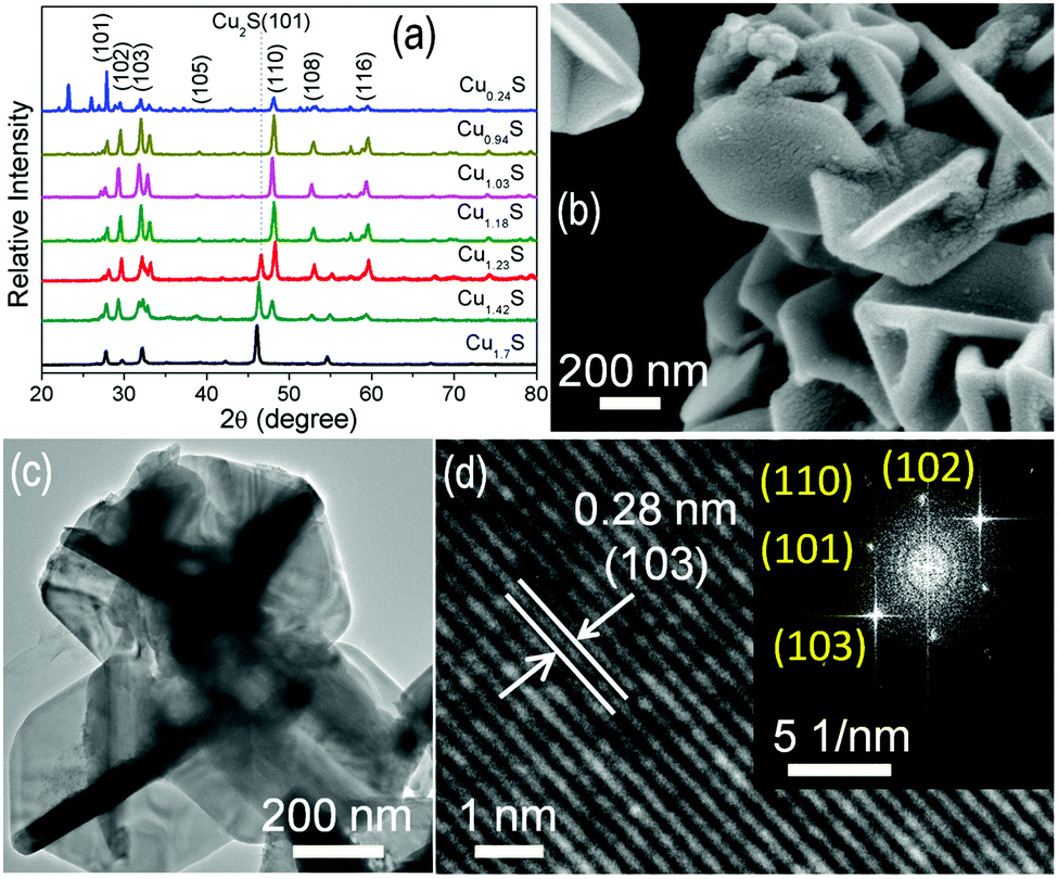
Fig. 1a shows the X-ray diffraction (XRD) patterns of seven different compositions of CuxS nanostructures. The XRD reflections match with the hexagonal crystal structure of covellite CuxS (JCPDS no. 06-0464). The reflection at 2θ = 46.5° for the (101) plane is observed for Cu/S ratio >1.18 due to the formation of the chalcocite Cu2S phase (JCPDS no. 01-0831462). In Cu1.42S the (101) reflection increases at the cost of the (110) reflection at 2θ = 47.9°, which is more prominent in Cu1.7S. The Cu:S stoichiometry was confirmed from the energy dispersive analysis of X-ray (EDAX) spectra recorded over 6 areas on each CuxS sample (ESI, Fig. S1†). Elemental mapping confirms the uniform composition of Cu:S over the entire nanostructure (Fig. S1h†). Fig. 1b shows a field emission scanning electron microscope (FESEM) 3-dimensional image of the representative Cu1.18S nanostructure. The CuxS nanostructures are composed of intersecting hexagonal nanoplates, the thickness of which increases from ∼50 nm for x = 0.24 to ∼80 nm for x = 1.03 and thereafter remains constant at 55–65 nm for x = 1.18–1.7 (Fig. S2†). Two of the major changes with increasing Cu/S ratio are the decrease in lateral dimension of the plates, say from >2 μm for Cu0.24S to ∼200 nm for Cu1.7S, and the gradual increase in roughness of the smaller plates at low S content. At a mean Cu/S ratio of x = 1.18, the diameter of the moderately rough surfaced plates is ∼600 nm and the thickness of the plates remains at ∼57 nm. Fig. 1c shows the transmission electron microscopy (TEM) image of Cu1.18S and the interplanar spacing corresponding to d102 = 0.28 nm is observed in Fig. 1d. The corresponding selected area electron diffraction (SAED) pattern (inset of Fig. 1d) shows the high crystallinity of Cu1.18S nanoplates.
 |
| | Fig. 1 (a) XRD spectra of CuxS nanostructures. (b) FESEM and (c) TEM images of Cu1.18S nanostructures. (d) High resolution TEM image showing the interplanar spacing of Cu1.18S and the inset shows the SAED pattern. | |
2.1.2 Carbon nanostructures: CNT, GO and GOR.
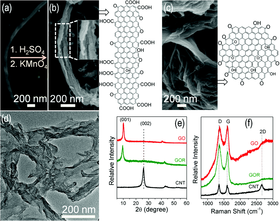
The commercial MWCNT (Fig. 2a) was unzipped by oxidative cleavage to produce GOR (Fig. 2b) whereas GO (Fig. 2c) was synthesized by the modified Hummers method. The TEM image of GOR (Fig. 2d) shows a multifaceted disordered structure with a distinct CNT wall opening giving rise to the ribbon-like morphology. As observed in the XRD patterns in Fig. 2e, the graphitic (002) reflection of the CNTs at 2θ ∼ 26.08° (JCPDS no. 75-1621) is absent in GO and GOR and a new peak at 2θ ∼ 9.56° corresponding to the (001) plane (JCPDS no. 89-8487) appears. The appearance of the (001) reflection and the absence of the (002) peak indicate the successful unzipping and exfoliation of the graphitic sheets into graphene oxide layers.40,50 Room temperature Raman spectra (Fig. 2f) show the disorder induced D-band at 1355 cm−1 and the G-band corresponding to the phonon stretching mode of the sp2 carbon atoms at 1599 cm−1. The intensity ratio of D and G bands increases from the CNTs towards GOR due to an increase in disorder due to unzipping. The 2D-band being an overtone of the D-band due to two-phonon scattering on a continuous graphene domain50,51 could be observed in CNTs but is absent after unzipping due to incomplete exfoliation of the staged graphene sheets. Both oxidative unzipping of CNTs to produce GOR and the modified Hummers method result in organic functionalities such as hydroxyl, carboxylic acid, keto, and epoxy groups attached to the carbon sheets, which result in structural defects as shown in the schematics of Fig. 2b and c. Fourier transform infrared (FTIR) spectra (Fig. S3†) confirm the above organic linkages. The carboxylic (–COOH) and hydroxyl (–OH) groups give rise to the broad peak at 3000–3700 cm−1, and the peak at 1600–1750 cm−1 is due to both ketonic C![[double bond, length as m-dash]](https://www.rsc.org/images/entities/char_e001.gif) O stretching and sp2 CC stretching frequencies. Due to the higher fraction of CO in GOR the peak intensity also increases compared to CNT. The vibration modes of epoxide (C–O–C) in GO and GOR are also observed at 1084 and 1387 cm−1.52,53 The optical band gap of GOR calculated from the UV-vis spectra (Fig. S4†) is ∼3.45 eV.
O stretching and sp2 CC stretching frequencies. Due to the higher fraction of CO in GOR the peak intensity also increases compared to CNT. The vibration modes of epoxide (C–O–C) in GO and GOR are also observed at 1084 and 1387 cm−1.52,53 The optical band gap of GOR calculated from the UV-vis spectra (Fig. S4†) is ∼3.45 eV.
 |
| | Fig. 2 (a) FESEM image of a single CNT. FESEM images and schematics of (b) GOR synthesized by unzipping CNT (enlarged view shown in the inset) and (c) GO. (d) TEM image of GOR. (e) XRD and (f) Raman spectra of CNT, GOR and GO. | |
2.2 Photovoltaic performance of CuxS nanostructure CEs
The CuxS nanostructures deposited on FTO substrates (Fig. S5†) were applied as CEs in the QDSSCs assembled with the CdS sensitized photoanode. Fig. 3a shows the current–voltage (J–V) characteristics for seven different CEs and Table 1 summarizes the photovoltaic parameters of the resulting QDSSCs. The device shows better performance with slightly copper rich films (Cu1.18S) than excess copper (Cu1.7S) or sulfur (Cu0.24S) containing films. Without the addition of carbon nanostructures, the CE prepared with Cu1.18S shows the highest efficiency of ∼3.05% with a corresponding open circuit voltage (Voc), short circuit current density (Jsc) and FF of ∼0.544 V, ∼10.68 mA cm−2 and ∼0.53, respectively. Electrochemical impedance spectroscopy (EIS) measurements on symmetric cells of the CE material were performed to analyze the photovoltaic performance. The Nyquist plots are shown in Fig. 3b and Rs and RCT extracted from the equivalent circuit (inset of Fig. 3b) are summarized in Table 1.37 Being a p-type material the electrical conductivity of CuxS is a signature of the free holes created by Cu-vacancies and, accordingly, with decreasing x, the electrical conductivity should also increase.54 According to the Rs and RCT values, although this analogy holds true for Cu/S ratios from Cu1.7S to Cu1.18S, an opposite trend is followed at higher Cu-deficiencies up to Cu0.24S. In fact, moving away from x = 1.18, Cu-rich films show better charge transfer kinetics than S-rich films. The nanoplate thickness does not show a systematic trend with Cu:S stoichiometry, still the roughness of these nanoplates with lower sulphur content can play a role in increasing the catalytically active sites. At x < 1.18, the smoother nanoplates cannot possess enough catalytic sites, which likely overrides the factor of the generation of a larger number of free holes from higher Cu-vacancies. Therefore, with decreasing Cu/S ratio the range of variation of Rs is only from 2.49 to 1.62 Ω and again rises with a high excess of sulphur, hence Rs does not have a great impact on the QDSSC performance. The main contribution towards PCE is from the interfacial charge transfer resistance (RCT) associated with the CE. Since RCT is the lowest for Cu1.18S, the charge transfer from the polysulfide electrolyte to the CE is faster, reflecting its superior PCE.
 |
| | Fig. 3 (a) J–V characteristics of a representative CdS sensitized photoanode with different CuxS CEs. (b) Nyquist plot and (c) Tafel polarization characteristics measured with a symmetric dummy cell. The inset of (b) shows the equivalent circuit where CPE stands for the constant phase element. (d) CV plots measured with a conventional three electrode cell for seven different CuxS nanostructures as the working electrode. | |
Table 1 QDSSC parameters derived from J–V, impedance and Tafel plots
| Sample name |
V
oc (V) |
J
sc (mA cm−2) |
FF |
η![[thin space (1/6-em)]](https://www.rsc.org/images/entities/char_2009.gif) a (%) a (%) |
R
s (Ω) |
R
CT (Ω) |
J
o (mA cm−2) |
J
lim (mA cm−2) |
|
The efficiency data shown are the average values obtained from 3 devices.
|
| Cu1.7S |
0.512 |
8.92 |
0.56 |
2.61 ± 0.08 |
2.49 |
18.01 |
0.49 |
5.52 |
| Cu1.42S |
0.540 |
10.07 |
0.50 |
2.74 ± 0.04 |
2.23 |
22.53 |
0.47 |
5.37 |
| Cu1.23S |
0.537 |
10.05 |
0.54 |
2.91 ± 0.05 |
1.93 |
11.5 |
0.57 |
7.58 |
| Cu1.18S |
0.544 |
10.68 |
0.53 |
3.05 ± 0.06 |
1.68 |
10.15 |
0.67 |
8.31 |
| Cu1.03S |
0.525 |
9.76 |
0.48 |
2.44 ± 0.10 |
1.65 |
15.93 |
0.39 |
3.71 |
| Cu0.94S |
0.519 |
7.91 |
0.55 |
2.41 ± 0.09 |
1.62 |
26.33 |
0.32 |
2.75 |
| Cu0.24S |
0.494 |
9.55 |
0.61 |
2.36 ± 0.11 |
2.83 |
27.48 |
0.23 |
1.44 |
2.3 Electrochemical analysis of CuxS nanostructures
The electrochemical activity of the CuxS nanostructures was investigated in terms of Tafel polarization and cyclic voltammetry (CV) studies. Fig. 3c shows the Tafel plots of symmetric cells for different CuxS films. The Tafel polarization measurement gives information on charge transfer kinetics. The exchange current density (Jo) estimated from the extrapolated intercepts of the anodic and cathodic branches of the Tafel polarization curves is related to RCT according to the following equation:28,37| |  | (1) |
where R is the gas constant, T is the absolute temperature, n is the number of electrons involved in the polysulfide reduction and F is the Faraday constant. The variation of Jo derived from the Tafel plots (Table 1) is in good agreement with the EIS results, reconfirming the smaller RCT for the Cu1.18S electrode. The catalytic properties can also be derived from the limiting current density (Jlim), which is mainly the diffusion velocity of the ionic carriers (polysulfide) between the two electrodes. Jlim is related to the diffusion coefficient (D) by the relation:36,38| |  | (2) |
where l is the electrolyte thickness, n is the number of electrons involved in the reduction of disulfide at CE, F is the Faraday constant, and C is the polysulfide concentration. Jlim is directly proportional to the diffusion of charge carriers, and the relatively high Jlim for the Cu1.18S CE (Table 1) relates to its higher catalytic activity towards polysulfide reduction. The electrocatalytic activity of the CuxS CEs was also cross-checked by the cyclic voltammetry (CV) analysis in a three electrode setup (Fig. 3d). Since the positive and negative currents correspond to the oxidation of S2− ions and the reduction of Sn2− ions in the redox polysulfide electrolyte,37 the relatively high reduction peak current densities of Cu1.18S electrodes reflect its agreement with the parameters obtained from Tafel plots. The next step will be to increase the FF of the QDSSCs with a composite CE of Cu1.18S and the carbon nanostructure.
2.4 Solar cell characteristics and EIS analysis of the composite CEs
For testing the most suitable carbon nanostructure, 10 wt% of either CNT, GO or GOR were mixed with Cu1.18S and the composite CEs termed as Cu1.18S–CNT, Cu1.18S–GO and Cu1.18S–GOR, respectively. Although the XRD patterns of the composites exhibit the hexagonal covellite structure of Cu1.18S, due to a low wt% of the carbon counterparts, a relatively weak (001) reflection is observed for CNT, GO and GOR (Fig. S6†). The CNT composite still exhibits the additional (002) hump. In contrast, the Raman spectra show the typical D- and G-bands of carbon without any observable vibration mode of Cu1.18S (Fig. S6†). The J–V characteristics of the FTO/TiO2/CdS photoanode show significant improvement in FF and PCE with both Cu1.18S–GO and Cu1.18S–GOR (Fig. 4a and Table 2), although CNTs did not help to increase the PCE. In the case of Cu1.18S–GOR CE, the maximum FF of 0.57 and increased device efficiency up to 3.38% are achieved. A possible reason for such an improvement in the device performance is the presence of highly catalytic crystalline edges of GOR (shown later in Fig. 5a), which effectively takes part in the polysulfide reduction process. Comparing this aspect with the morphology of CNT and GO (Fig. 2c and d), GOR has larger edge sites with attached functional groups (Fig. S3†) than CNT and GO, which helps faster reduction kinetics.55,56 From a general perspective, the presence of edge sites in GOR is apparent since unzipping along the cylindrical morphology of the CNT will result in a higher number of dangling bonds at the unzipped edges which should be catalytically active. Also the quasi-1D structure of GOR helps in faster electron transfer through the 1D channel and therefore increases the device efficiency. The performance of the composite CEs was found to be far superior with respect to the conventional Pt and brass/Cu2S CE (Table 2). The Rs values in Table 2 obtained from the Nyquist plots (Fig. 4b) of EIS measurements for the three composite CEs are slightly higher than those for Pt and brass/Cu2S CEs (Fig. 4b, inset), due to the metallic nature of the latter. In fact, the large RCT of Pt (1988 Ω) and brass/Cu2S (1588 Ω) has a huge impact on the QDSSC performance. Interestingly, Cu1.18S–GOR shows the lowest RCT (1.26 Ω) as compared to Cu1.18S–GO (1.63 Ω) and Cu1.18S–CNT (3.17 Ω) which is also less than Cu1.18S (10.15 Ω) itself. Since RCT is related to the charge transfer at the CE–electrolyte interface, lower RCT signifies higher charge transfer kinetics, which helps in improving the PCE with the Cu1.18S–GOR CE.
 |
| | Fig. 4 (a) J–V characteristics, (b) Nyquist plot and (c) Tafel polarization characteristics of Pt, brass/Cu2S and Cu1.18S composites with 10 wt% CNT, GOR and GO. (d) Stability tests: 50 cycles of CV plots for Cu1.18S–GOR as the working electrode in a three electrode cell. The insets show the CV plots of the Cu1.18S working electrode for 50 cycles and Pt for 10 cycles, with an arrow showing the downward shift in current density over an increasing number of cycles. (e) Energy band diagram (not to scale) of Cu1.18S–GOR CE showing electron transfer. The energy levels of GO are also indicated. | |
 |
| | Fig. 5 (a) TEM micrograph of the 15 wt% GOR and Cu1.18S composite. The magnified regions show inter-planar spacing. (b) SAED patterns from the composite show the indexed crystallographic planes of Cu1.18S. The (002) plane corresponds to GOR. (c) J–V characteristics for different GOR wt% in the Cu1.18S–GOR composite CE. (d) Photogenerated charge carrier lifetime from Voc decay for the composite CEs, Cu1.18S and Pt. The inset shows the IPCE spectra. | |
Table 2 QDSSC parameters for five different CEs derived from J–V, impedance and Tafel plots. The composite CEs with Cu1.18S are with 10 wt% CNT, GOR and GO
| Sample name |
V
oc (V) |
J
sc (mA cm−2) |
FF |
ηa (%) |
R
s (Ω) |
R
CT (Ω) |
J
o (mA cm−2) |
J
lim (mA cm−2) |
|
The efficiency data shown are the average values obtained from 3 devices.
|
| Cu1.18S–CNT |
0.543 |
11.35 |
0.50 |
3.1 ± 0.04 |
11.16 |
3.17 |
0.274 |
7.24 |
| Cu1.18S–GOR |
0.543 |
10.92 |
0.57 |
3.38 ± 0.05 |
11.02 |
1.26 |
0.467 |
9.12 |
| Cu1.18S–GO |
0.543 |
11.08 |
0.53 |
3.22 ± 0.02 |
10.95 |
1.63 |
0.350 |
8.12 |
| Pt |
0.458 |
5.34 |
0.21 |
0.5 ± 0.02 |
9.95 |
1988 |
0.005 |
0.24 |
| Brass/Cu2S |
0.514 |
9.6 |
0.50 |
2.48 ± 0.10 |
10.58 |
1544 |
0.010 |
0.14 |
2.5 Tafel plots and CV analysis of the composite CEs
Fig. 4c shows the Tafel plots of symmetric cells for all the CEs discussed above. The Cu1.18S–GOR CE shows the highest Jo (Table 2), which according to eqn (1) relates to the lowest RCT obtained from EIS measurements. Also Jlim is higher for the Cu1.18S–GOR CE (Table 2), which implies larger diffusion of charge carriers following eqn (2). Since stability has been a major issue of CE for several years,36 consecutive 50 CV cycles were tested in a conventional three electrode cell with the polysulfide electrolyte for both Cu1.18S and Cu1.18S–GOR electrodes with excellent results (Fig. 4d). In contrast, a Pt working electrode with only 10 cycles shows a downward shift indicating poor stability owing to the poisoning effect of sulfur ions on Pt.22 The superiority of the Cu1.18S–GOR CE over Pt or Cu1.18S–CNT, Cu1.18S–GO composite CEs could be better understood from the parameters obtained from CV analyses (Table S7†). The electrochemical active surface area (ECSA) was calculated from the CV plots (Discussion and Fig. S7†) according to a standard protocol.57,58 Cu1.18S–GOR exhibits an ECSA of 575.2 cm2 mg−1, higher than 538.0, 420.2 and 264.7 cm2 mg−1 for Cu1.18S–GO, Cu1.18S–CNT and Pt, respectively. Higher ECSA of Cu1.18S–GOR CE results in better interaction with the polysulfide electrolyte. Moreover, the relatively high negative current density (−25.35 mA cm−2) of Cu1.18S–GOR CE also confirms its superior catalytic activity towards the reduction process.
So far it has been obvious that the higher electrocatalytic activity of Cu1.18S–GOR CE is the prime reason for its superior performance in increasing the PCE. In fact, the band diagram explanation (Fig. 4e) based on the available literature also justifies the experimental observations.16,40 The standard reduction potential for polysulfide (Sn2−/S2−) is −5.0 eV from the vacuum level.16 The valence band maximum for CuS is −5.3 eV and the highest occupied molecular orbital (HOMO) level of GOR as measured by Dai et al.40 is −5.0 eV. Because of the p-type nature of CuxS the Fermi level (EF) will be close to the valence band and will be in equilibrium with the reduction potential of the polysulfide under dark conditions. Since the HOMO level of GOR also matches with the reduction potential of polysulfide, it closely overlaps with the EF of CuxS (Fig. 4e). GOR helps in increasing the number of available states close to its HOMO level facilitating the electron transfer, whereas CuxS actually participates in the reduction process. In QDSSCs, when the electron will move from the photoanode to CE to reduce the polysulfide, if the redox potential for polysulfide is closer to the HOMO level of GOR it will supply the electrons faster than Cu1.18S itself. This particular band alignment will therefore reduce the carrier loss and hence increase the PCE. The optical band gap of GO calculated from the UV-vis spectra is ∼3.65 eV (Fig. S8†). Although the HOMO level of GO matches well with the Fermi level of the FTO contact (both ∼−4.7 eV),59,60 it is slightly higher above the redox potential of the polysulfide, which makes the transfer of electrons a little stringent compared to GOR.
2.6 Optimization of the composite CE and photovoltaic parameters
A TEM image of a representative composite with 15 wt% GOR and Cu1.18S is shown in Fig. 5a, where GOR wraps around the Cu1.18S plates shown by undulated nanoplate edges with lower contrast. GOR is observed to have graphitic edges and the composite as a whole is perfectly crystalline (Fig. 5a and b). The following step to improve the device performance is to alter the GOR wt% in the Cu1.18S–GOR CEs. Fig. 5c shows the J–V curves for four different GOR percentages wherein PCE increases up to 15 wt% GOR and then decreases (Table 3). Since Cu1.18S is the reducing agent of oxidized polysulfide and GOR acts as the medium of charge transport between the Cu1.18S domains and then to FTO, excess GOR will block the active catalytic sites and decrease the electrochemical activity of Cu1.18S. With 15 wt% GOR, the maximum PCE of 3.55% with a Voc of ∼0.551 V and a Jsc of ∼11.33 mA cm−2 was achieved. The photogenerated charge carrier lifetime (τn) can be extracted from Voc decay measurements using the equation:14| |  | (3) |
where k is the Boltzmann constant, q the elemental charge, and T the absolute temperature. Fig. 5d shows the plots of τnversus Voc, and the actual decay curves of QDSSCs with CEs consisting of a composite of Cu1.18S with 15 wt% each of CNT, GO and GOR are shown in Fig. S9.† The faster decay corresponds to fast recombination kinetics and higher loss of charge carriers. From the decay curve it is evident that Cu1.18S–GOR has the slowest decay owing to less recombination kinetics whereas Pt has the fastest decay. Considering the maximum power point condition (where Voc∼0.4 V) in Fig. 5d, τn is also maximum for the Cu1.18S–GOR CE. With 15 wt% GOR, the catalytically active Cu1.18S–GOR CE experiences less RCT in polysulfide reduction and hence it can reduce Sn2− to S2− ions faster leading to lower recombination losses and higher τn. The incident photon-to-current conversion efficiency (IPCE) spectra for different CEs exhibit a superior photoresponse over the range from 300 to 550 nm (Fig. 5d, inset). Although the IPCE spectra for Cu1.18S and its composites with 15 wt% CNT, GO and GOR do not show significant differences, they are far superior with respect to Pt or brass/Cu2S CEs. Since IPCE is directly related to the photocurrent and there are minimal differences in Jsc with different composite CEs, IPCE also follows suit.
Table 3 QDSSC parameters for Cu1.18S–GOR CEs with different weight percentages of GOR
| Cu1.18S–GOR |
V
oc (V) |
J
sc (mA cm−2) |
FF |
ηa (%) |
| GOR (wt%) |
|
The efficiency data shown are the average values obtained from 3 devices.
|
| 5 |
0.538 |
10.94 |
0.55 |
3.22 ± 0.09 |
| 10 |
0.543 |
10.98 |
0.57 |
3.38 ± 0.05 |
| 15 |
0.551 |
11.33 |
0.58 |
3.55 ± 0.07 |
| 20 |
0.548 |
11.51 |
0.44 |
2.80 ± 0.12 |
2.7 Improvement of previously reported PCEs
When Cu1.18S–GOR is used as the CE on previously reported CdTe/CdS/CdS core/shell/quasi-shell QDSSC,14 the PCE improves to 6.81% from earlier reported 6.32% using the brass/Cu2S CE (Fig. 6a) due to a proportionate increase in FF from 0.50 to 0.53 (Table 4). Fig. 6a (inset) shows the corresponding IPCE characteristics with brass/Cu2S and Cu1.18S–GOR CEs, where the photo-response is observed from 300 to 750 nm with a small enhancement in IPCE. In addition, the PCE of 1.44 at% Mn doped CdS QDSSC61 is also improved from 2.08 to 2.42% with an increase in FF from 0.49 to 0.56 (Fig. 6b and Table 4). The performance of Cu1.18S–GOR CE is also evaluated with respect to other reported CEs with a common SILAR deposited CdS/CdSe co-sensitized photoanode. The Cu1.18S–GOR CE achieves a record PCE of 5.42% with a Voc of ∼0.52 V and a Jsc of ∼18.04 mA cm−2 (Fig. 6c), higher than the PCEs reported with other CEs (Table 4).18,25,30,62–64
 |
| | Fig. 6
J–V characteristics showing the improvement of PCE with Cu1.18S–GOR CE for (a) CdTe/CdS/CdS core/shell/quasi-shell, (b) Mn-doped CdS, and (c) CdS/CdSe photoanodes. The inset in (a) shows the corresponding IPCE spectra. | |
Table 4 Photovoltaic parameters for different photoanodes and CEs providing a comparison of Cu1.18S–GOR with the literature reported CEs. Here Cu1.18S–GOR represents a composite with 15 wt% GOR
| QDs |
CEs |
V
oc (V) |
J
sc (mA cm−2) |
FF |
η (%) |
Ref. |
| CdTe/CdS/CdS |
Brass/Cu2S |
0.630 |
20.31 |
0.50 |
6.32 |
14
|
| Cu1.18S–GOR |
0.626 |
20.55 |
0.53 |
6.81 |
This work |
| |
| Mn:CdS |
Brass/Cu2S |
0.501 |
8.39 |
0.49 |
2.08 |
60
|
| Cu1.18S–GOR |
0.501 |
8.64 |
0.56 |
2.42 |
This work |
| |
| CdS/CdSe |
CuS |
0.42 |
9.38 |
0.37 |
1.47 |
30
|
| Cu2S |
0.54 |
11.70 |
0.48 |
3.04 |
25
|
| Carbon–Cu2S |
0.49 |
10.70 |
0.58 |
3.08 |
62
|
| Cu2S |
0.60 |
11.69 |
0.44 |
3.18 |
63
|
| Cu1.8S/CuS |
0.54 |
14.50 |
0.41 |
3.22 |
64
|
| RGO–Cu2S |
0.52 |
18.40 |
0.46 |
4.40 |
18
|
| Brass/Cu2S |
0.504 |
15.66 |
0.56 |
4.50 |
This work |
| Cu1.18S–GOR |
0.520 |
18.04 |
0.58 |
5.42 |
This work |
3. Conclusions
In conclusion, a highly efficient electrocatalytic CE for QDSSCs was designed through a composite of microwave synthesized Cu1.18S nanostructures and 15 wt% GOR, prepared by unzipping MWCNTs. Seven different compositions of CuxS were tested by photovoltaic and electrocatalytic performance to determine the best performing stoichiometry of x = 1.18. The composite of GOR with Cu1.18S was found to be superior with respect to those with CNT and GO, due to higher functionalization, graphitic edge sites and quasi-1D structure in GOR. The reasons pertaining to better photovoltaic performance have been elucidated by EIS analysis and the electrocatalytic performance by Tafel polarization and CV studies. The high electrocatalytic activity of GOR could be retained after several cycles, maintaining a low RCT for efficient electron transfer from the CE to the polysulfide electrolyte. A record PCE ∼3.55% for SILAR deposited CdS sensitized QDSSC was achieved, apart from a significant increase in PCE of reported QDSSCs. Moreover, the comparison of different CEs in the case of CdS/CdSe co-sensitized QDSSC has demonstrated the superiority of Cu1.18S–GOR CE. However, there is enough room to further optimize the CEs for improved QDSSC performance and research is underway in this direction.
4. Experimental details
4.1 Materials
Cu(NO3)2·3H2O (≥99%), sulfur powder (99%), ethylene glycol (≥99%), cadmium acetate dihydrate (≥99%), potassium chloride (99%), sodium sulphide flakes (≥50%), zinc acetate dihydrate (≥99%), sulfuric acid (98%), ethanol (absolute), and titanium tetrachloride (99%) were purchased from Merck, India. MWCNT (≥95%) was purchased from SRL Pvt. Ltd, Mumbai, India. 1-Methyl-2-pyrrolidone (NMP, 99%), Zn(NO3)2·6H2O (98%), and polyvinylidene fluoride (PVDF) powder were purchased from Sigma Aldrich. F:SnO2 (FTO)-coated glass (TCO 22-7), TiO2 paste (Ti-nanoxide T/SP, average size ∼20 nm), and scattering TiO2 paste (Ti-nanoxide R/SP, average size >100 nm) were purchased from Solaronix. Graphite powder (99.99%) and a brass foil (0.25 mm thick) were purchased from Alfa Aesar. All reagents were used as received.
4.2 Methods
4.2.1. Synthesis of CuxS nanostructures.
A flux of 5 mmol Cu(NO3)2·6H2O and 5 mmol sulfur powder in 100 mL ethylene glycol was irradiated in a microwave chamber for 12 min at 600 W and 125 °C with constant stirring at 2000 rpm. For different Cu/S ratios the solution turned from deep green to greyish and the grey product was centrifuged at 6500 rpm for 15 min, washed with ethanol and dried at 80 °C for 1.5 h to obtain the CuxS nanostructures.
4.2.2. Synthesis of GOR and GO.
GOR was synthesized modifying the procedure of Dai et al.40 50 mg MWCNTs was bath sonicated in 50 mL 98% H2SO4 for 30 min and stirred overnight at room temperature. 250 mg 500 wt% KMnO4 was added to this dispersion slowly over 2 h under constant stirring at room temperature followed by additional stirring at 70 °C for 2 h during which 100 mg KMnO4 was added slowly and the colour changed from grey to dark brown indicating completion of the reaction. The reaction mixture was allowed to cool to room temperature and poured into a mixture of 150 ml ice and 2.5 mL H2O2 under stirring. Finally, the resultant dispersion was diluted by adding 2 L of cold distilled ionized (DI) water and vacuum filtered through a PTFE membrane (0.45 μm pore size). The product was removed and stirred in 100 mL ethanol for 30 min. The final product was washed twice with ethanol to obtain the dried GOR as a dark precipitate.
GO was prepared by a modified Hummers method.65 The mixture of 1 g graphite powder, 0.5 g NaNO3 and 25 mL conc. H2SO4 was cooled to 0 °C in an ice bath and stirred for 2 h. 3 g KMnO4 was added slowly with continuous stirring for another 1 h, followed by 30 mL DI water at room temperature. The temperature of the mixture was then increased to 90 °C in an oil bath and 100 mL DI water was added under continuous stirring for 1 h until the colour of the mixture turned to mud brown. The mixture was further treated with 10 mL 30% H2O2, diluted with 2.5 L excess DI water, filtered and, after drying overnight under vacuum conditions, GO was collected.
4.2.3. Preparation of QD-sensitized photoanodes.
FTO coated glass was washed in a soap solution, DI water, and ethanol under sonication for 20 min each. A compact TiO2 layer was deposited on the FTO glass by dip coating in 40 mM TiCl4 solution at 80 °C for 40 min and washed with DI water and ethanol. A 8 μm thick mesoporous TiO2 active layer was then doctor-bladed onto the compact TiO2 layer coated FTO and dried at 80 °C for 30 min, followed by annealing at 500 °C for 1 h in a box furnace. A 4 μm thick scattering TiO2 layer was similarly doctor bladed on top of the active layer and annealed at 500 °C for 1 h.
CdS photoanode.
CdS QDs were deposited through the SILAR process. In brief, the TiO2 films were dipped alternately in a 0.1 M methanol solution of Cd(OAc)2·2H2O and a 0.1 M methanol:water (1:1) solution of Na2S·9H2O for 1 min. The films were washed with DI water and dried in air between each step to complete one SILAR cycle. The total number of CdS SILAR cycles was 8, followed by two SILAR cycles of a ZnS passivation layer deposited from 0.1 M aqueous solutions of Zn(NO3)2·6H2O and Na2S·9H2O, respectively. Finally, the films were washed with excess amounts of DI water and allowed to dry in air at room temperature.
The CdTe/CdS/CdS core/shell/quasi-shell sensitizer.
CdTe/CdS core/shell QDs were prepared according to our previous report.14 In brief, the mercaptopropionic acid capped core/shell QDs were deposited on TiO2 coated FTO glass by dipping into this sensitizing solution with pH ∼ 13 for 12 h and washed sequentially with DI water and ethanol. After loading the pre-synthesized core/shell QDs, the CdS quasi-shell was deposited through 4 cycles of SILAR and 2 cycles of ZnS and finally washed and dried at room temperature.
The Mn-doped CdS QD sensitizer.
Mn-doped CdS QDs were also prepared according to our previous report.61 In brief, the degassed solution of 1 mmol CdCl2·H2O, MnCl2·4H2O (Mn:Cd at% 1.44) and 5 mL of oleylamine was heated and aged at 100 °C for 30 min followed by the injection of sulfur–oleylamine solution at 170 °C. The colloidal solution was aged at 170 °C for 70 min followed by cooling the flask to room temperature, precipitation of the product with isopropanol, centrifugation, and washing with ethanol to obtain the QDs finally suspended in chloroform. The Mn-doped CdS QDs were deposited on the FTO/TiO2 photoanode by electrophoretic deposition.61
CdS/CdSe photoanode.
Firstly, the TiO2 film on FTO was alternately dipped into a 0.1 Cd(OAc)2 and 0.1 M Na2S solution five times for 1 min in each dipping cycle. Secondly, the TiO2/CdS film was dipped into a bath of 0.1 M Cd(OAc)2 and Na2SeSO3 solution for 3 h. The aqueous solution of Na2SeSO3 was prepared by refluxing 0.1 M Se and 0.2 M Na2SO3 in an aqueous solution at 100 °C for 3 h. Finally, the TiO2/CdS/CdSe electrodes were passivated with ZnS by alternate dipping into 0.1 M Zn(NO3)2 and 0.1 M Na2S solutions two times for 2 min for each dipping cycle.
4.2.4. Preparation of CE.
A paste of CuxS was prepared by stirring overnight a mixture of 100 mg of CuxS nanostructures, 10 mg PVDF and 300 μL NMP. The paste was doctor bladed on pre-cleaned FTO with a layer thickness of ∼2 μm, dried and annealed at 80 °C for 1.5 h. The composites of CuxS with GOR, GO and MWCNT were prepared by making a similar paste of CuxS and GOR/GO/MWCNT in different proportions. The Pt electrode was prepared by doctor blading platisol (T/SP, Solaronix) on cleaned FTO followed by annealing at 500 °C for 1 h. The Cu2S on brass foil counter electrodes (named as brass foil) were prepared by immersing a brass foil in HCl solution at 70 °C for 10 min followed by vulcanization using the polysulfide electrolyte solution. The polysulfide electrolyte solution consists of 2 M Na2S, 2 M S, and 0.2 M KCl in a methanol–water (1:1, v/v) solution.
4.2.5. Device fabrication.
The solar cell devices were prepared by assembling the QD-sensitized photoanode and counter electrode using a parafilm spacer (50 μm) in a sandwich configuration. A 80 μL droplet of the polysulfide electrolyte was employed between the two electrodes before assembling them. The active area of the working electrodes was 0.23 cm2.
4.2.6. Characterization.
The FESEM images were recorded using a Carl Zeiss SUPRA 55VP FESEM. EDAX spectra were recorded with an Oxford Instruments X-Max with the INCA software coupled to the FESEM. The XRD measurements were carried out with a Rigaku (Mini Flex II, Japan) powder X-ray diffractometer having Cu Kα = 1.54059 Å radiation. TEM images were obtained with a UHR-FEG TEM system (JEOL, Model JEM 2100 F) using a 200 kV electron source. A LABRAM HR800 Raman spectrometer was employed using the 633 nm line of a He–Ne ion laser as the excitation source to analyze the nanostructures. FTIR studies were performed with a Perkin Elmer spectrum RX1 with KBr pellets. UV-vis absorbance spectra were recorded using a Jasco Model V-670 spectrophotometer equipped with an integrating sphere. Photovoltaic performances (J–V curves) of QDSSCs were measured using an electrochemical workstation (CH Instruments, Model CHI604D), and the illumination source was a 150 W AM 1.5G solar simulator (Newport, Model no. 65007). The intensity of the simulated solar light was calibrated to 100 mW cm−2 using a standard silicon solar cell (NREL). EIS measurements were performed by making a symmetric dummy cell on a workstation (CH Instruments, Model CHI604D) in the dark at zero bias, with a frequency ranging from 1 MHz to 0.1 Hz. Tafel polarization characteristics were also measured on the dummy cells with a scan rate of 50 mV s−1. Open circuit voltage decay measurements were performed by illuminating the cell until a steady photovoltage was registered, after which the illumination was instantaneously switched off while monitoring the change in Voc with time. IPCE spectra were recorded using a Newport Apex monochromator illuminator. The CV studies were performed with a conventional three electrode cell in a CHI604D electrochemical workstation. The three electrode cell was assembled by using a Pt wire as the counter electrode, a saturated Ag/AgCl as the reference electrode and a CuxS or CuxS–GOR on FTO as the working electrode. The scanning rate was maintained constant at 50 mV s−1.
Acknowledgements
D. G. acknowledges the Department of Science and Technology (DST) India-Israel S&T Cooperation for his fellowship. The Department of Science and Technology (DST) Indo-Israeli S&T Programme of Cooperation is duly acknowledged for the financial support, under Sanction No. DST/INT/ISR/P-11/2014. GH thanks University Grants Commission (UGC), New Delhi for her fellowship. A. S. thanks the academic and research funding of IISER Kolkata for his fellowship.
References
- K. Zhao, Z. Pan, I. Mora-Seró, E. Cánovas, H. Wang, Y. Song, X. Gong, J. Wang, M. Bonn, J. Bisquert and X. Zhong, J. Am. Chem. Soc., 2015, 137, 5602–5609 CrossRef CAS PubMed.
- A. Loiudice, A. Rizzo, G. Grancini, M. Biasiucci, M. R. Belviso, M. Corricelli, M. L. Curri, M. Striccoli, A. Agostiano, P. D. Cozzoli, A. Petrozza, G. Lanzanidh and G. Gigli, Energy Environ. Sci., 2013, 6, 1565–1572 CAS.
- J. A. Christians, R. C. M. Fung and P. V. Kamat, J. Am. Chem. Soc., 2014, 136, 758–764 CrossRef CAS PubMed.
- Z. X. Pan, I. Mora-Sero, Q. Shen, H. Zhang, Y. Li, K. Zhao, J. Wang, X. H. Zhong and J. Bisquert, J. Am. Chem. Soc., 2014, 136, 9203–9210 CrossRef CAS PubMed.
- P. V. Kamat, K. Tvrdy, D. R. Baker and J. G. Radich, Chem. Rev., 2010, 110, 6664–6688 CrossRef CAS PubMed.
- I. Mora-Seró, S. Giménez, T. Moehl, F. Fabregat-Santiago, T. Lana-Villareal, R. Gómez and J. Bisquert, Nanotechnology, 2008, 19, 424007 CrossRef PubMed.
- J. Xu, Z. H. Chen, J. A. Zapien, C. S. Lee and W. J. Zhang, Adv. Mater., 2014, 26, 5337–5367 CrossRef CAS PubMed.
- Z. Pan, K. Zhao, J. Wang, H. Zhang, Y. Feng and X. Zhong, ACS Nano, 2013, 7, 5215–5222 CrossRef CAS PubMed.
- C. Giansante, L. Carbone, C. Giannini, D. Altamura, Z. Ameer, G. Maruccio, A. Loiudice, M. R. Belviso, P. D. Cozzoli, A. Rizzo and G. Gigli, J. Phys. Chem. C, 2013, 117, 13305–13317 CAS.
- Z. Ren, J. Wang, Z. Pan, K. Zhao, H. Zhang, Y. Li, Y. Zhao, I. Mora-Sero, J. Bisquert and X. Zhong, Chem. Mater., 2015, 27, 8398–8405 CrossRef CAS.
- G.-H. Kim, F. P. G. de Arquer, Y. J. Yoon, X. Lan, M. Liu, O. Voznyy, Z. Yang, F. Fan, H. Ip. Alexander, P. Kanjanaboos, S. Hoogland, J. Y. Kim and E. H. Sargent, Nano Lett., 2015, 15, 7691–7696 CrossRef CAS PubMed.
- P. K. Santra and P. V. Kamat, J. Am. Chem. Soc., 2013, 135, 877–885 CrossRef CAS PubMed.
- J. Wang, I. Mora-Seró, Z. X. Pan, K. Zhao, H. Zhang, Y. Y. Feng, G. Yang, X. H. Zhong and J. Bisquert, J. Am. Chem. Soc., 2013, 135, 15913–15922 CrossRef CAS PubMed.
- A. Sahasrabudhe and S. Bhattacharyya, Chem. Mater., 2015, 27, 4848–4859 CrossRef CAS.
- A. Loiudice, G. Grancini, A. Taurino, M. Corricelli, M. R. Belviso, M. Striccoli, A. Agostiano, M. L. Curri, A. Petrozza, P. D. Cozzoli, A. Rizzo and G. Gigli, ACS Appl. Mater. Interfaces, 2014, 6, 5026–5033 CAS.
- V. Chakrapani, D. Baker and P. V. Kamat, J. Am. Chem. Soc., 2011, 133, 9607–9615 CrossRef CAS PubMed.
- J. Xu, X. Yang, Q. D. Yang, W. J. Zhang and C. S. Lee, ACS Appl. Mater. Interfaces, 2014, 6, 16352–16359 CAS.
- J. G. Radich, R. Dwyer and P. V. Kamat, J. Phys. Chem. Lett., 2011, 2, 2453–2460 CrossRef CAS.
- V. González-Pedro, X. Xu, I. Mora-Seró and J. Bisquert, ACS Nano, 2010, 4, 5783–5790 CrossRef PubMed.
- F. Liu, J. Zhu, Y. Li, J. Wei, M. Lv, Y. Xu, L. Zhou, L. Hu and S. Dai, J. Power Sources, 2015, 292, 7–14 CrossRef CAS.
- M. Seol, D. H. Youn, J. Y. Kim, J.-W. Jang, M. Choi, J. S. Lee and K. Yong, Adv. Energy Mater., 2014, 4, 1300775 Search PubMed.
- Z. Yang, C.-Y. Chen, C.-W. Liu, C.-L. Li and H.-T. Chang, Adv. Energy Mater., 2011, 1, 259–264 CrossRef CAS.
- M. D. Ye, C. Chen, N. Zhang, X. Wen, W. Guo and C. J. Lin, Adv. Energy Mater., 2014, 4, 1301564 Search PubMed.
- J. Xu, X. Yang, T. L. Wong and C. S. Lee, Nanoscale, 2012, 4, 6537–6542 RSC.
- Y. Jiang, X. Zhang, Q. Q. Ge, B. B. Yu, Y. G. Zou, W. J. Jiang, W. G. Song, L. J. Wan and J. S. Hu, Nano Lett., 2014, 14, 365–372 CrossRef CAS PubMed.
- G. S. Selopal, I. Concina, R. Milan, M. M. Natile, G. Sberveglieri and A. Vomiero, Nano Energy, 2014, 6, 200–210 CrossRef CAS.
- C. Shen, L. Sun, Z. Y. Koh and Q. Wang, J. Mater. Chem. A, 2014, 2, 2807–2813 CAS.
- M. L. Que, W. X. Guo, X. J. Zhang, X. Y. Li, Q. L. Hua, L. Dong and C. F. Pan, J. Mater. Chem. A, 2014, 2, 13661–13666 CAS.
- M. S. Faber, K. Park, M. Cabán-Acevedo, P. K. Santra and S. Jin, J. Phys. Chem. Lett., 2013, 4, 1843–1849 CrossRef CAS PubMed.
- H. N. Chen, L. Q. Zhu, H. C. Liu and W. P. Li, J. Phys. Chem. C, 2013, 117, 3739–3746 CAS.
- C. Y. Lin, C. Y. Teng, T. L. Li, Y. L. Lee and H. S. Teng, J. Mater. Chem. A, 2013, 1, 1155–1162 CAS.
- Z. Tachan, M. Shalom, I. Hod, S. Rühle, S. Tirosh and A. Zaban, J. Phys. Chem. C, 2011, 115, 6162–6166 CAS.
- H. J. Yu, H. L. Bao, K. Zhao, Z. L. Du, H. Zhang and X. H. Zhong, J. Phys. Chem. C, 2014, 118, 16602–16610 CAS.
- M.-S. Fan, J.-H. Chen, C.-T. Li, K.-W. Cheng and K.-C. Ho, J. Mater. Chem. A, 2015, 3, 562–569 CAS.
- H. Zhang, H. Bao and X. Zhong, J. Mater. Chem. A, 2015, 3, 6557–6564 CAS.
- F. Liu, J. Zhu, L. Hu, B. Zhang, J. Yao, Md. K. Nazeeruddin, M. Gratzel and S. Dai, J. Mater. Chem. A, 2015, 3, 6315–6323 CAS.
- M. Ye, X. Wen, N. Zhang, W. Guo, X. Liua and C. Lin, J. Mater. Chem. A, 2015, 3, 9595–9600 CAS.
- V.-D. Dao, P. Kim, S. Baek, L. L. Larina, K. Yong, R. Ryoo, S. H. Ko and H.-S. Choi, Carbon, 2016, 96, 139–144 CrossRef CAS.
- M. J. Allen, V. C. Tung and R. B. Kaner, Chem. Rev., 2010, 110, 132–145 CrossRef CAS PubMed.
- J. Liu, G.-H. Kim, Y. Xue, J. Y. Kim, J.-B. Baek, M. Durstock and L. Dai, Adv. Mater., 2014, 26, 786–790 CrossRef CAS PubMed.
- D. V. Kosynkin, A. L. Higginbotham, A. Sinitskii, J. R. Lomeda, A. Dimiev, B. K. Price and J. M. Tour, Nature, 2009, 458, 872–877 CrossRef CAS PubMed.
- A. Giuri, S. Rella, M. Corricelli, S. Colella, A. Listorti, G. Gigli, A. Rizzo, P. D. Cozzoli, R. M. Acocella, G. Guerra and E. C. Corcione, Sci. Adv. Mater., 2015, 7, 2445–2451 CrossRef CAS.
- Md. A. Hossain, J. R. Jennings, C. Shen, J. H. Pan, Z. Y. Koh, N. Mathews and Q. Wang, J. Mater. Chem., 2012, 22, 16235–16242 RSC.
- L. Mu, C. Liu, J. Jia, X. Zhou and Y. Lin, J. Mater. Chem. A, 2013, 1, 8353–8357 CAS.
- K. Zhao, H. Yu, H. Zhang and X. Zhong, J. Phys. Chem. C, 2014, 118, 5683–5690 CAS.
- L. Li, P. Zhu, S. Peng, M. Srinivasan, Q. Yan, A. S. Nair, B. Liu and S. Samakrishna, J. Phys. Chem. C, 2014, 118, 16526–16535 CAS.
- J. Zhang, H. Feng, J. Yang, Q. Qin, H. Fan, C. Wei and W. Zheng, ACS Appl. Mater. Interfaces, 2015, 7, 21735–21744 CAS.
- Y. Shengyuan, A. S. Nair, Z. Peining and S. Ramakrishna, Mater. Lett., 2012, 76, 43–46 CrossRef.
- Y. Xie, L. Carbone, C. Nobile, V. Grillo, S. D'Agostino, F. D. Sala, C. Giannini, D. Altamura, C. Oelsner, C. Kryschi and P. D. Cozzoli, ACS Nano, 2013, 7, 7352–7369 CrossRef CAS PubMed.
- A. Z. Yazdi, K. Chizari, A. S. Jalilov, J. Tour and U. Sundararaj, ACS Nano, 2015, 9, 5833–5845 CrossRef PubMed.
- K.-H. Wu, D.-W. Wang and I. R. Gentle, Carbon, 2015, 81, 295–304 CrossRef CAS.
- L. Jiang, L. Sheng, C. Long, T. Wei and Z. Fan, Adv. Energy Mater., 2015, 5, 1500771 Search PubMed.
- Sudesh, N. Kumar, S. Das, C. Bernhard and G. D. Varma, Supercond.
Sci. Technol., 2013, 26, 095008 CrossRef.
- C. S. Kim, S. H. Choi and J. H. Bang, ACS Appl. Mater. Interfaces, 2014, 6, 22078–22087 CAS.
- S. Fujii, M. Ziatdinov, M. Ohtsuka, K. Kusakabe, M. Kiguchia and T. Enoki, Faraday Discuss., 2014, 173, 173–199 CAS.
- J. Benson, Q. Xu, P. Wang, Y. Shen, L. Sun, T. Wang, M. Li and P. Papakonstantinou, ACS Appl. Mater. Interfaces, 2014, 6, 19726–19736 CAS.
- H.-Y. Du, C.-H. Wang, C.-S. Yang, H.-C. Hsu, S.-T. Chang, H.-C. Huang, S.-W. Lai, J.-C. Chen, T. L. Yu, L.-C. Chenc and K.-H. Chen, J. Mater. Chem. A, 2014, 2, 7015–7019 CAS.
- A. L. Ong, K. K. Inglis, D. K. Whelligan, S. Murphy and J. R. Varcoe, Phys. Chem. Chem. Phys., 2015, 17, 12135–12145 RSC.
- J. Liu, M. Durstock and L. Dai, Energy Environ. Sci., 2014, 7, 1297–1306 CAS.
- I. Mora-Sero, L. Bertoluzzi, V. Gonzalez-Pedro, S. Gimenez, F. Fabregat-Santiago, K. W. Kemp, E. H. Sargent and J. Bisquert, Nat. Commun., 2013, 4, 2272 Search PubMed.
- G. Halder and S. Bhattacharyya, J. Phys. Chem. C, 2015, 119, 13404–13412 CAS.
- M. H. Deng, Q. X. Zhang, S. Q. Huang, D. M. Li, Y. H. Luo, Q. Shen, T. Toyoda and Q. B. Meng, Nanoscale Res. Lett., 2010, 5, 986–990 CrossRef CAS PubMed.
- H. Salaramoli, E. Maleki, Z. Shariatinia and M. Ranjbar, J. Photochem. Photobiol., A, 2013, 271, 56–64 CrossRef CAS.
- L. Quan, W. Li, L. Zhu, X. Chang and H. Liu, RSC Adv., 2014, 4, 32214–32220 RSC.
- N. A. Kumar, S. Gambarelli, F. Duclairoir, G. Bidan and L. Dubois, J. Mater. Chem. A, 2013, 1, 2789–2794 CAS.
Footnote |
| † Electronic supplementary information (ESI) available. See DOI: 10.1039/c6nr01161f |
|
| This journal is © The Royal Society of Chemistry 2016 |
Click here to see how this site uses Cookies. View our privacy policy here. 
 Open Access Article
 This Open Access Article is licensed under a Creative Commons Attribution-Non Commercial 3.0 Unported Licence