
 Open Access Article
 This Open Access Article is licensed under a Creative Commons Attribution-Non Commercial 3.0 Unported Licence
Tuneable light-emitting carbon-dot/polymer flexible films prepared through one-pot synthesis†
Susanta Kumar
Bhunia
a,
Sukhendu
Nandi
a,
Rafi
Shikler
b and
Raz
Jelinek
*ac
aDepartment of Chemistry, Ben Gurion University of the Negev, Beer Sheva 84105, Israel. E-mail: razj@bgu.ac.il
bDepartment of Electrical and Computer Engineering, Ben Gurion University of the Negev, Beer Sheva 84015, Israel
cIlse Katz Institute for Nanotechnology, Ben Gurion University of the Negev, Beer Sheva 84105, Israel
First published on 6th January 2016
Abstract
Development of efficient, inexpensive, and environmentally-friendly light emitters, particularly devices that produce white light, have drawn intense interest due to diverse applications in the lighting industry, photonics, solar energy, and others. We present a simple strategy for the fabrication of flexible transparent films exhibiting tuneable light emission through one-pot synthesis of polymer matrixes with embedded carbon dots assembled in situ. Importantly, different luminescence colours were produced simply by preparing C-dot/polymer films using carbon precursors that yielded C-dots exhibiting distinct fluorescence emission profiles. Furthermore, mixtures of C-dot precursors could be also employed for fabricating films exhibiting different colours. In particular, we successfully produced films emitting white light with attractive properties (i.e. “warm” white light with a high colour rendering index) – a highly sought after goal in optical technologies.
Introduction
Tuneable light emitters, particularly producing white light, have been a focus of intense research-and-development efforts.1–7 The technological challenges in this field are significant because light-emitting devices need to be optically-stable and energy-efficient. Moreover, the fabrication methods of light emitters should be simple, scalable, based upon inexpensive chemical reagents, and not harmful to the environment. Many strategies have been introduced for the construction of optically tuneable light sources, based upon a variety of light-emitting materials,2–11 and exploiting different luminescence and phosphorescence phenomena.1–13 Luminescent nanoparticles such as semiconductor quantum dots have gained interest in recent years as a promising new platform for light-emitting devices, as these particles enable tuning of the emission colours through modulating particle sizes and chemical properties.14,15 Despite the progress in this field, however, current technologies still have drawbacks, specifically elaborate synthesis schemes that are difficult to scale up and which use toxic substances, limitations regarding light emitter configurations and morphologies, and in many instances insufficient optical and energy performance.15Carbon dots (C-dots), newly-discovered carbon nanoparticles comprising crystalline graphitic cores, are considered as potential luminescent sources in light-emitting devices and photonic systems and as substitutes for inorganic quantum dots.7 C-dots exhibit attractive photophysical properties, in particular a broad range of emission wavelengths (i.e. different colours) achieved through simple chemical modifications of the dot surfaces.6,7,16–18 C-dots could be useful as light emitters due to their brightness, high thermal stability, “green energy” properties, inexpensive and readily-available reagents, and simple synthesis procedures.7 However, applications of C-dots in solid-state illumination devices have been limited in color tunable and particularly white light fabrication and development due to luminescence quenching induced by aggregation of the particles leading to electron–hole radiative recombination interruption.6,19
Recent studies have shown that specific types of C-dots could be immobilized within transparent matrixes, epoxy resin, agarose gel and ionogels generating different luminescent colours as well as white light either independently or upon co-addition of phosphors.19–26 Generally in most of these cases, different emissive C-dots have been prepared separately and then mixed with transparent polymers, either independently or in mixtures with additional emissive phosphors. These multistep processes are often elaborate, and it should be further noted that as-prepared C-dots are not readily and seamlessly encapsulated within host matrixes.
Here we demonstrate the fabrication of flexible transparent films exhibiting tuneable light emission through one-pot synthesis of polymer matrixes with embedded carbon dots assembled in situ – i.e., the C-dots were prepared simultaneously with the polymer host matrix. Importantly, distinct luminescence colours could be produced simply by preparing the C-dot/polymer films using carbon precursors which formed C-dots exhibiting different emission profiles. Furthermore, by using mixtures of C-dot precursors, we constructed films comprising C-dots exhibiting different colours, and successfully generated white light emission exhibiting attractive properties (i.e. “warm” white light with high colour rendering index) – a highly sought after goal in optical technologies.
Experimental
Materials
D-(+)-Glucose, L-(+)-ascorbic acid, vitamin B1, oleic acid, sodium sulfate and pyridine were purchased from Sigma Aldrich, USA. L-(+)-Tartaric acid was purchased from Alfa-Aesar, England. Lauroyl chloride was bought from TCI, Japan. Chloroform and n-hexane were bought from Daejung chemicals, Korea. Tetrahydrofuran was purchased from Acros Organics, USA. Dimethyl formamide (DMF) and acetone were purchased from Frutarom (Haifa, Israel). Ethyl acetate and concentrated hydrochloric acid were purchased from Bio-Lab Ltd (Jerusalem, Israel). Precursors for PDMS film formation (Sylgard 184 silicone elastomer base and Sylgard 184 silicone elastomer curing agent) were purchased from Dow Corning Co., USA. All chemicals were used without further purification. UV-LED of wavelength 403 nm was purchased from Farnell Co., UK.Synthesis
![[thin space (1/6-em)]](https://www.rsc.org/images/entities/char_2009.gif) :1 (precursor 1:precursor 2).
:1 (precursor 1:precursor 2).
Characterization
C-dot/PDMS films were immerged in ethyl acetate for extraction of carbon dots from the polymer films, the solution was concentrated by evaporation of the solvent in a rotary evaporator. High resolution transmission electron microscopy (HRTEM) samples were prepared by placing a drop of C-dot solution on a graphene-coated copper grid and observed with a 200 kV JEOL JEM-2100F microscope (Japan). Emission spectra at different excitation wavelengths were recorded on a FL920 spectrofluorimeter (Edinburgh Instruments, UK).Optical measurements
For optical measurements, different emissive films were placed upon a UV-LED (403 nm wavelength). The fiber optic cable connected to a BLACK-Comet spectrometer (StellerNet Inc., Tampa, Florida, USA) was placed above the films to record the different emissions. Emission measurements were carried out using the spectrometer by excitation with LED with current controlled by a Keithley SourceMeter instrument.Optical parameters
Chromaticity points (X,Y) were collected by the SpectraWiz software and the correlated colour temperature (CCT) was obtained from the CIE 1931 coordinate diagram. The colour rendering index (CRI) was measured by recording the ratio of the integrated area of carbon dot emission and total area of the UV and carbon dot emission.Results and discussion
Fig. 1 depicts the synthesis procedure and presents photographs of the resultant luminescent films. We selected polydimethylsiloxane (PDMS) as the host matrix since this widely-used elastomer can be easily molded into flexible transparent films.27 The single-step preparation scheme simply consisted of mixing the precursor substances for both PDMS and C-dots (Fig. 1A). Flexible and thin PDMS films were formed through gradually increasing the temperature of the solution to 75 °C. Subsequent heating of the PDMS films (of which the duration and temperature were dependent upon the type of C-dots selected, see the Experimental section) produced the composite films comprising C-dots embedded within the polymer matrix. Table 1 outlines the different carbon precursors employed and spectroscopic/optical properties of the resultant C-dots produced.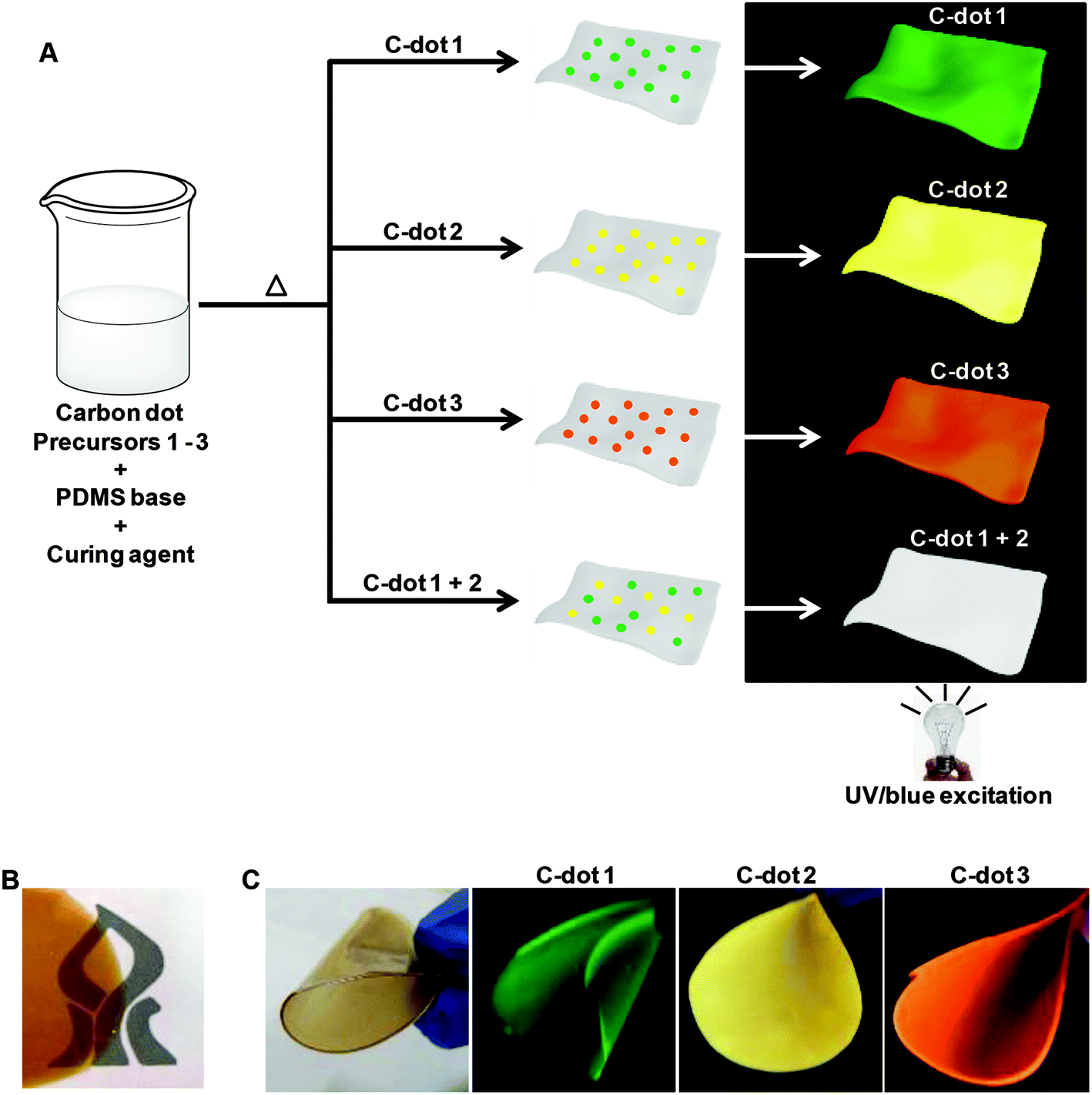 | ||
| Fig. 1 Luminescent C-dot/PDMS films. (A) Scheme depicting the preparation of the mixed films by a one-pot thermal synthesis route. Distinct-coloured C-dots embedded in the films were prepared by using different precursors. The numbers 1–3 correspond to the C-dots indicated in Table 1. (B) C-dot/PDMS film placed on the left side of a dark logo, underscoring film transparency. (C) Photographs of C-dot/PDMS films prepared using different C-dot precursors, yielding distinct colours upon illumination with an ultraviolet lamp (365 nm). The picture on the left depicts the C-dot/PDMS film in regular light. | ||
| Carbon precursor | Maximal emission (excitation wavelength) | Emission coloura | |
|---|---|---|---|
| a Colour observed upon illumination with an UV lamp (365 nm). | |||
| 1 | 6-O-(O-O′-Di-lauroyl-tartaryl)-D-glucose16 | 525 nm (450 nm) | Green |
| 2 | 6-O-(O-O′-Di-lauroyl-tartaryl)-L-ascorbic acid17 | 560 nm (475 nm) | Yellow |
| 3 | Vitamin B1 + oleic acid | 585 nm (475 nm) | Orange |
The image in Fig. 1B shows a C-dot/PDMS film placed upon a paper clip displaying a black-ink logo, demonstrating optical transparency of the film. Fig. 1C highlights the flexibility of the films and the remarkable film colours achieved through synthesis of different PDMS-embedded C-dots. The photographs in Fig. 1C capture the colours recorded upon illuminating the C-dot/PDMS films, prepared using the different precursors outlined in Table 1, with a UV lamp (365 nm excitation). The distinct luminescent colours shown in Fig. 1C further confirm that distinct C-dots exhibiting different emission properties were formed within the polymer films through the one-pot synthesis scheme (Fig. 1A).
The emission characteristics of C-dots are ascribed to the properties of the surface states associated with these nanoparticles.6,7 Indeed, the properties, particularly the energy levels, of the surface states are determined in large part by the carbonaceous building blocks used in the synthesis of the C-dots. Accordingly, the use of different carbon sources as precursors is expected to generate distinct emission properties, such as those shown in Fig. 1 and Table 1.
Fig. 2 presents the microscopy and spectroscopy data characterizing the structural and spectroscopic properties of the PDMS-embedded C-dots. The transmission electron microscopy (TEM) images in Fig. 2A were recorded after extraction of the C-dots from the PDMS matrix by placing in an organic solvent. The C-dots appeared relatively uniform in size, exhibiting diameters of 4.2 ± 0.6 nm (Fig. S1, ESI†). The homogeneity of the C-dots, apparent in Fig. 2A and S1,† is notable since the synthesis strategy (Fig. 1A) did not involve any purification or annealing steps, which are usually required in solution-based procedures in order to attain uniform size and shape distributions of C-dots.6,7 Indeed, the mono-dispersity of the particles suggests that the assembly process of the C-dots was intimately affected by the polymer matrix.
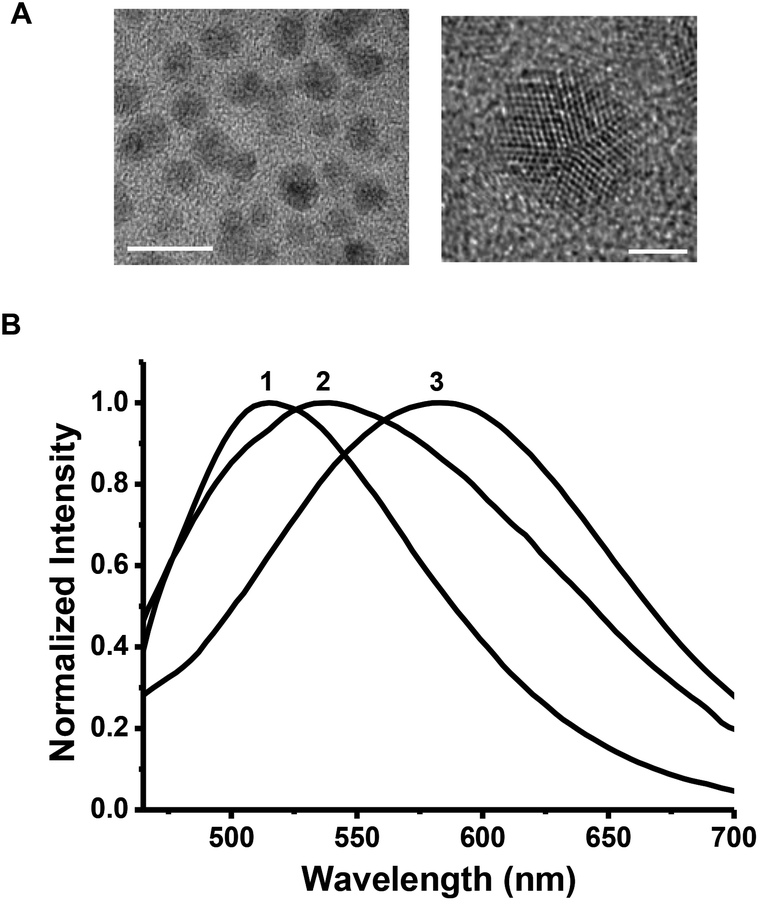 | ||
| Fig. 2 Microscopic and spectroscopic characterization of PDMS-embedded C-dots. (A) High resolution transmission electron microscopy (HR-TEM) images of C-dots (prepared from precursor 1, Table 1) after extraction from the PDMS film. The bars correspond to 10 nm (left image) and 2 nm (right image). (B) Normalized photoluminescence spectra of the C-dots/PDMS films upon excitation at 400 nm. The numbers correspond to the different C-dot precursors indicated in Table 1. | ||
The high resolution TEM (HR-TEM) image depicted in Fig. 2A (right) displays the crystal planes of the graphitic core of the C-dots, providing additional evidence for the integrity of the nanoparticles.16,28 It should be noted that the C-dots analyzed in the TEM experiments in Fig. 2A were prepared from the 6-O-(O-O′-di-lauroyl-tartaryl)-D-glucose precursor (e.g. green luminescent C-dots, Table 1); similar TEM results were obtained in the case of C-dots constructed from the other precursors indicated in Table 1 and synthesized in situ within the PDMS film. X-ray photoelectron spectroscopy (XPS, Fig. S2†), Raman spectroscopy (Fig. S3A†), and Fourier transform infrared (FTIR, Fig. S3B†) analyses confirmed that C-dots exhibiting different surface functional groups (and the corresponding different luminescence properties) were formed within the PDMS matrix according to the synthesis scheme in Fig. 1A.
Fig. 2B depicts the normalized photoluminescence (PL) spectra recorded for C-dot/PDMS films prepared using different C-dot precursors, upon excitation at 400 nm. The PL spectra in Fig. 2B are notable, since the different peak positions associated with each C-dot species indicate that the distinctive colours (i.e. luminescence properties) of the C-dots were retained in the composite C-dot/PDMS films. Indeed, the excitation-dependent emission spectra recorded for the C-dot/PDMS films (Fig. 3) were similar to the corresponding spectra of C-dots in solution, thus confirming that the polymer matrix did not disrupt the photo-physical properties of the embedded C-dots. Similar to other C-dot systems recently reported, the absence of excitation-dependent emission shifts, apparent in Fig. 3iii, might be attributed to a relative uniformity of surface functionalization resulting in narrow distribution of surface energy states.29,30 Quantum yields of 6.5%, 7.4%, and 14.5% were calculated for the green, yellow, and orange C-dots, respectively.
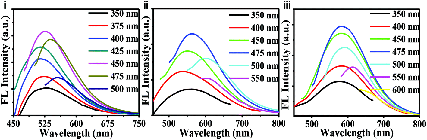 | ||
| Fig. 3 Photoluminescence spectra of C-dot/PDMS films excited at different wavelengths. (i) Green C-dots (precursor 1); (ii) yellow C-dots (precursor 2); and (iii) orange C-dots (precursor 3). | ||
As indicated in Fig. 1 and 2, the PL properties of the C-dot/PDMS films were determined by the C-dot precursors used in the synthesis. Fig. 4 depicts the chromatic properties of C-dot/PDMS films prepared by using different carbon precursors upon illumination with a UV light emitting diode (UV-LED, excitation 403 nm).
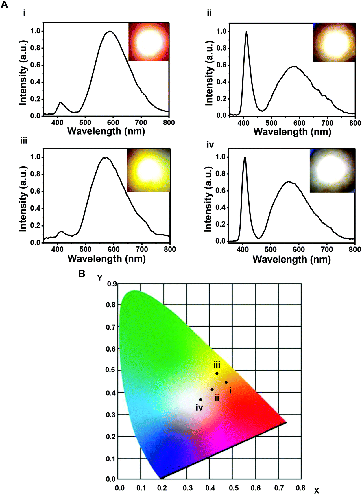 | ||
| Fig. 4 Chromaticity of C-dot/PDMS films. (A) Emission spectra recorded upon illumination of different C-dot/PDMS films with a UV-LED (403 nm) operating at a current of 10 mA. The insets depict the digital images of the light generated. (i) Orange C-dots (precursor 3); (ii) yellow C-dots (precursor 2); (iii) stacking a green C-dot/PDMS (precursor 1) film upon an orange C-dot/PDMS (precursor 3) film; (iv) film containing a mixture of green and yellow C-dots (1 + 2) (white light generated). (B) CIE diagram showing the light coordinates of the respective C-dot/PDMS films depicted in A. | ||
The optical data in Fig. 4 dramatically illustrate the light-tuning capabilities available through either embedding different individual C-dots within the films, stacking two films together, as well as utilizing mixtures of distinct C-dots within single films. Specifically, Fig. 4 presents the emission spectra, respective coordinates in the Commission Internationale de l'Eclairage (CIE) diagram, and digital images recorded upon illuminating the C-dot/PDMS films with the UV-LED. The generation of light exhibiting different emission wavelengths (i.e. different colours) is apparent both in the shifts in the emission spectra (Fig. 4A), the corresponding digital light emission images (insets in Fig. 4A), and CIE coordinates (Fig. 4B). The optical parameters recorded for the different films are summarized in Table 2.
| Film number | C-dots embeddeda | (X,Y)b | CRIc | CCT (K)d |
|---|---|---|---|---|
| a The numbers in parentheses indicate the carbon precursors used, Table 1. b X,Y coordinates in the CIE diagram, Fig. 4B. c Colour rendering index. d Correlated colour temperature. | ||||
| i | Orange C-dots (3) | 0.47, 0.46 | 93 | 2900 |
| ii | Yellow C-dots (2) | 0.41, 0.41 | 74 | 3500 |
| iii | Green C-dots (1); orange C-dots (3) | 0.43, 0.48 | 91 | 3700 |
| iv | Green + yellow C-dots (1 + 2) | 0.36, 0.37 | 80 | 4500 |
Notably, the distinct shifts between the emission peaks reported in Fig. 2B and 3 on the one hand, and Fig. 4 on the other hand, reflect the different setups in the two experiments. Specifically, the data reported in Fig. 2B and 3 were recorded on a spectrofluorimeter. This means that the excitation source was used only for generating emission spectra of the samples (without any transmittance of the irradiating light). In contrast to the emission data, Fig. 4 depicts the results obtained upon illuminating the sample with an LED. Due to the significant intensity of the irradiating light emitted from the LED source, the emission spectra recorded comprise a combination of the emissions of the LED source and sample.
Fig. 4A shows that C-dot/PDMS films prepared using precursors 3 and 2 (Table 1) that were embedded separately produced orange (Fig. 4Ai) and yellowish-white (4Aii) emissions, respectively. The light generated in both cases featured a low light temperature (e.g. “warm” light) and a relatively high colour rendering index (CRI), as outlined in Table 2. Fig. 4Aiii reveals that illuminating with the UV-LED a stack of two PDMS films comprising green C-dots (precursor 1) and orange C-dots (precursor 3), respectively, produced a warm yellow light exhibiting a very high CRI of 91% (Table 2).
CRI of light emitters reflects both the properties of the color-modulating species (C-dots in the case here) and the extent of absorbance/transformation of the illuminating light. In the C-dot/PDMS system, each C-dot exhibits distinct emission properties (e.g. emission maxima and intensity), as well as different interactions with the irradiating light, thereby producing a variety of CRI values. Notably, the full width at half maxima of the C-dot spectra are broad (Fig. 4), providing a further contribution to the potential fabrication of light emitters having high CRI.
A notable feature of the new C-dot/PDMS films, apparent in Fig. 4Aiv, the CIE diagram in Fig. 4B, and Table 2, is the feasibility of generating white light through UV-LED illumination of a PDMS film comprising a mixture of green (1) and yellow (2) C-dots at a weight ratio of 3:1. Importantly, the spectral data in Fig. 4B and the optical parameters in Table 2 indicate that the white light recorded exhibited favorable optical properties, which are comparable or better than other reported white light emitters. In particular, the mixed C-dot/PDMS film emitted warm white light (colour temperatures of <5000 K), and a high CRI (80%). The digital image of the LED-illuminated mixed C-dot/PDMS film in Fig. 4Aiv visually depicts the white light generated. It should be emphasized that while Fig. 4 demonstrates white light emission using the embedded mixture of green and yellow C-dots, it is conceivable that other C-dot combinations would also produce white light [akin to the red-blue-green (RGB) scheme commonly used for generating white light].20,23 Fig. S4,† for example, depicts “cool” white light generated by illumination of a PDMS film comprising green C-dots (e.g. precursor 1, Table 1).
Fig. 4Aiv clearly shows that the emitted light from a PDMS film containing mixed C-dots exhibited chromatic properties, particularly color, that were distinct compared to films comprising the separate C-dots. While we cannot determine unequivocally the extent of interaction between the optical transitions of the C-dots in the mixtures, it is conceivable that this interaction is minimal since the emitted light from the mixed-C-dot/PDMS film exhibits a color that essentially combines the luminescence emission properties of both C-dot species.
The possibility to control the chromatic properties of the emitted light (e.g. CRI, color, temperature) through proper selection of the embedded C-dots and their concentrations within the PDMS matrix is one of the attractive practical features of the new C-dot/PDMS system. Specifically, high CRI values point to the potential of the films for solid state lighting applications providing “true color” capabilities. Moreover, the generation of warm white light is important as such light is the most widely used in consumer applications.
Conclusions
In conclusion, we demonstrate a new strategy for constructing tuneable light emitters, based upon transparent and flexible carbon dot/polymer films prepared through a simple one-pot process. The single step synthetic route constitutes mixing the precursors of both the PDMS host matrix and the C-dots, followed by thermal treatment. This strategy is advantageous compared to other reported approaches in which C-dots are first prepared, then subsequently embedded within transparent matrixes. Indeed, in many instances, already-prepared C-dots cannot be incorporated within host matrixes or their optical properties are adversely affected, for example due to aggregation processes. Moreover, the use of natural, “green” sources for fabrication of the light emitters is important because environmental concerns are currently a major issue in the production processes of light emitting devices.Illuminating the C-dot/PDMS films with ultraviolet light produces emission at different colours, depending upon the types and concentrations of C-dots embedded within the films. Importantly, the synthetic strategy facilitates tuning of light properties and colours through selection of the C-dot precursors. In particular, we demonstrated generation of warm white light through utilization of a PDMS film encapsulating green-emitting and yellow-emitting C-dots. The C-dot/PDMS films can be fabricated in varied sizes and thicknesses, and the technology is scalable.
The new method for construction of tunable, transparent color films could be employed in diverse optical and photonic applications. In particular, the technology might be readily employed for fabrication of solid-state light emitters and photonic devices exhibiting controlled light properties. The variety of C-dots reported in recent years exhibiting a wide luminescence spectral range might enable generation of diverse light colours by the C-dot/PDMS film platform. The C-dot/PDMS system might be also used to produce infrared (IR) (rather than visible) light through embedding pertinent C-dots,31 and possibly even transform light from IR to visible light using up-conversion processes demonstrated for several C-dot species.32
Acknowledgements
Dr Susanta Kumar Bhunia is grateful to the Planning and Budgeting Committee (PBC) of the Israeli Council for Higher Education for an Outstanding Post-doctoral Fellowship. Kreitman School of Advanced Graduate Studies is acknowledged (S. N.). We are also grateful to Shiran Nabha and Roi Pinhas for help with the spectroscopic measurements.Notes and references
- H. S. Jang, H. Yang, S. W. Kim, J. Y. Han, S. G. Lee and D. Y. Jeon, Adv. Mater., 2008, 20, 2696 CrossRef CAS PubMed.
- E. Jang, S. Jun, H. Jang, J. Llim, B. Kim and Y. Kim, Adv. Mater., 2010, 22, 3076 CrossRef CAS PubMed.
- T. Erdem and H. V. Demir, Nat. Photonics, 2011, 5, 126 CrossRef CAS.
- E. Mutlugun, P. L. Hernandez-Martinez, C. Eroglu, Y. Coskun, T. Erdem, V. K. Sharma, E. Unal, S. K. Panda, S. G. Hickey, N. Gaponik, A. Eychmuller and H. V. Demir, Nano Lett., 2012, 12, 3986 CrossRef CAS PubMed.
- S. K. Panda, S. G. Hickey, H. V. Demir and A. Eychmuller, Angew. Chem., Int. Ed., 2011, 50, 4432 CrossRef CAS PubMed.
- W. Kwon, S. Do, J. Lee, S. Hwang, J. K. Kim and S. W. Rhee, Chem. Mater., 2013, 25, 1893 CrossRef CAS.
- X. Y. Zhang, Y. Zhang, Y. Wang, S. Kalytchuk, S. V. Kershaw, Y. H. Wang, P. Wang, T. Q. Zhang, Y. Zhao, H. Z. Zhang, T. Cui, Y. D. Wang, J. Zhao, W. W. Yu and A. L. Rogach, ACS Nano, 2013, 7, 11234 CrossRef CAS PubMed.
- S. Nakamura, T. Mukai and M. Senoh, Appl. Phys. Lett., 1994, 64, 1687 CrossRef CAS.
- H. S. Jang, W. Bin Im, D. C. Lee, D. Y. Jeon and S. S. Kim, J. Lumin., 2007, 126, 371 CrossRef CAS.
- R. J. Xie, N. Hirosaki, M. Mitomo, K. Takahashi and K. Sakuma, Appl. Phys. Lett., 2006, 88, 101104 CrossRef.
- J. K. Park, K. J. Choi, J. H. Yeon, S. J. Lee and C. H. Kim, Appl. Phys. Lett., 2006, 88, 043511 CrossRef.
- J. Lee, H. F. Chen, T. Batagoda, C. Coburn, P. I. Djurovich, M. E. Thompson and S. R. Forrest, Nat. Mater., 2016, 15, 92 CrossRef CAS PubMed.
- M. A. Baldo, D. F. O'Brien, Y. You, A. Shoustikov, S. Sibley, M. E. Thompson and S. R. Forrest, Nature, 1998, 395, 151 CrossRef CAS.
- H. S. Chen, S. J. J. Wang, C. J. Lo and J. Y. Chi, Appl. Phys. Lett., 2005, 86, 131905 CrossRef.
- H. Song and S. Lee, Nanotechnology, 2007, 18, 255202 CrossRef.
- S. Nandi, R. Malishev, K. P. Kootery, Y. Mirsky, S. Kolusheva and R. Jelinek, Chem. Commun., 2014, 50, 10299 RSC.
- S. Nandi, H. J. Altenbach, B. Jakob, K. Lange, R. Ihizane and M. P. Schneider, Org. Lett., 2011, 13, 1980 CrossRef CAS PubMed.
- S. K. Bhunia, A. Saha, A. R. Maity, S. C. Ray and N. R. Jana, Sci. Rep., 2013, 3, 1473 Search PubMed.
- P. K. Sarswat and M. L. Free, Phys. Chem. Chem. Phys., 2015, 17, 27642 RSC.
- K. Jiang, S. Sun, L. Zhang, Y. Lu, A. G. Wu, C. Z. Cai and H. W. Lin, Angew. Chem., Int. Ed., 2015, 54, 5360 CrossRef CAS PubMed.
- B. Chen and J. C. Feng, J. Phys. Chem. C, 2015, 119, 7865 CAS.
- L. Ma, W. D. Xiang, H. H. Gao, L. Pei, X. Ma, Y. Y. Huang and X. J. Liang, J. Mater. Chem. C, 2015, 3, 6764 RSC.
- C. Sun, Y. Zhang, K. Sun, C. Reckmeier, T. Q. Zhang, X. Y. Zhang, J. Zhao, C. F. Wu, W. W. Yu and A. L. Rogach, Nanoscale, 2015, 7, 12045 RSC.
- C. Sun, Y. Zhang, S. Kalytchuk, Y. Wang, X. Y. Zhang, W. Z. Gao, J. Zhao, K. Cepe, R. Zboril, W. W. Yu and A. L. Rogach, J. Mater. Chem. C, 2015, 3, 6613 RSC.
- W. L. Wei, C. Xu, L. Wu, J. S. Wang, J. S. Ren and X. G. Qu, Sci. Rep., 2014, 4, 3564 Search PubMed.
- Y. Wang, S. Kalytchuk, Y. Zhang, H. C. Shi, S. V. Kershaw and A. L. Rogach, J. Phys. Chem. Lett., 2014, 5, 1412 CrossRef CAS PubMed.
- S. Y. Lien, A. Nautiyal and S. J. Lee, Jpn. J. Appl. Phys., 2013, 52, 115801 CrossRef.
- L. B. Tang, R. B. Ji, X. K. Cao, J. Y. Lin, H. X. Jiang, X. M. Li, K. S. Teng, C. M. Luk, S. J. Zeng, J. H. Hao and S. P. Lau, ACS Nano, 2012, 6, 5102 CrossRef CAS PubMed.
- P. Dubey, K. M. Tripathi and S. K. Sonkar, RSC Adv., 2014, 4, 5838 RSC.
- S. J. Zhuo, M. W. Shao and S. T. Lee, ACS Nano, 2012, 6, 1059 CrossRef CAS PubMed.
- H. Q. Tao, K. Yang, Z. Ma, J. M. Wan, Y. J. Zhang, Z. H. Kang and Z. Liu, Small, 2012, 8, 281 CrossRef CAS PubMed.
- Y. Guo, P. J. Yao, D. Q. Zhu and C. Gu, J. Mater. Chem. A, 2015, 3, 13189 CAS.
Footnote |
| † Electronic supplementary information (ESI) available. See DOI: 10.1039/c5nr08400h |
| This journal is © The Royal Society of Chemistry 2016 |