
 Open Access Article
 This Open Access Article is licensed under a Creative Commons Attribution-Non Commercial 3.0 Unported Licence
A pneumatic pressure-driven multi-throughput microfluidic circulation culture system†
T.
Satoh
a,
G.
Narazaki
b,
R.
Sugita
b,
H.
Kobayashi
b,
S.
Sugiura
*a and
T.
Kanamori
a
aBiotechnology Research Institute for Drug Discovery, National Institute of Advanced Industrial Science and Technology (AIST), Central 5th, 1-1-1 Higashi, Tsukuba, Ibaraki 305-8565, Japan. E-mail: shinji.sugiura@aist.go.jp
bDaiichi Sankyo Co. Ltd., 1-2-58, Hiromachi, Shinagawa-ku, Tokyo 140-8710, Japan
First published on 13th May 2016
Abstract
Here, we report a pneumatic pressure-driven microfluidic device capable of multi-throughput medium circulation culture. The circulation culture system has the following advantages for application in drug discovery: (i) simultaneous operation of multiple circulation units, (ii) use of a small amount of circulating medium (3.5 mL), (iii) pipette-friendly liquid handling, and (iv) a detachable interface with pneumatic pressure lines via sterile air-vent filters. The microfluidic device contains three independent circulation culture units, in which human umbilical vein endothelial cells (HUVECs) were cultured under physiological shear stress induced by circulation of the medium. Circulation of the medium in the three culture units was generated by programmed sequentially applied pressure from two pressure-control lines. HUVECs cultured in the microfluidic device were aligned under a one-way circulating flow with a shear stress of 10 dyn cm−2; they exhibited a randomly ordered alignment under no shear stress and under reciprocating flow with a shear stress of 10 dyn cm−2. We also observed 2.8- to 4.9-fold increases in expression of the mRNAs of endothelial nitric oxide synthase and thrombomodulin under one-way circulating flow with a shear stress of 10 dyn cm−2 compared with conditions of no shear stress or reciprocating flow.
Introduction
Blood vessels are the part of the circulatory system which transports nutrients and oxygen in the body and exchanges waste. Endothelial cells (ECs) form the inner walls of blood vessels and are continuously exposed to shear stress induced by blood flow. In the past few decades, the effects of this shear stress on vascular ECs have been investigated.1,2 It has been revealed that shear stress affects various physiological responses mediated by ECs, including vasodilation,3 vasoconstriction,4 anticoagulant activity,5 and vascular permeability.6 In these studies, ECs were cultured under shear stress by using characteristic culture systems including a cone-plate,7–9 parallel plates,10–12 or capillaries.13,14 These traditional culture systems can provide physiological shear stress conditions in vitro. However, they have not been used extensively in drug discovery research because they are both cumbersome and complicated. For example, most of these systems consist of a culture vessel, pump, medium reservoir, and transfer tubes. This configuration challenges the operator when performing even mandatory routine tasks such as cell observation and medium exchange. More importantly, throughput in these complicated culture systems has been a bottleneck in their application to drug discovery. Ideally, it should be possible to evaluate multiple drug candidates simultaneously in a physiological culture system when screening for superior drug candidates.Recently, there has been interest in the use of microfluidic devices as novel cell-culture platforms to reduce time and costs in drug discovery owing to their advantages of small-scale analysis and efficient liquid handling.15,16 More recently, microfluidic devices called “organs-on-a-chip,” which provide physiological and pathological tissue or organ functions in vitro, have been attracting attention as next-generation research tools for performing reliable assays using human-derived cells.17–20 Microfluidic devices that can provide multiple shear stresses over a wide range of ECs have been reported.21–24 The use of microfluidic devices for culturing kidney epithelial cells or brain ECs under physiological shear stress has also been reported in the evaluation of drug toxicity and permeability.25,26 In some of these studies, the medium is circulated through the microfluidic device by using a rotary peristaltic pump,21–25 which requires a large volume of medium due to the off-chip nature of the medium circulation apparatus and tube connections. The circulating medium volume has been reduced by using an integrated pneumatic peristaltic micropump27 or piezoelectric Braille pins.28 However, these microfluidic devices are used in the analysis of only one drug candidate by a single device: complicated operation of microvalves is required to analyze multiple drug candidates. Therefore, parallelization of medium circulation on the microfluidic device is the key to applying microfluidic circulation culture systems to drug discovery research.
As other examples, multi-organs-on-a-chip and body-on-a-chip have recently been developed for culture of multiple types of cells in discrete microchambers connected to each other via microchannels to circulate medium.29–33 The medium is circulated on these devices by using an off-chip peristaltic pump,29 integrated pneumatic peristaltic micropump,30–32 or integrated stirrer-based micropump.34 Notably, none of these studies have demonstrated the circulation of multiple liquids without increasing the number of pumps or tube connections. As far as we know, only one example has been reported for the parallelization of medium circulation in microfluidic devices; in this example, two sets of exactly the same microfluidic networks and pneumatic peristaltic micropumps were fabricated on a single device.30 Further increases in the number of parallelizations by simply increasing the number of circulation microfluidic networks and pumps will require complicated system setups and operation. Therefore, parallelization of medium circulation in microfluidic devices with simple system setups is still a big challenge.
In our previous study, we developed a pressure-driven perfusion-culture microchamber array device,35 in which pneumatic pressure was applied to a reservoir and multiple liquids in multiple wells were simultaneously delivered into the microchamber array. The advantage of using pneumatic pressure to deliver multiple liquids is that the pressure can be easily distributed to multiple wells in a reservoir with a common gas-phase space without increasing tube connections (Fig. S1 in the ESI†). In this study, we expanded this idea to parallelization of medium circulation: multiple medium-circulation units on a single microfluidic device were driven by only two pneumatic pressure lines. This new microfluidic device contained three independent culture units, in which cells were cultured under medium circulation flow. One-way circulation of medium was generated by applying sequential pressure to the microfluidic network by using passive one-way check valves. We used our system to culture human umbilical vein endothelial cells (HUVECs) under physiological shear stress caused by circulation of the medium. We also compared the cellular functions of HUVECs under one-way circulating flow with those under reciprocating flow and static culture conditions.
Experimental
Pneumatic pressure-driven circulation culture system
A schematic of the concept used to parallelize the circulation culture units driven by pneumatic pressure is shown in Fig. 1. Compressed air containing 5% CO2, from a gas cylinder, was used as a pressure source to provide the same atmospheric conditions as in a cell-culture incubator. A pressure line from the gas cylinder was branched to two lines in a sequential pressure-control system (ASTF0301, Engineering System Co., Ltd., Matsumoto, Japan), and the two lines were connected to a single culture device placed in a cell-culture incubator maintained at 37 °C under 5% CO2. Sequential processes of pressurization to a predetermined pressure and pressure release to atmospheric pressure were generated in a programmed manner. The applied pressure was controlled within the range of 5 kPa to 20 kPa using the sequential pressure-control system. The three circulation culture units on the single device were simultaneously driven by two pneumatic pressure lines. The pneumatic pressure lines could be branched to increase the number of culture devices (Fig. 1). The pneumatic pressure-driven circulation culture device was composed of air vent filters (Ekicrodisc 3CR, Gelman Sciences Japan Ltd., Tokyo, Japan), polytetrafluoroethylene screw caps, a polycarbonate reservoir cover, a polycarbonate reservoir box, an aluminum holder, and a microfluidic polystyrene culture plate (Fig. 2a). These components were assembled with stainless-steel clips and tightly sealed against each other with elastomeric O-rings to form the circulation culture device. The screw caps, reservoir cover, reservoir box, and holder were fabricated by machining. Three sets of storage and feed reservoirs were fabricated in the reservoir box. The volume of each reservoir was 3.5 mL; this determined the maximum volume of medium in each circulation unit. Each circulation culture unit was composed of microfluidic channels in the culture plate, reservoirs, and check valves (IMCD116P, International Scientific Instruments Supply Co., Ltd., Osaka, Japan) (Fig. 2b). Each storage reservoir and each feed reservoir had two open holes at the bottom and were connected to the culture and return channels via these holes. A one-way passive check valve on one of the open holes in the storage or feed reservoir allowed unidirectional (clockwise direction, as shown in Fig. 2b) medium circulation by sequential application of pressure. Three sets of circulation microfluidic networks were fabricated on the culture plate (Fig. 2c). Each circulation microfluidic network was composed of culture channels, a return channel, and connecting channels. Each culture channel was designed to have a cell-culture surface area of 4.2 cm2, which corresponds to the culture area for 2.0 × 10⁵ cells in conventional static culture. The ratio of medium volume to culture surface area in the culture device was 0.71 mL cm−2, which is comparable to that in conventional cell-culture dishes. For example, the medium volume and culture area for the 35 mm dish are usually 2 mL and 9 cm2, and the ratio of medium volume to culture surface area is 0.22 mL cm−2. The width and height of the culture channels were designed to be 1500 and 200 μm, respectively, and those of the connecting channels were designed to be 250 and 200 μm, respectively, to provide physiological shear stress, which is in the order of 10 dyn cm−2 in human carotid arteries,36 by application of a physiological pressure of 14.4 kPa. To determine the relationship between shear stress and applied pressure, analytical solutions to Navier–Stokes equations in a microchannel with a rectangular cross-section37 were used, as described in our previous study.22 Fluid shear stress was calculated according to the equation for a microchannel with a rectangular cross-section: τ = 6μQC/wchC2, where μ is the fluid viscosity of water, QC is the flow rate in the culture channel, and wC and hC are the width and height, respectively, of the culture channel.37 In addition, 24 shallow microchannels, which we called “Laplace valves,” were radially arranged around the storage reservoir–side aperture of the return channel (Fig. 2b, c and S2a in the ESI†) to prevent introduction of the gas phase into the return channel (Fig. S2b and c in the ESI†). The width and height of the shallow microchannels in the Laplace valve were designed to be 200 and 25 μm, respectively. The break pressure of the Laplace valve (ΔPLap) was calculated to be 5.4 kPa from the Young–Laplace equation: ΔPLap = 2γ(1/wL + 1/hL), where γ is the interfacial tension of the medium (60 mN m−1) and wL and hL are the width and height, respectively, of the shallow microchannels in the Laplace valve. The culture plate was fabricated by injection molding, bonded with a 50 μm-thick polystyrene film (Enplas Corp., Saitama, Japan), and sterilized with gamma-ray irradiation. The width and height of the fabricated channels were measured using microscopy and laser interferometry, respectively. Fig. 2d displays an external view of the assembled device.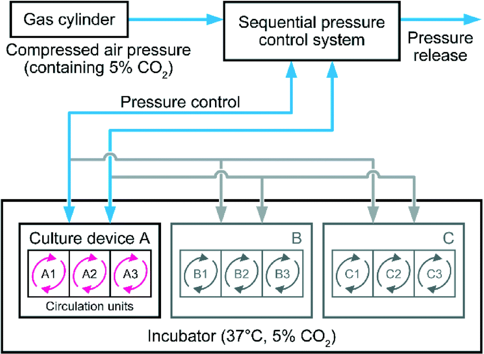 | ||
| Fig. 1 Pressure-driven circulation culture system for parallelized medium circulation. Air under pressure and containing 5% CO2 was introduced into a sequential pressure-control system from a gas cylinder. Sequential pressure was applied to the cell-culture device, which was placed in a thermostatic incubator at 37 °C under 5% CO2, via two pressure lines. Blue arrows indicate the pneumatic lines and direction of gas flow. Red arrows indicate circulation of medium in the culture units on the culture device. Gray arrows indicate additional pneumatic lines and medium circulation for additional culture devices, which can be added as necessary. | ||
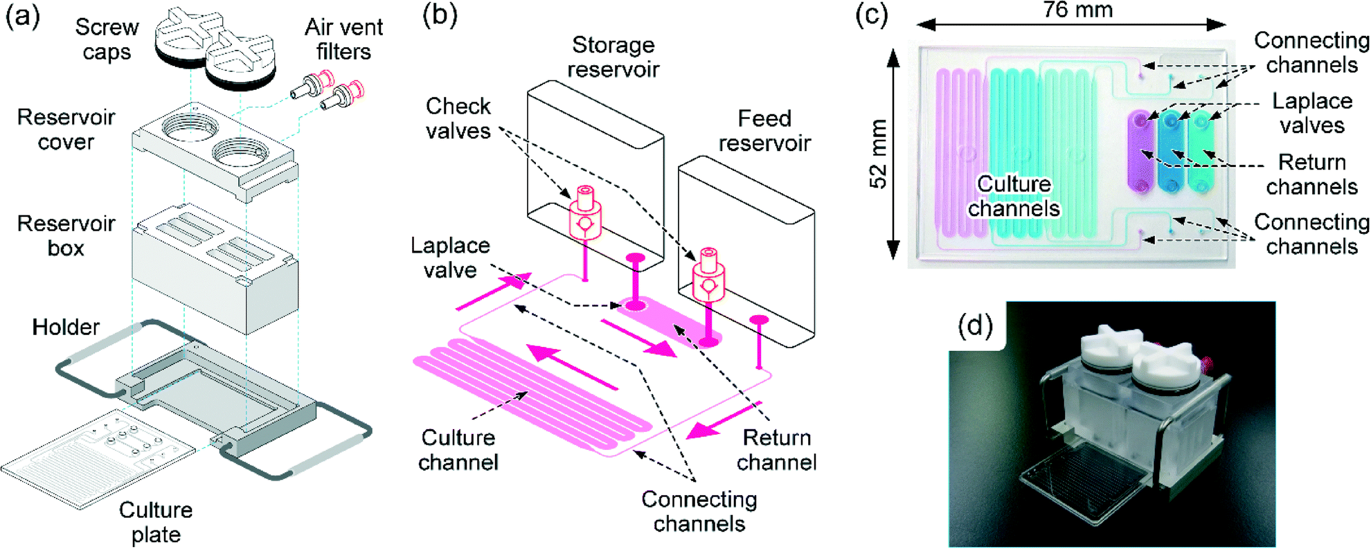 | ||
| Fig. 2 Schematic of the pneumatic pressure-driven circulation culture device. (a) Components of the cell culture device. (b) Layout of a single circulation unit. Pink arrows indicate the direction of medium circulation flow. (c) Top view of a culture plate. The three circulation units are indicated by different-colored solutions. (d) External view of an assembled device. | ||
Operating procedure for circulation of medium
The medium was circulated by a sequential pressurizing process of feed and return steps (Fig. 3). In the feed step, pneumatic pressure applied to the feed reservoir induces flow of the medium from the feed reservoir to the storage reservoir via the culture channel, while the check valve in the feed reservoir prevents flow into the return channel (Fig. 3a). At the end of the feed step, some of the medium should remain in the feed reservoir to avoid introduction of air into the culture channel (Fig. 3b). In the return step, pneumatic pressure lower than ΔPLap is applied to the storage reservoir. This induces flow of medium from the storage reservoir to the feed reservoir via the return channel, while the check valve in the storage reservoir prevents flow into the culture channel (Fig. 3c). At the end of the return step, all of the medium in the storage reservoir is transferred into the feed reservoir (Fig. 3d). At this point, the Laplace valve prevents the introduction of air and the formation of bubbles in the return channel (details of the working mechanism of the Laplace valve are shown in Fig. S2b and c in the ESI†). Repeating the feed and return steps provides intermittent unidirectional circulating flow in the culture channel.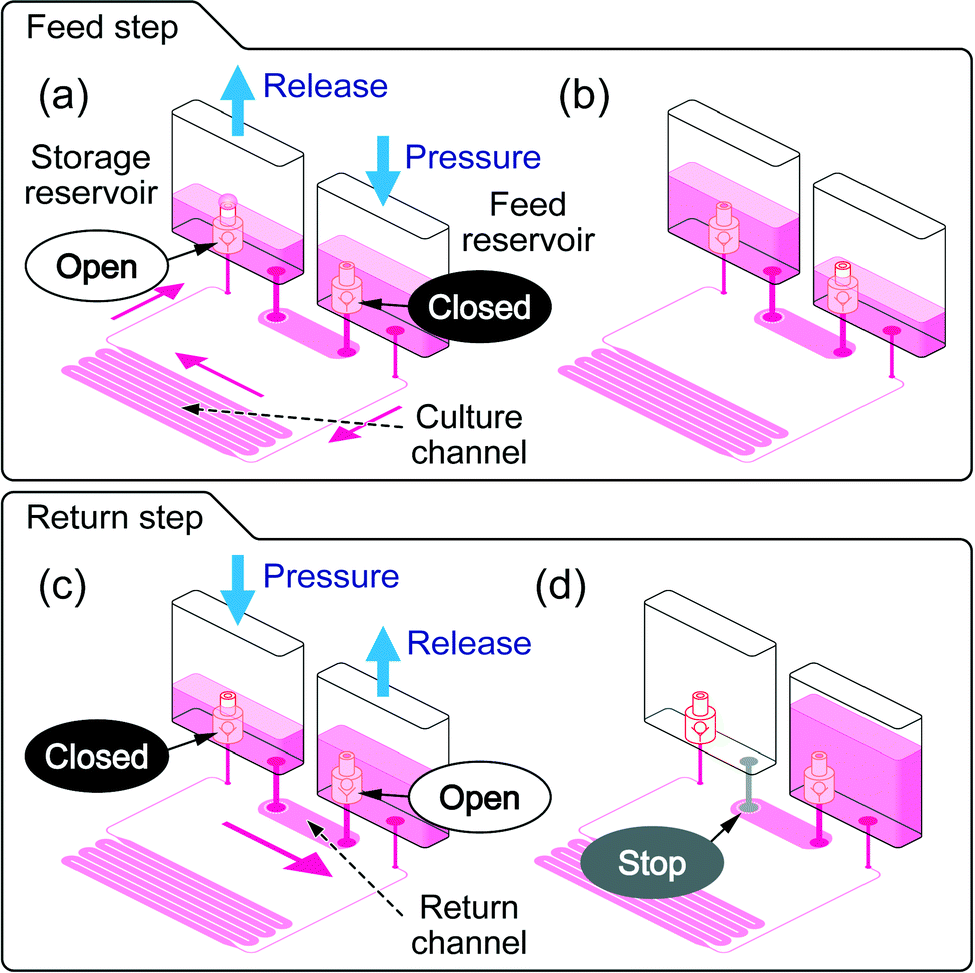 | ||
| Fig. 3 Operating procedure for circulation of medium by using a sequential pressurizing process. During (a) and at the end of (b) the feed step. During (c) and at the end of (d) the return step. Blue arrows indicate pressurization of, and pressure release from, the reservoirs. Red arrows indicate the direction of flow of the medium. “Open” and “Closed” in (a) and (c) indicate check valve functions. “Stop” in (d) indicates the Laplace valve function. | ||
In the return step, the cells are not exposed to shear stress. To shorten the return step, we incorporated as many as 24 Laplace valves and a return channel as thick as 6500 × 500 μm2 measuring in cross-section to increase the flow rate during this step.
The pneumatic pressure applied to the reservoir box is distributed to the three sets of circulation culture units via the common gas-phase space in the reservoir cover (Fig. S1 in the ESI†). Therefore, three sets of circulation culture units are operated by a single set of two pneumatic pressure lines.
Flow simulation
We performed a computational analysis of shear stress by using simulation software (COMSOL Multiphysics 3.3a, COMSOL AB, Stockholm, Sweden). The Navier–Stokes equations were solved in a finite element model built for a culture channel with the following boundary conditions: a homogeneous flow rate of 950 μL min−1 at the channel inlet, zero pressure at the channel exit, and no slip boundary on the walls. The cross-sectional dimensions of the culture channels were 1500 × 200 μm2.Flow characterization
To estimate the working pressure range in the return step, we experimentally measured the actual value of ΔPLap for the microfluidic device. The storage reservoir and return channel were filled with medium and a pressure of 4 kPa was applied to the storage reservoir. After the transfer of all of the medium to the feed reservoir, the pressure applied was gradually increased. The pressure at which the air phase passed through the return channel was determined as the ΔPLap. Experiments were performed in triplicate by using three different culture plates at room temperature.To estimate the actual shear stress in the culture channel during the feed step, we experimentally measured the flow rate of the medium in the culture channels. The flow rate was measured by determining the weight of medium accumulated in the storage reservoir after applying pressure to the feed reservoir. Experiments were performed nine times using three different culture plates at room temperature. The shear stress at 37 °C was estimated from the measured flow rate at room temperature by correcting for the temperature dependency of the viscosity of water.
We measured the flow rate of medium in the return channel when we applied 4.4 kPa of pressure to the storage reservoir at room temperature. Twenty-seven experiments were performed using three different culture plates. The flow rate per unit pressure in a return channel was calculated, and the flow rate at 37 °C was estimated from the data obtained at room temperature by correcting for the temperature dependency of the viscosity of water.
Cell culture in the circulation culture system
HUVECs were obtained from Kurabo Industries, Ltd. (Osaka, Japan) and maintained in growth medium (HuMedia-EB2, Kurabo) containing 2% fetal bovine serum and growth supplements (KE-6150, Kurabo). The air vent filters, screw caps, reservoir cover, reservoir box, and holder were autoclaved and dried in advance. The circulation culture device was assembled in a tissue culture hood. The culture channel was coated with type I collagen (Cellmatrix type I-C, Nitta Gelatin Co., Ltd., Osaka, Japan), as follows: the culture channel was filled with collagen solution (0.3 mg mL−1 in aqueous HCl, pH 3.0) and kept at room temperature for 1 h. After replacement of the collagen solution with growth medium, a suspension of HUVECs (2.0 × 10⁶ cells per mL, 300 μL) was injected into the culture channels and kept static for cell adhesion for 2 h in the cell-culture incubator. Excess suspension containing unattached cells and debris was removed by changing the growth medium in the culture channel. Growth medium (3 mL) was poured into the storage reservoir; most of the medium was then transferred to the feed reservoir via the return channel to remove the air in the return channel. The process of medium circulation under one-way flow conditions was performed by repeating the (i) feed and (ii) return steps, as follows: (i) pressure was applied to the feed reservoir at 3.7 or 18.7 kPa, which correspond to a shear stress of 2 or 10 dyn cm−2, and pressure to the storage reservoir was released to atmospheric pressure for 45 s to induce flow of medium from the feed reservoir to the storage reservoir via the culture channels. After 45 s, some amount of medium remained in the feed reservoir, which prevents the introduction of air into the culture channel. (ii) Pressure was applied to the storage reservoir at 5.5 kPa and pressure to the feed reservoir was released to atmospheric pressure for 15 s to induce flow of medium from the storage reservoir to the feed reservoir via the return channel. In a human body, shear stress in blood vessels depends on the type of vessel, and is 10–20 dyn cm−2 in aorta, 5 dyn cm−2 in small arteries, 2.5 dyn cm−2 in capillary vessels, and 1–5 dyn cm−2 in venous systems.38 Based on this information, we adopted 10 dyn cm−2 as the high shear stress condition in aortic vessels and 2 dyn cm−2 as the low shear stress condition in microvessels and veins.Static culture was performed as a control experiment. A 12-well plate was coated with type I collagen (Nitta Gelatin). HUVECs were seeded onto the 12-well plate at the same number of cells per unit area as in the culture channels. The volume of medium per unit area was also adjusted to make it the same as in the circulation culture.
Reciprocating perfusion culture was performed as a comparison. For reciprocating perfusion, the check valves in the reservoir box were removed and both ends of the return channel were plugged with epoxy resin. Collagen coating and cell seeding were performed in the same manner as in the circulation culture. In the reciprocating perfusion culture, we repeated the following four steps: (i) pressure was applied to the feed reservoir at 18.7 kPa and pressure to the storage reservoir was released to atmospheric pressure for 45 s to induce flow of medium from the feed reservoir to the storage reservoir via the culture channels. (ii) The pressure applied was released to atmospheric pressure for 15 s to keep the medium in the culture channels without flow. (iii) Pressure was applied to the storage reservoir at 18.7 kPa and pressure to the feed reservoir was released to atmospheric pressure for 45 s to induce flow of medium from the storage reservoir to the feed reservoir via the culture channels. (iv) The pressure applied was released for 15 s to keep the medium in the culture channels without flow.
Cell count and morphological analysis
After 2 days of one-way circulation, static, or reciprocating perfusion culture, cells were collected by trypsin–EDTA treatment. The number of cells was counted in triplicate for each experiment and the number of cells per unit area was calculated.Cell orientation was evaluated by measuring the angle between the flow direction and the long axis of the cell. After 2 days of culture, phase-contrast microscopy images of cells were taken under an inverted microscope (IX71, Olympus, Tokyo, Japan). In each experiment, images of 100 cells were analyzed to determine the cell orientation. For the static culture control, an arbitrary line was chosen as the zero line, instead of using the flow direction in circulation or reciprocating culture.
Fluorescence staining of HUVECs in the circulation culture system
In order to demonstrate cell staining in the circulation culture system, the HUVECs in the circulation culture system were stained with calcein-AM (Life Technologies, California, USA), or with fluorescent phalloidin (Alexa Fluor 594 Phalloidin, Life Technologies, California, USA) and 4′,6-diamidino-2-phenylindole (DAPI) (Dojindo Laboratories, Kumamoto, Japan). All procedures were conducted in the circulation culture system.For live cell staining, the cells were stained with calcein-AM solution in phosphate buffer saline (PBS) (1 μM, 300 μL) for 30 min in the dark at room temperature after washing the feed and storage reservoirs with PBS (3 mL). Then, the cells were washed with PBS (300 μL) and fluorescence images were taken with a fluorescence microscope (Biorevo BZ9000, Keyence, Osaka, Japan).
For filamentous actin (F-actin) staining, after washing the feed and storage reservoirs with PBS (3 mL), the cells were fixed with 4% paraformaldehyde in PBS (300 μL) for 15 min and then washed with PBS (300 μL). The cells were permeabilized with 0.5% Triton X-100 in PBS (300 μL) for 15 min and washed with PBS (300 μL). Then, the cells were stained with fluorescent phalloidin and DAPI in PBS (100 nM each, 300 μL) for 30 min in the dark at room temperature, and washed with PBS (300 μL). The fluorescence images were taken with a fluorescence microscope (Biorevo BZ9000). As a comparison, static culture was performed in a 24-well plate and live cells, F-actin, and nucleus were stained using the same solutions and incubation time described above.
Gene expression analysis
After 2 days of one-way circulation, static, or reciprocating perfusion culture, cells were collected by trypsin–EDTA treatment. Expression levels of mRNAs for endothelial nitric oxide synthase (eNOS) and thrombomodulin (THBD) in the collected cells were measured by real-time PCR as described in our previous report.22 Both of these proteins are associated with endothelial functions and their production is upregulated by fluid shear stress; eNOS is an enzyme involved in the production of nitric oxide, which is an important mediator of the induction of vasodilation,39 and THBD is a membrane glycoprotein that plays an important role in blood coagulation.40 Experiments were performed in triplicate for each set of experimental conditions.Statistical analysis
Experimental data were analyzed by using one-way ANOVA with post-hoc Tukey–Kramer testing using GraphPad Prism software (GraphPad Software, Inc., La Jolla, USA).Results
Circulation culture system
The circulation culture device that we developed was an assemblable type; the culture plate was a single-use product and the other components were autoclavable and reusable (Fig. 2a). Device assembly was a simple process in which the holder, culture plate, reservoir box, and reservoir cover were positioned and then fastened with clips. Tight sealing with O-rings between each component enabled us to avoid leakage of pressure or liquid. Solutions for coating, washing, and culture could be loaded into the circulation culture device with a micropipette in a tissue culture hood by opening the screw caps on the reservoir cover. The culture device was connected to the pressure lines via Luer fittings on the air vent filters. These connections were detachable for liquid manipulation in the tissue culture hood and observation under a microscope. Pneumatic pressure was applied via the sterile air vent filter. Therefore, sterile conditions could be maintained throughout the entire cell-culture procedure.Characterization of flow in the circulation culture device
The widths and heights of the culture channels, return channels, connecting channels, and the thin microchannels in the Laplace valves in the fabricated device are shown in Table 1. The actual value of ΔPLap in the device was measured experimentally. Bubbles appeared in the feed reservoir during the return step at pressures higher than 5.6 ± 0.0 kPa (mean ± S.D., n = 3), which was determined as the ΔPLap of the Laplace valves. This pressure value was within a tractable range for operating the circulation culture device.| Designed value [μm] | Measured value [μm] | |||
|---|---|---|---|---|
| Width | Height | Width | Height | |
| a Width was measured at the narrow end of the shallow microchannels in the Laplace valves. | ||||
| Culture channels | 1500 | 200 | 1485 ± 5 | 209 ± 1 |
| Return channels | 6500 | 500 | 6491 ± 17 | Not measured |
| Connecting channels | 250 | 200 | 256 ± 3 | 186 ± 19 |
| Laplace valvesa | 200 | 25 | 201 ± 3a | 29 ± 1 |
The average height of the culture channels in the fabricated device was 209 ± 1 μm (Table 1); the uniform height of the culture channels was expected to induce a homogeneous shear stress in these channels in the feed step. Flow simulation also indicated that most of the cell growth area was exposed to homogeneous shear stress during the feed step (Fig. 4a). Variations in shear stress appeared mainly in the curved areas of the culture channels (enlargement in Fig. 4a).
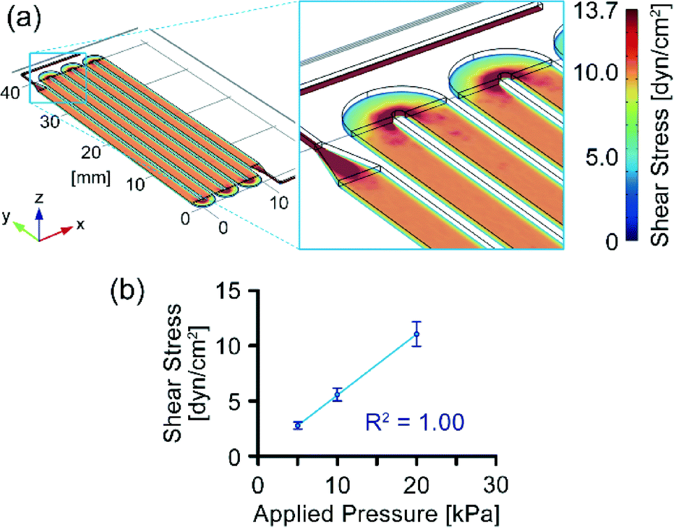 | ||
| Fig. 4 Characterization of flow in the circulation culture device. (a) Distribution of shear stress in the culture channels. (b) Relationship between gas pressure applied in the feed reservoir and fluid shear stress in the cell-culture channel at 37 °C. Error bars indicate standard deviations (n = 9). | ||
Flow rates in the culture channels were measured at applied pressures of 5, 10, and 20 kPa, and shear stress was calculated from the flow rates in, and actual dimensions of, the culture channels (Table 1 and Fig. 4b). The linear relationship between the pressure applied and shear stress indicated that shear stress in the culture channels could be controlled by varying the pressure applied. The coefficient of variation of flow rate for the three culture units under each of the three experimental conditions was within 12%. This variation was likely due to variations in height of the connecting channels caused by the limited accuracy of machining fabrication of the master mold for the injection molding (Fig. S3 in the ESI†). Flow rates in the return channels per unit pressure were measured at room temperature and calculated to be 2.0 ± 0.2 mL min−1 kPa−1 at 37 °C (mean ± S.D., n = 27).
Flow rates in the culture and return channels differed with a 12% coefficient of variation between each culture unit in the culture device. If medium were left in both of the reservoirs after both the feed step and the return step, the remaining amount of medium would be expected to differ up to 12% of the flow volume after each step, and this difference would therefore accumulate with repetition of the circulation sequence. To avoid the accumulation of this difference, we adopted an operational sequence that transferred all of the medium from the storage reservoir to the feed reservoir in the return step. This sequence enabled stable and simultaneous operation of the three circulation units by using a single set of two pneumatic pressure lines.
One-way circulation culture
We investigated the effects of shear stress on HUVECs in the one-way circulation culture system (Fig. 5). Circulation culture with a shear stress of 2 or 10 dyn cm−2 was performed by repeating the feed step to apply a pressure of 3.7 or 18.7 kPa for 45 s and the return step for 15 s (Fig. 5a and b). After 2 days of culture, HUVECs covered the bottom surface of the culture channel. HUVECs under shear stress had a spindle-shaped morphology compared with those under static culture conditions (0 dyn cm−2) and the long axes of 80% of them were aligned within ±30° of the direction of flow under shear stresses of 2 and 10 dyn cm−2. No orientation of cells was observed without shear stress (Fig. 5c and d). Relative expression levels of the mRNAs for eNOS and THBD in the cultured cells increased significantly with increasing shear stress, and 2.8- and 4.9-fold increases for eNOS and THBD were observed under shear stresses of 10 dyn cm−2 (Fig. 5e). Cell density after 2 days of culture did not differ markedly among 0, 2, and 10 dyn cm−2 (Fig. 5f). These results indicate that our pneumatic pressure-driven circulation culture system is capable of inducing endothelial responses to shear stress.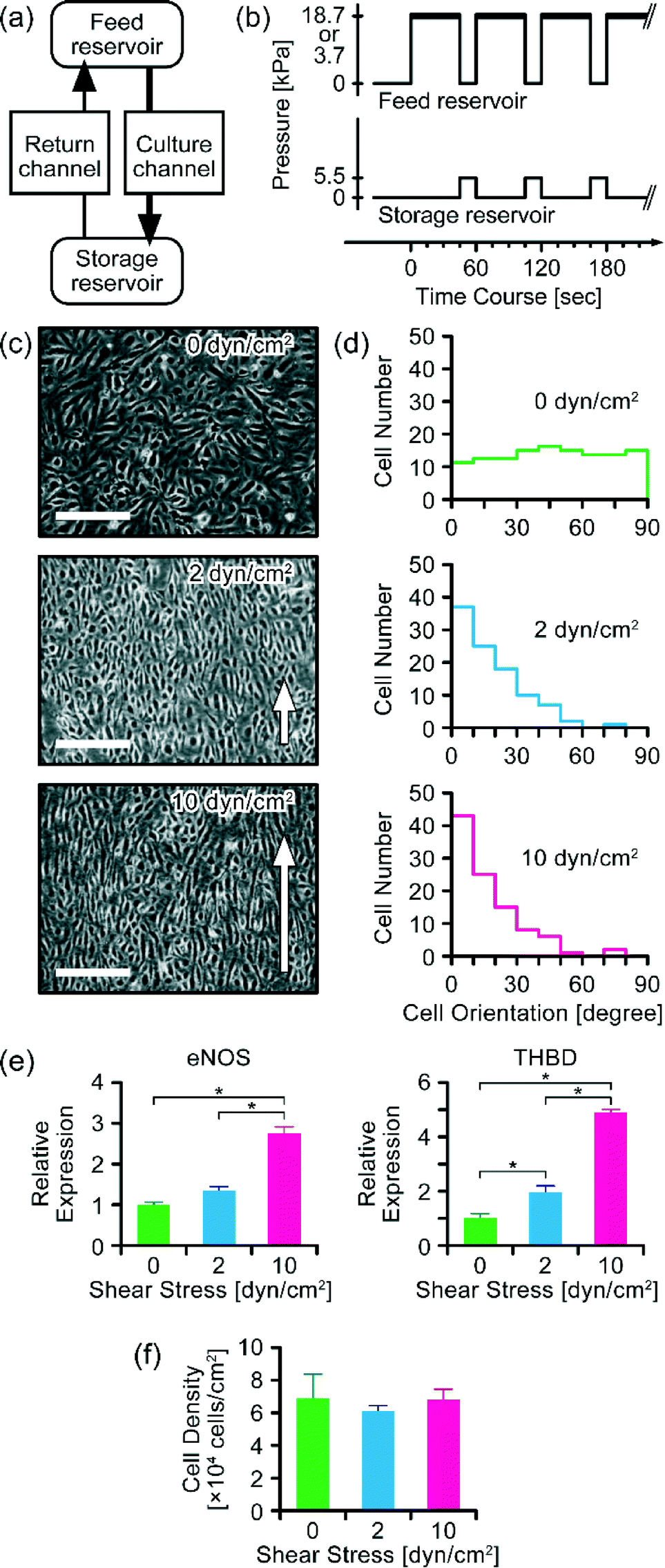 | ||
| Fig. 5 Effects of shear stress on HUVECs in one-way circulation culture. (a) Schematic of flow of medium in the circulation culture device. (b) Schematic of the pressure sequence used for circulation culture. Thick lines in the pressure sequence applied to the feed reservoir indicate periods of exposure of cells to fluid shear stress. (c) Microscopy images of HUVECs after 2 days of culture. Scale bars: 300 μm. (d) Orientation of HUVECs after 2 days of culture. (e) Relative mRNA expression levels of eNOS and THBD after 2 days of culture. (f) Cell densities after 2 days of culture. Error bars indicate standard deviations. Asterisks in (e) indicate significant differences (P < 0.05), (n = 2 or 3). | ||
Reciprocating perfusion culture
We investigated the effects of reciprocating flow on HUVECs and compared them with the effects of circulating flow (Fig. 5 and 6). Reciprocating perfusion culture with a shear stress of 10 dyn cm−2 was performed by repeating alternate pressurization of the feed and storage reservoirs at 18.7 kPa for 45 s, with 15 s intervals (Fig. 6a and b). This operational sequence provided the same shear stress and intermittence of flow as in the circulation culture with a shear stress of 10 dyn cm−2. After 2 days of culture, HUVECs covered the bottom surface of the culture channel. Unlike one-way circulating flow, reciprocating flow induced slight morphological changes but no obvious change in cell orientation after 2 days of culture (Fig. 6c and d). Furthermore, no significant increases in expression levels of the mRNAs of eNOS and THBD were observed under reciprocating flow (Fig. 6e). In addition, no significant differences in cell density were observed between one-way circulating flow and reciprocating flow (Fig. 6f). These results indicate that one-way flow is the key to inducing endothelial responses to shear stress. | ||
| Fig. 6 Comparison of effects of one-way circulating flow and reciprocating flow on HUVECs. (a) Schematic of reciprocating flow in the culture channels. (b) Schematic of the pressure sequence used in reciprocating flow culture. Thick lines in the pressure sequence applied to the feed reservoir indicate periods of flow in the forward direction. Broken lines in the pressure sequence applied to the storage reservoir indicate periods of flow in the reverse direction. (c) Microscopy images of HUVECs after 2 days of culture. Scale bars: 300 μm. (d) Orientation of HUVECs after 2 days of culture. (e) Relative mRNA expression levels of eNOS and THBD after 2 days of culture. (f) Cell densities after 2 days of culture. Asterisks in (e) and (f) indicate significant differences (P < 0.05), (n = 3). | ||
Fluorescence staining of HUVECs in the circulation culture system
The connections between our circulation culture system and the pressure lines are detachable. These convenient interfaces enabled fluorescence staining after cultivation as well as microscopic observation. Living HUVECs were stained with calcein-AM (Fig. 7a). HUVECs were fixed in the culture channels and stained with fluorescent phalloidin for F-actin and DAPI for the nucleus (Fig. 7b). We could observe aligned cells under a shear stress of 10 dyn cm−2 in the circulation culture system using a fluorescence microscope. | ||
| Fig. 7 Fluorescence staining of HUVECs cultured under static culture conditions (0 dyn cm−2) and under shear stress in the circulation culture system (10 dyn cm−2). (a) HUVECs stained with calcein-AM for living cell staining. (b) HUVECS stained with fluorescent phalloidin for F-actin in red and DAPI for the nucleus in blue. Scale bars are 300 μm in (a) and 100 μm in (b). | ||
Discussion
The use of pneumatic pressure to drive multiple liquids enabled us to parallelize circulation of medium in a single microfluidic device with a simple setup (Fig. 1 and S1 in the ESI†). Intermittent one-way circulating flow was generated by programmed sequential pressure applied to a culture device equipped with passive one-way check valves. A further increase in the number of devices is possible by branching the pneumatic pressure lines without increasing the number of pressure control systems (Fig. 1). Our pneumatic pressure-driven circulation culture system also has the advantage of using a small volume of medium compared with that required by circulation systems with off-chip peristaltic pumps that have often been used in previous studies,21–25 because our system does not require connection to a tube filled with medium. In fact, the medium volume per unit growth area in our circulation culture was comparable to that in conventional culture dishes. The circulation culture device could be assembled easily with clips (Fig. 2). The medium and cell suspension could be loaded in a tissue culture hood via micropipettes by opening the screw caps. The device was easily attached to and detached from the pneumatic pressure lines via Luer fittings on the sterile air vent filters. The pipette-friendly liquid manipulation and the detachable connections to the pneumatic lines dramatically improved usability compared with that of previous microfluidic devices that have used rotary peristaltic pumps.21–25 The detachable connection enabled microscopic observation, aseptic manipulation for medium exchange, and cell staining in the context of microplate use. This convenient system setup has already enabled biologists in the pharmaceutical industry to use microfluidic devices routinely.Unlike previous microfluidic circulation culture devices using integrated pneumatic peristaltic micropumps,27,30–32 piezoelectric Braille pins,28 or integrated stirrer-based micropumps,34 our system has the advantages of parallelization of circulation units in addition to the simple and convenient setup mentioned above. For example, we achieved medium circulation of three culture units with only two pneumatic tube connections. Theoretically, the number of circulation units can be increased by simply increasing the number of microfluidic networks and reservoirs without increasing the number of any tube connections. This advantageous medium circulation platform was developed based on the pressure-driven perfusion culture system, which we have developed previously.35 Furthermore, the culture plate in our system contains a single microchannel layer, while the device using integrated pneumatic peristaltic micropumps requires an actuator layer in addition to the microchannel layer.27,30–32 Therefore, our culture plate could be fabricated by injection molding and bonding without alignment. However, circulation of medium by directly applying pneumatic pressure to the medium could potentially cause introduction of the gas phase into the circulation microchannels. We addressed this issue by using a Laplace valve on one aperture of the microchannel. The Laplace valve enabled an operational sequence that transferred all of the medium from the storage reservoir to the feed reservoir in the return step without introduction of the gas phase into the circulation microchannels.
The morphology and RNA expression of the HUVECs cultured in our one-way circulation culture system responded to shear stress as expected (Fig. 5). The observed changes in morphology and orientation and the increase in expression of the mRNAs of eNOS and THBD were typical response of HUVECs to shear stress, as reported in previous studies,13,22,39–42 where increases of 150% to 600% in eNOS mRNA expression and of 100% to 800% in THBD mRNA expression have been reported under shear stresses of 0.5 to 60 dyn cm−2. The induction ratios of eNOS and THBD varied in each report. The difference in induction ratio might reflect the shear stress duration time and endothelial cell type. We observed morphological changes and increases in eNOS and THBD mRNA expression only under one-way circulating flow, not under reciprocating flow (Fig. 6d and e). These results are also consistent with those of a previous report, in which the effects of reciprocating flow on the ECs were examined in a flow chamber with parallel plates.11
Compared to the previous microfluidic circulation culture devices, our system adopted pressure changes. It is known that ECs respond to pressure and mechanical strain as well as shear stress.43 The pressure-driven circulation system is potentially applied to the parallelized platform to investigate the effect of shear stress, pressure, and mechanical strain simultaneously. In addition, the sequential pressure-control system can generate pulsatile flow in a programmed manner. The effect of pulsatile flow on the circulatory organs and tissues is another subject of interest in biology.44 Our circulation culture device can theoretically generate different shear stresses with different pulses, although the effects of pulsatile flow on HUVECs were beyond the scope of this study.
Conclusions
We developed a pneumatic pressure-driven microfluidic circulation culture system. The system enabled us to simultaneously generate circulation of medium in three culture units by delivering a programmed sequence of pneumatic pressure via two pressure-control lines. HUVECs were cultured in the system under shear stresses of up to 10.0 dyn cm−2 and exhibited appropriate biological responses to shear stress. Our circulation system possesses the following advantages for use in drug discovery: (i) simultaneous medium circulation in multiple culture units; (ii) use of a small volume of medium for circulation; (iii) pipette-friendly liquid handling; (iv) a convenient interface detachable from the pneumatic pressure lines. Therefore, we believe that our system will be an advantageous cell-culture platform for drug discovery in the near future.Acknowledgements
TS, SS, and HK conceived the ideas and planned the research. TS and SS designed the microfluidic devices. TS, SS, and HK designed the experimental protocols. TS, GN, and RS performed the experiments and analyzed the experimental data. TS and SS wrote the paper. HK and TK supervised the research. All authors reviewed the manuscript and agreed on its final contents. This work was supported by KAKENHI (26106726).Notes and references
- P. F. Davies, J. A. Spaan and R. Krams, Ann. Biomed. Eng., 2005, 33, 1714–1718 CrossRef PubMed.
- Y.-S. J. Li, J. H. Haga and S. Chien, J. Biomech., 2005, 38, 1949–1971 CrossRef PubMed.
- P. Butler, S. Weinbaum, S. Chien and D. E. Lemons, Microcirculation, 2000, 7, 53–65 CrossRef CAS PubMed.
- M. Hecker, A. Mulsch, E. Bassenge and R. Busse, Am. J. Physiol., 1993, 265, H828–H833 CAS.
- A. E. Ensley, R. M. Nerem, D. E. J. Anderson, S. R. Hanson and M. T. Hinds, Tissue Eng., Part A, 2012, 18, 127–136 CrossRef CAS PubMed.
- N. Sakamoto, T. Ohashi and M. Sato, JSME Int. J., Ser. C, 2004, 47, 992–999 CrossRef.
- M. Noris, M. Morigi, R. Donadelli, S. Aiello, M. Foppolo, M. Todeschini, S. Orisio, G. Remuzzi and A. Remuzzi, Circ. Res., 1995, 76, 536–543 CrossRef CAS PubMed.
- S. Dimmeler, B. Assmus, C. Hermann, J. Haendeler and A. M. Zeiher, Circ. Res., 1998, 83, 334–341 CrossRef CAS PubMed.
- O. Yalcin, P. Ulker, U. Yavuzer, H. J. Meiselman and O. K. Baskurt, Am. J. Physiol., 2008, 294, H2098–H2105 CrossRef CAS PubMed.
- M. J. Levesque and R. M. Nerem, J. Biomech. Eng., 1985, 107, 341–347 CrossRef CAS PubMed.
- N. Kataoka, S. Ujita and M. Sato, Med. Biol. Eng. Comput., 1998, 36, 122–128 CAS.
- K. Nishida, D. G. Harrison, J. P. Navas, A. A. Fisher, S. P. Dockery, M. Uematsu, R. M. Nerem, R. W. Alexander and T. J. Murphy, J. Clin. Invest., 1992, 90, 2092–2096 CrossRef CAS PubMed.
- Y. Li, J. Zheng, I. M. Bird and R. R. Magness, Biol. Reprod., 2004, 70, 785–796 CrossRef CAS PubMed.
- N. Bergh, E. Ulfhammer, K. Glise, S. Jern and L. Karlsson, Biochem. Biophys. Res. Commun., 2009, 385, 314–318 CrossRef CAS PubMed.
- P. Neuzil, S. Giselbrecht, K. Lange, T. J. Huang and A. Manz, Nat. Rev. Drug Discovery, 2012, 11, 620–632 CrossRef PubMed.
- D. Lombardi and P. S. Dittrich, Expert Opin. Drug Discovery, 2010, 5, 1081–1094 CrossRef CAS PubMed.
- S. N. Bhatia and D. E. Ingber, Nat. Biotechnol., 2014, 32, 760–772 CrossRef CAS PubMed.
- E. W. Esch, A. Bahinski and D. Huh, Nat. Rev. Drug Discovery, 2015, 14, 248–260 CrossRef CAS PubMed.
- A. Polini, L. Prodanov, N. S. Bhise, V. Manoharan, M. R. Dokmeci and A. Khademhosseini, Expert Opin. Drug Discovery, 2014, 9, 335–352 CrossRef CAS PubMed.
- C. Y. Chan, P.-H. Huang, F. Guo, X. Ding, V. Kapur, J. D. Mai, P. K. Yuen and T. J. Huang, Lab Chip, 2013, 13, 4697–4710 RSC.
- R. Booth, S. Noh and H. Kim, Lab Chip, 2014, 14, 1880–1890 RSC.
- K. Hattori, Y. Munehira, H. Kobayashi, T. Satoh, S. Sugiura and T. Kanamori, J. Biosci. Bioeng., 2014, 118, 327–332 CrossRef CAS PubMed.
- M. Y. Rotenberg, E. Ruvinov, A. Armoza and S. Cohen, Lab Chip, 2012, 12, 2696–2703 RSC.
- P. A. Galie, A. van Oosten, C. S. Chen and P. A. Janmey, Lab Chip, 2015, 15, 1205–1212 RSC.
- R. Booth and H. Kim, Lab Chip, 2012, 12, 1784–1792 RSC.
- K.-J. Jang, A. P. Mehr, G. A. Hamilton, L. A. McPartlin, S. Chung, K.-Y. Suh and D. E. Ingber, Integr. Biol., 2013, 5, 1119–1129 RSC.
- J. Shao, L. Wu, J. Wu, Y. Zheng, H. Zhao, Q. Jin and J. Zhao, Lab Chip, 2009, 9, 3118–3125 RSC.
- J. W. Song, W. Gu, N. Futai, K. A. Warner, J. E. Nor and S. Takayama, Anal. Chem., 2005, 77, 3993–3999 CrossRef CAS PubMed.
- J. H. Sung and M. L. Shuler, Lab Chip, 2009, 9, 1385–1394 RSC.
- I. Wagner, E. M. Materne, S. Brincker, U. Sussbier, C. Fradrich, M. Busek, F. Sonntag, D. A. Sakharov, E. V. Trushkin, A. G. Tonevitsky, R. Lauster and U. Marx, Lab Chip, 2013, 13, 3538–3547 RSC.
- Y. Imura, E. Yoshimura and K. Sato, Anal. Chem., 2013, 85, 1683–1688 CrossRef CAS PubMed.
- I. Maschmeyer, A. K. Lorenz, K. Schimek, T. Hasenberg, A. P. Ramme, J. Hubner, M. Lindner, C. Drewell, S. Bauer, A. Thomas, N. S. Sambo, F. Sonntag, R. Lauster and U. Marx, Lab Chip, 2015, 15, 2688–2699 RSC.
- H. Nakayama, H. Kimura, K. Komori, T. Fujii and Y. Sakai, J. Rob. Mechatronics, 2007, 19, 544–549 CrossRef.
- H. Kimura, T. Yamamoto, H. Sakai, Y. Sakai and T. Fujii, Lab Chip, 2008, 8, 741–746 RSC.
- S. Sugiura, J. Edahiro, K. Kikuchi, K. Sumaru and T. Kanamori, Biotechnol. Bioeng., 2008, 100, 1156–1165 CrossRef CAS PubMed.
- A. D. Augst, B. Ariff, S. A. G. McG. Thom, X. Y. Xu and A. D. Hughes, Am. J. Physiol., 2007, 293, H1031–H1037 CrossRef CAS PubMed.
- F. M. White, Viscous Fluid Flow, McGraw-Hill Companies, Inc, Boston, 2006 Search PubMed.
- A. Kamiya, R. Bukhari and T. Togawa, Bull. Math. Biol., 1984, 46, 127–137 CrossRef CAS PubMed.
- M. Noris, M. Morigi, R. Donadelli, S. Aiello, M. Foppolo, M. Todeschini, S. Orisio, G. Remuzzi and A. Remuzzi, Circ. Res., 1995, 76, 536–543 CrossRef CAS PubMed.
- Y. Takada, F. Shinkai, S. Kondo, S. Yamamoto, H. Tsuboi, R. Korenaga and J. Ando, Biochem. Biophys. Res. Commun., 1994, 205, 1345–1352 CrossRef CAS PubMed.
- S. Dimmeler, I. Fleming, B. Fisslthaler, C. Hermann, R. Busse and A. M. Zeiher, Nature, 1999, 399, 601–605 CrossRef CAS PubMed.
- A. Ishibazawa, T. Nagaoka, T. Takahashi, K. Yamamoto, A. Kamiya, J. Ando and A. Yoshida, Invest. Ophthalmol. Visual Sci., 2011, 52, 8496–8504 CAS.
- R. Estrada, G. A. Giridharan, M. D. Nguyen, T. J. Roussel, M. Shakeri, V. Parichehreh, S. D. Prabhu and P. Sethu, Anal. Chem., 2011, 83, 3170–3177 CrossRef CAS PubMed.
- S.-J. Kim, R. Yokokawa, S. Cai Lesher-Perez and S. Takayama, Nat. Commun., 2015, 6, 7301 CrossRef PubMed.
Footnote |
| † Electronic supplementary information (ESI) available: Schematics of pressure-driven liquid delivery (Fig. S1), Laplace valve function (Fig. S2), and dimensions of microchannels (Fig. S3) are available in the ESI. See DOI: 10.1039/c6lc00361c |
| This journal is © The Royal Society of Chemistry 2016 |