Gotta catch ‘em all: the microscale quest to understand cancer biology
Zhenwei
Ma
and
Christopher
Moraes
*
Department of Chemical Engineering, McGill University, Canada. E-mail: chris.moraes@mcgill.ca
First published on 15th November 2016
Abstract
Developing an improved understanding of the processes that drive cancer initiation and progression has been the focus of intense research in recent years. Here, we highlight recent advances in the innovative use of microscale engineered technologies to gain new insight into the integrative biophysical mechanisms that drive these processes.
The recent frenzy of finding virtual Pokémon on smartphones seems to have abated, but various aspects of playing the game are reminiscent of our current understanding of cancer. Pokémon appear (or in game parlance, ‘spawn’) in unexpected places and at unpredictable times. While this works out well for those who happen to be within range and are actively engrossed in the game at the right time, it's easy to miss the event. It is also clear that this is not entirely random, as an algorithm biases when and where the next Jigglypuff will appear. If Pokémon spawned at only one site in the world at a random time, the chances of being present to observe the event would be minuscule. This is a similar challenge to determining when, where and under what conditions cancer is initiated within the billions of cells present in the body. High-precision observations must be made at the right time in the right location to understand the initiation process, and although we now have a general understanding of risk factors that contribute to the formation of several cancers, determining precisely why, how and when the disease strikes and progresses remains elusive. Similarly, when designing therapeutic strategies, one has to make sure that every cancer cell is eliminated (i.e., gotta catch ‘em all), or the cancer may recur.
While we remain unable to watch these processes occur in the body, recent advances in microengineered cultured systems give us the ability to better understand how and when cancers initiate and progress. Here, we highlight the recent use of innovative technologies to shed light on the origin of cancer stem cells, the progression of the disease, and the development of advanced culture platforms that will help monitor and understand tissue changes during cancer progression.
Understanding the interactions that govern cancer initiation at the single-cell level requires exquisite spatial control over these cells. Although microcontact printing was developed nearly two decades ago, these simple strategies to manipulate the shape of single cells and small colonies of cells continues to provide critical insight that connects the cellular microenvironment to cell function. In general, this technique involves establishing a surface material that does not allow cell adhesion, and adhesive micro-patterns are either stamped, etched or activated to ensure that cells adopt a pre-defined shape. Although well-established, the original technique does require considerable experimental skill, and recent technological developments in this area have successfully improved the production throughput and ease of use of this technique. For example, in Biomaterials Science Yang et al. recently applied ‘projection microstereolithography’ to create adhesive patterns to study breast cancer cell behaviours (DOI: 10.1039/c6bm00103c). Recognizing that physical handling of the ‘stamps’ used for microcontact printing often results in experimental pattern blurs in untrained hands, this technique applies a patterned UV illumination, such as those now commonly found in photo-resin based 3D printers, to photo-pattern a non-adhesive layer of poly(ethylene) glycol, over which cell adhesion is prevented. This technique hence allows rapid and robust production of micropatterned substrates, and the authors used this technology to demonstrate that cancer cells are stiffer and migrate more rapidly in narrow adhesive patches, while normal cells migrate more rapidly in wider channels.
More advanced insight into the stem-cell like nature of cancer cells was recently obtained using other microcontact printing methods by Kilian and co-workers, writing in Nature Materials (DOI: 10.1038/NMAT4610). While their impact in vivo remains the subject of considerable debate, “cancer stem cells” are defined as a sub-population of a heterogeneous tumor that exhibit self-renewal, proliferative and differentiation behaviours typically associated with stem cells, and can give rise to all cell types found in a tumor. Hence, treatments that do not specifically target these cancer stem-cell (CSC) cells would make the patient susceptible to relapse, and the presence and activity of these cells may be a predictive indicator of cancer severity. Study of these cells is challenging, because only a small percentage of the billions of cells in a tumor possess these characteristics, and understanding how these phenotypes arise and can be targeted is hence an area of critical research interest.
Rather than focusing on identifying existing CSCs, Kilian and co-workers asked whether tumor cells show sufficient plasticity to exhibit CSC phenotypes in response to the microenvironment. Using microcontact printing techniques on stiffness-tunable polyacrylamide hydrogels enabled them to identify specific geometric and mechanical cues that promote CSC phenotypes. Melanoma cells cultured on various patterns (Fig. 1) showed spatial patterns of activation of stem-cell biomarkers, suggesting that perimeter tension of these features plays a critical role in driving CSC differentiation. These results were replicated in multiple human cancer cell types, and were shown to persist beyond culture on the micropatterns. A spiral geometry was created to produce a large fraction of cells exhibiting CSC markers and cells cultured on glass and on the patterned surfaces were harvested and tested in migration assays and in an in vivo mouse model. Cells that had been cultured on the patterns exhibited greater migration in a scratch wound assay, and proliferated an order of magnitude faster when injected subcutaneously, indicating that this CSC plasticity persists for at least a few weeks. Taken together, these findings suggest that a growing tumor can leverage the microenvironmental geometry to advance oncogenesis, and the specific signaling pathways identified using this model system may prove suitable targets for therapies that specifically target the induction of pluripotency in cancer cell populations. The idea that interfacial constraints imposed by perimeter geometric features may also provide valuable insight into the clinical analysis of patient-specific biopsies.
Understanding the contributions of heterogeneous cell cultures themselves to the emergence of CSC populations requires an alternative strategy which may also benefit from microscale-engineered approaches. Reporting in Lab on a Chip, Chen et al. developed a novel co-culture device able to simultaneously culture both adherent stromal fibroblast cells and suspended single cancer cells (DOI: 10.1039/c6lc00062b), to determine the effects of co-culture on the formation and activation of CSCs on a single-cell basis. A novel polyHEMA-based device and protocol was developed to perform this culture (Fig. 2), in which two chambers are separated by microfluidic channels too small to allow the passage of cells. Adherent cells are first loaded into one chamber by applying a negative pressure, and the constricted channels prevent them from entering the central chamber. The flow direction is then reversed, and a single cancer cell is positioned in suspension in the central chamber, and allowed to grow under various culture conditions. Although initially demonstrated with cell lines, the group found a rapid increase in sphere formation rate, and in sphere size over time, when the single cell was co-cultured with cancer-associated fibroblasts. Consistent with other studies of fibroblast–tumor interactions, these results suggest that factors secreted by each cell population synergistically regulate the growth of the tumor. To identify the genetic mechanism that gave rise to this behaviour, the group leveraged a unique feature of their device: the ability to remove the cultured sphere, for off-chip analysis using advanced genomic technologies. These single-cell analyses revealed that co-culture has a significant influence on a number of markers related to proliferation, apoptosis and epithelial–mesenchymal transition. These pathways are critical to CSC activation, and the results demonstrate that this combination of microtechnology and single cell PCR can be used to functionally select CSCs as clonal spheres, and investigate clonal heterogeneity in populations derived from a CSC.
Other factors in the microenvironment may also play a critical role in CSC regulation, and the importance of hydrodynamics has largely been neglected due to the relative difficulty of manipulating and investigating these parameters. Reasoning that CSC phenotypes may be affected by extra-tumoral flow, Ip et al. present a recent Scientific Reports paper in which a customized microfluidic model of the peritoneum is developed with 3D ovarian cancer spheroid cultures to simulate a tumor (DOI: 10.1038/srep26788). This simple microfluidic device allows the precise application of a shear stress to spheroids, and shear stresses as low as 0.02 dyn cm−2 were shown to dramatically improve viability of cells within the growing tumor spheroid. By assessing pluripotency markers, the group showed that shear stresses significantly upregulate the CSC phenotype, and that tumors grown under these conditions retain a heightened CSC function when injected subcutaneously into mice, where they formed substantially larger tumors after 20 days of growth, compared to tumors formed from ovarian spheroids cultured under static conditions. In addition to the large up-regulation in CSC and epithelial-to-mesenchymal markers, the cultures also displayed a remarkable resistance to clinically-relevant chemotherapeutic drugs, and that this is regulated through activation of the well-known PI3K/Akt pathway. Hence, shear stress may also play a role in CSC formation, and taken together, these three studies highlight how simple microscale engineered platforms may be used to better understand how cancers function as stem cells, and ultimately leverage this knowledge to design better therapeutic strategies.
In addition to the factors that promote the formation of cancer, the factors that mediate critical processes of tumor progression including invasion and metastasis are also critically important to understand. Deciding between an aggressive treatment approach and a ‘wait-and-see’ strategy has significant repercussions on patient quality of life, and particularly for breast cancers, recent epidemiological studies have suggested that aggressive treatments of early-stage cancers have little to no impact on overall survival, while subjecting the patient to harsh treatments rife with side-effects. However, our inability to predict when a cancer will transition from an early-stage pre-cancer towards a malignant phenotype makes these decisions uncertain. Microtechnologies may be applied to gain a better understanding of these processes, and ultimately impact clinical strategies.
One of the core challenges in understanding what regulates this transition is heterogeneity. Tumor composition is inherently heterogeneous, as is patient-to-patient variability, and variations in types of cancer between patients. Microscale systems offer the opportunity to create a large number of samples with which to study these systems, and this ‘big data’ approach may be able to address this issue of heterogeneity. However, ensuring that this big data is translational requires engineering realistic culture environments to understand how and when a cancer might progress. Multicellular spheroids mimic the intercellular interactions within the tumor, and have been considered a more physiologically relevant model to study processes such as cell invasion, where a cell leaves the tumor and migrates into the surrounding extracellular matrix for dissemination to form secondary tumors. Spheroids are typically formed through some variation of the hanging drop method, in which cells are placed in a non-adherent environment and form aggregates which develop into compact spheroids. Migration assays are then conducted by embedding the spheroid within an enzymatically cleavable hydrogel, and determining under which conditions cells at the spheroid edge will migrate into the matrix.
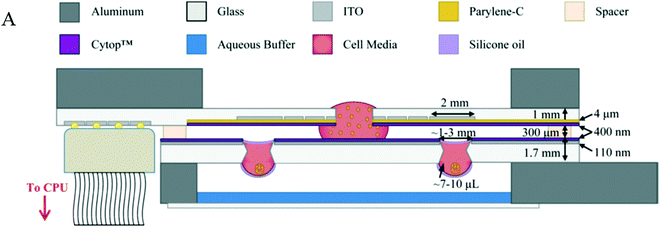
Although extremely useful for lab-scale development and research activities, this culture system is particularly challenging to implement at the industrial scale of throughput needed for drug discovery, primarily because there is no fully automated technique to culture and maintain these organized microtissues. Writing in Lab on a Chip, Bender et al. developed a powerful digital microfluidic platform (Fig. 3), which enables the automated manipulation of spheroid formation, encapsulation of the spheroid within a small hydrogel, and application of exogenous agents to determine what might trigger invasion (DOI: 10.1039/c5lc01569c). The digital microfluidics platform uses computer control to apply electrical charge to micropatterned electrodes, which can be used to actuate droplets of liquid through the system. Droplets containing cells, media or extracellular matrix can hence be added to and removed from a reservoir on demand, allowing for sequential construction, maintenance and stimulation of an embedded spheroid tissue. Although broadly applicable to a variety of materials, in this demonstration, Bender et al. cultured HT-29 spheroids, and fibroblast spheroids within a collagen matrix. Collagen solutions up to 4 mg mL−1 could be dispensed and used to encapsulate produced spheroids. They first used the platform to assay fibroblast spheroids with exogenous migration modulating agents including bone morphogenetic protein 2 or prostaglandin E2. Spheroids exposed to BMP-2 increased invasion by ∼85% compared to spheroids in standard growth medium, while PGE2 spheroids exhibited an ∼33% decrease in invasion. Furthermore, because the system allows precise manipulation of fluids, culture media from a cancer spheroid could be transferred to a fibroblast culture, allowing scalable studies of the effects of secreted factors on biological function. Treating fibroblasts with media from HT-29 cancer cells resulted in an increase of several kinds of migration of cells from the quiescent spheroid including mesenchymal, amoeboid and collective migration, demonstrating the complexity of this issue and emphasizing the need for high-throughput approaches to tease these distinct modes of migration apart. These studies are only the first step in leveraging the automation capabilities of similar digital microfluidics platforms: the potential exists to integrate sample treatment and analytical capabilities, including localized heating, sorting, electrochemical and fluorescence detection methods, that will provide levels of in situ analysis difficult or impossible to achieve using robotic liquid handling systems.
While the digital microfluidics platform is useful to construct relatively unstructured tissues such as spheroids, other advanced microtissue engineering methods have recently been used to better reconstruct the tissue environment to understand angiogenesis in liquid tumors. Zheng et al. (DOI: 10.1002/adhm.201501007), writing in Advanced Healthcare Materials, apply previously-developed microfluidic culture technologies to create a functional endothelial monolayer to study angiogenesis in response to leukemic cell lines cultured in suspension, with and without co-cultures of human fibroblasts. The degree of complexity achieved with this system enabled the researchers to identify distinct modes of angiogenesis driven by various leukemic cell populations, which was influenced by fibroblast co-culture in a cell-type dependent fashion. The platform simultaneously allows advanced analysis of secreted factors, which proved to be distinct for each culture condition tested, once again demonstrating the complexity of this biological system and emphasizing the need to adequately replicate critical features of the environment in understanding these culture systems.
Given the powerful techniques available to manipulate, control and reconstruct cells and tissues to further our understanding of biological systems, these technological approaches hold strong promise to provide unique insight into biology. Development of these culture technologies has been matched with corresponding developments in analytical techniques, which can provide faster and more precise analysis of ever-smaller quantities of biological material, and the integrative synergy between these two innovative types of technologies will dramatically alter our insight into biology. Specific to cancer, Bhola et al. recently outlined a technique in The Journal of Clinical Investigation to identify subpopulations of cells that exhibit apoptotic sensitivity using synthetic peptides (DOI: 10.1172/JCI82908). These precision techniques allow them to identify chemo-resistance at the level of single cells, which will ultimately provide insight into the role of heterogeneity of apoptotic resistance in cancer chemo-resistance and dormancy. Alternatively, the ability to rapidly measure multiple biomarkers from patient samples will be of particular relevance in cancer diagnosis and precision medicine decisions. Kim et al. present a new technique in ACS Nano in which a homogeneous entropy-driven biomolecular assay (HEBA) creates a simple transduction mechanism where a protein is transduced into an amplified nucleic acid output (DOI: 10.1021/acsnano.6b02060). This simple one-pot reaction allows homogeneous and rapid detection of analytes within complex samples such as whole blood. By integrating this technology into a microfluidic digital assay format, sensitivity can approach single-molecule levels, as demonstrated in the detection of nanoparticles at the attomolar scale. Advanced techniques such as these will play an important complementary role to the biology-on-a-chip revolution, in which the reduction of sample sizes is simultaneously a blessing and a curse.
| This journal is © The Royal Society of Chemistry 2016 |


