
 Open Access Article
 This Open Access Article is licensed under a
 Creative Commons Attribution 3.0 Unported Licence
Highly selective and sensitive fluorescence detection of Zn2+ and Cd2+ ions by using an acridine sensor†
A.
Visscher
a,
S.
Bachmann
a,
C.
Schnegelsberg
b,
T.
Teuteberg
c,
R. A.
Mata
 c and
D.
Stalke
*a
c and
D.
Stalke
*a
aInstitut für Anorganische Chemie der Universität Göttingen, Tammannstraße 4, 37077 Göttingen, Germany. E-mail: dstalke@chemie.uni-goettingen.de
bInstitut für Organische und Biomolekulare Chemie der Universität Göttingen, Tammannstraße 2, 37077 Göttingen, Germany
cInstitut für Physikalische Chemie der Universität Göttingen, Tammannstraße 6, 37077 Göttingen, Germany
First published on 11th February 2016
Abstract
Fluorescence spectroscopy investigations of the new acridine derivative bis(N,N-dimethylaminemethylene)acridine (3) show remarkable selectivity and sensitivity towards Zn2+ and Cd2+ ions in methanol and for the latter even in water. Through the chelation of the metal ions the present PET effect is quenched, significantly enhancing the emission intensity of the fluorophore. In solution, the bonding situation is studied by fluorescence and NMR spectroscopy, as well as ESI-TOF mass-spectrometry measurements. The solid state environment is investigated by X-ray diffraction and computational calculations. Here, we can show the complexation of the zinc and cadmium ions by the methylene bridged amine receptors as well as by the nitrogen atom of the acridine system.
Introduction
The detection of metal ions in solution has always been an important topic in analytical chemistry, environmental protection and medicinal applications. Metal ions like magnesium, calcium, or zinc are essential components of many enzymes in the human body.1 Especially Zn2+ plays a fundamental role in many different areas; e.g. in the emergence of Alzheimer's disease.2 Moreover, its level of concentration could help in diagnosing the growth of tumour cells in the prostate.3 Zinc deficiency increases the susceptibility to a variety of pathogens and is of central importance for the immune system.4The heavier homologue Cd2+ is known to be a very toxic metal ion. Over several decades in the first half of the 20th century, hundreds of people in Japan were affected by the deadly itai-itai disease due to a cadmium polluted river.5 Therefore, it is vital to have a fast working and very sensitive method for the detection of metal ions in e.g. blood or water supplies. Fluorescence spectroscopy combined with suitable fluorescent sensors is a well-recognized method for such analysis. Sensor molecules should be able to switch instantaneously between fluorescent on/off states or change their emission wavelength significantly upon addition of an analyte. To achieve this response several different mechanisms can be employed. Besides intramolecular charge transfer (ICT),6 metal–ligand charge transfer (MLCT)7 or excimer formation,8 an extensively investigated concept is the photoinduced electron transfer (PET)9 effect. Here a poor or non-fluorescent ligand starts to emit light upon coordination of an analyte under UV light irradiation.
The great interest in molecular sensors is reflected by the multitude of publications.10 Acridine derivatives in particular are commonly applied in the detection of bioorganic compounds. For example, it is frequently used as a fluorescent dye for DNA intercalation.11 Acridine based sensors for cationic analytes are less commonly used12 since they often do not show a strong enhancement of the fluorescence emission.13 In the following, we present the characterization in the solid state and in solution of an interesting new acridine based sensor.
Results and discussion
Synthesis
Many of the molecular sensors reported in the literature contain elaborated side arms working as the receptor unit.10a,14 However, another requirement to be met by a sensor is the economic aspect of feasibility and cheap availability. Therefore, we chose a short and effective synthetic route to produce derivatives of acridine systems with small amine side arms (Scheme 1). The desired sensor system with the concept “fluorophore–spacer–receptor” can be obtained in a quick two-step synthesis. The two bromomethylene units can be easily introduced at the 4- and 5-positions employing bromomethyl methyl ether (BMME) and a strong acid like conc. H2SO4.15 The bromine atoms can straightforwardly be substituted by a variety of amines. In this publication we present two new acridine derivatives with distinct amine moieties: 4,5-bis(N-methyl-N-phenylaminemethylene)acridine (2) and 4,5-bis(N,N-dimethylaminemethylene)acridine (3). Although the two molecules are very similar, only the latter shows extraordinary fluorescence properties.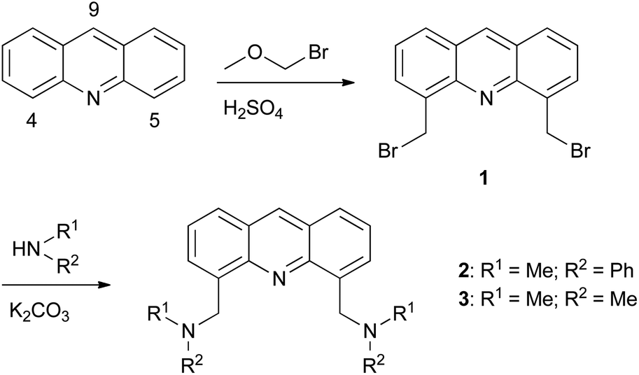 | ||
| Scheme 1 Reaction pathway to 4,5-substituted acridine derivatives. | ||
The introduction of two receptor units directly neighboured to the aromatic nitrogen atom holds the option to coordinate a target cation with the side arms as well as with the fluorophore. This is a great advantage compared to its lighter congener, the widely used fluorophore anthracene.13,14b
Fluorescence studies
The fluorescence measurements were carried out in methanol in which both the ligands and the metal salts are readily soluble. Moreover, this protic and very polar solvent exhibits similar properties to water, which is important for possible future applications. The employed structural design (fluorophore–spacer–receptor) results in a very efficient electron transfer which is favourable for a molecular off/on switch: the methylene units (spacer) allow the amines (receptor) to rotate easily about their N–C and C–C bonds. This provides the conformational freedom to interact electronically with the π-system of the acridine (fluorophore) and results in an effective quenching of the fluorescence.16 At the same time, the amines are able to employ their lone pair in complexation of analytes. If this is the case, the PET is hindered and the emission of light is facilitated. Fig. 1 shows the excitation and emission spectra of the ligands 2 and 3 in methanol.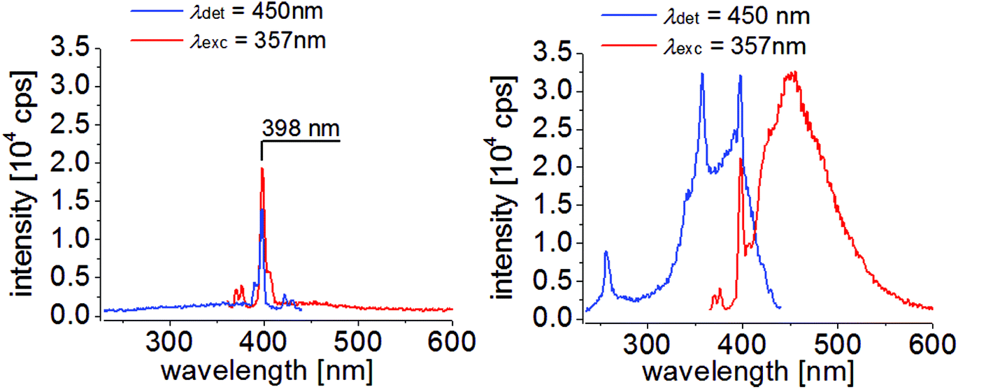 | ||
| Fig. 1 Excitation (blue, λdet = 450 nm) and emission spectra (red, λexc = 357 nm) of 2 (left) and 3 (right) in a 10−5 M methanol solution. | ||
The emission intensities of both ligands are very weak, whereas they show a strong absorption behaviour (ESI Fig. S4.1†) which hints at a working PET. Interestingly, the intensity of the methyl derivative 3 is considerably higher than the phenyl derivative 2. This observation could indicate impurities and will be discussed later in full detail. In Fig. 1, left, the highest peak at 398 nm in both spectra is attributed to Raman scattering of the used solvent methanol.17
In the next step, a metal ion screening was conducted to check the sensing ability of the ligands. Here, sixteen different metal salts were dissolved in methanol and each was titrated to the 1 × 10⁻⁵ M concentrated solutions of the ligands, respectively. To afford a good comparability, the metal ion concentrations were increased stepwise in the same manner (1, 5, 15, 100 eq.). Additionally, only bromides were used to standardize the possible interference of the counter ion. With 2 as the ligand system, no significant change in the emission intensity is observed for the tested metal salts (ESI Fig. S5.1†). To check whether the structural motif nevertheless is suitable as a sensor in general, we used the less polar and donating solvent dichloromethane. In this solution, we observed a massive fluorescence enhancement by adding ZnBr2 to the ligand (ESI Fig. S5.2†). Since this solvent is not in the focus of our interest, we concentrated on the dimethylamine system 3. In Fig. 2, we compare the intensity maxima of its emission spectra during the metal ion screening. Among the tested sixteen metal salts, this ligand shows a very high selectivity towards Zn2+ and Cd2+ ions. The emission intensity increases enormously. The only minute emission enhancement with AlBr3 and SnBr2 is solely due to the strong Lewis acidity of these metal ions which leads to slight protonation of the ligand. This can be evidenced by the different resulting emission wavelengths. If the ligand is protonated, the wavelength of the emission maximum is blue-shifted to 425 nm (ESI Fig. S5.4†), whereas in the case of Zn2+ or Cd2+ coordination, the maximum is observed at 457 nm and 445 nm, respectively. In this context it should be mentioned that although the emission shift between the two metal ions is only 12 nm, this is sufficient to differentiate the metals by fluorescence spectroscopy. A closer look at the detailed emission spectra depicted in Fig. 3 additionally demonstrates the high sensitivity of this sensor molecule.
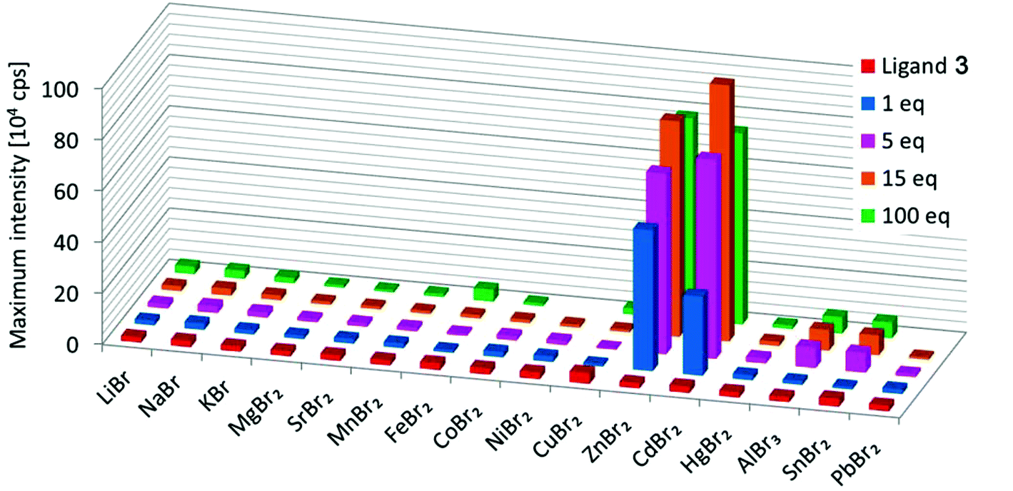 | ||
| Fig. 2 Metal ion screening with ligand 3 in methanol (c = 1 × 10⁻⁵ M). The maximum emission intensity is depicted for each metal ion addition (λexc = 357 nm). | ||
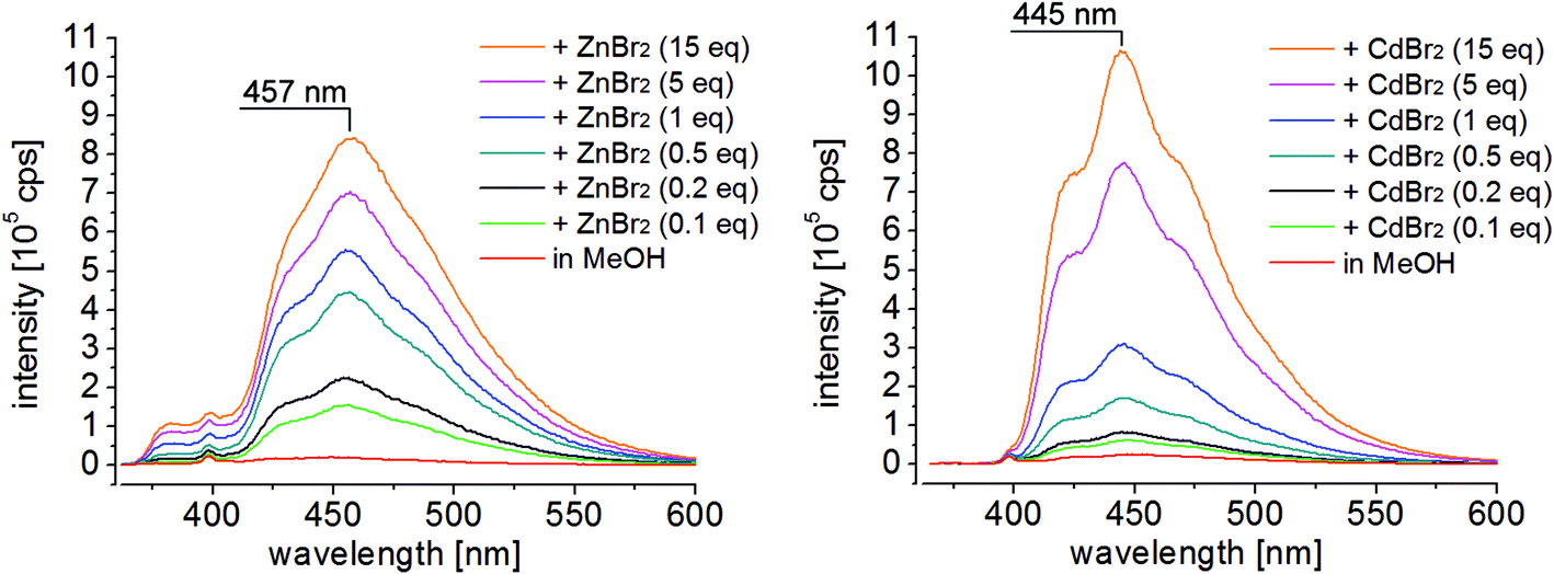 | ||
| Fig. 3 Emission spectra of 3 titrated with ZnBr2 (left) and CdBr2 (right) in methanol; both were irradiated at 357 nm. | ||
After adding only 0.1 eq. of ZnBr2 to the ligand solution, a significant increase of emission intensity is observed. Besides the standard titration of the analytes, we checked the reversibility of the metal complexation. Therefore, we alternately added a specified amount of metal salt and ethylenediamine (en) to the ligand (CdBr2 is depicted in Fig. 5, for ZnBr2 see the ESI Fig. S5.6†). With 1 eq. of CdBr2 in the cuvette, the emission intensity rises from 0.2 to 3.5 × 10⁵ cps and is afterwards quenched to 0.1 × 10⁵ cps when 10 eq. of the chelating amine are added.
This experiment can be repeated several times, confirming the great reversibility of the complexation of the two metal ions. Moreover, the starting intensity of the pure ligand (red line) is about twice as high as the intensity after adding ethylenediamine. This indicates possible traces of impurities by preparing the ligand solutions. The methanol was purchased from VWR Chemicals® (AnalaR NORMAPUR) which provides analytical data for their solvents. Each kilogram contains impurities of <0.2 mg Zn2+ and <0.01 mg Cd2+ ions.
Converted to the employed volume and concentration for the measurements, every sample could contain an amount of <0.3 eq. Zn2+ ions and <0.009 eq. Cd2+ ions with respect to the ligand. This is a significant amount one has to consider when comparing the results. But again, this underlines the sensitivity of the synthesized sensor system which enables even to detect the traces of impurities. In addition, this could explain the higher excitation and emission spectrum of 3 in comparison with 2 (Fig. 1), which is not sensitive to the mentioned metal ions.
In addition to the bromide salt, we investigated the influence of Zn(OAc)2 and Zn(NO3)2 on the fluorescence properties of 3. With small additions of the nitrate salt, the emission intensity rises as usual whereas an excess quickly leads to its reduction. However, using acetate as the counter anion, the fluorescence is increased strongly even resulting in an eight times higher emission maximum than with ZnBr2 (ESI Fig. S5.5†).
For a better assessment of the measured fluorescence values, the relative emission intensities are compared with the typical fluorescent compound anthracene. In Table 2, the maximum intensities of the used compounds and their relative intensities divided by the fluorescence of anthracene are shown.
Investigations of the metal complexes
To gain a better understanding of the observed fluorescence properties, it is necessary to know how the metal ions are linked to the ligand. Therefore, we synthesized the two metal complexes and investigated them with different analytical methods. For the synthesis of 4, the ligand and ZnBr2 were dissolved in THF whereas MeOH was used for the preparation of 5 (Scheme 2). In both cases, one equivalent of the metal salts was added dropwise to 3, resulting in an immediate precipitation of the metal complexes. The crystallization of 3 with zinc acetate to give 7 was endeavoured to check whether complexes with different zinc salts behave the same which is indeed the case here. The comparison of the crystal structures containing zinc ions is provided in the ESI.† The single crystals of 4a and 5 were obtained from crystallization in a THF/DMF mixture. By X-ray diffraction experiments, we could determine their solid state structures (Fig. 4). The zinc complex 4a crystallizes in the monoclinic space group P21/c and the cadmium derivative in the chiral orthorhombic space group P212121. Both complexes are almost isostructural. In the zinc structure one bromide ion is substituted by a coordinating solvent molecule DMF. The positively charged complex is counterbalanced by a solvent-separated [ZnBr3]− anion in the asymmetric unit (ESI Fig. S7.1†).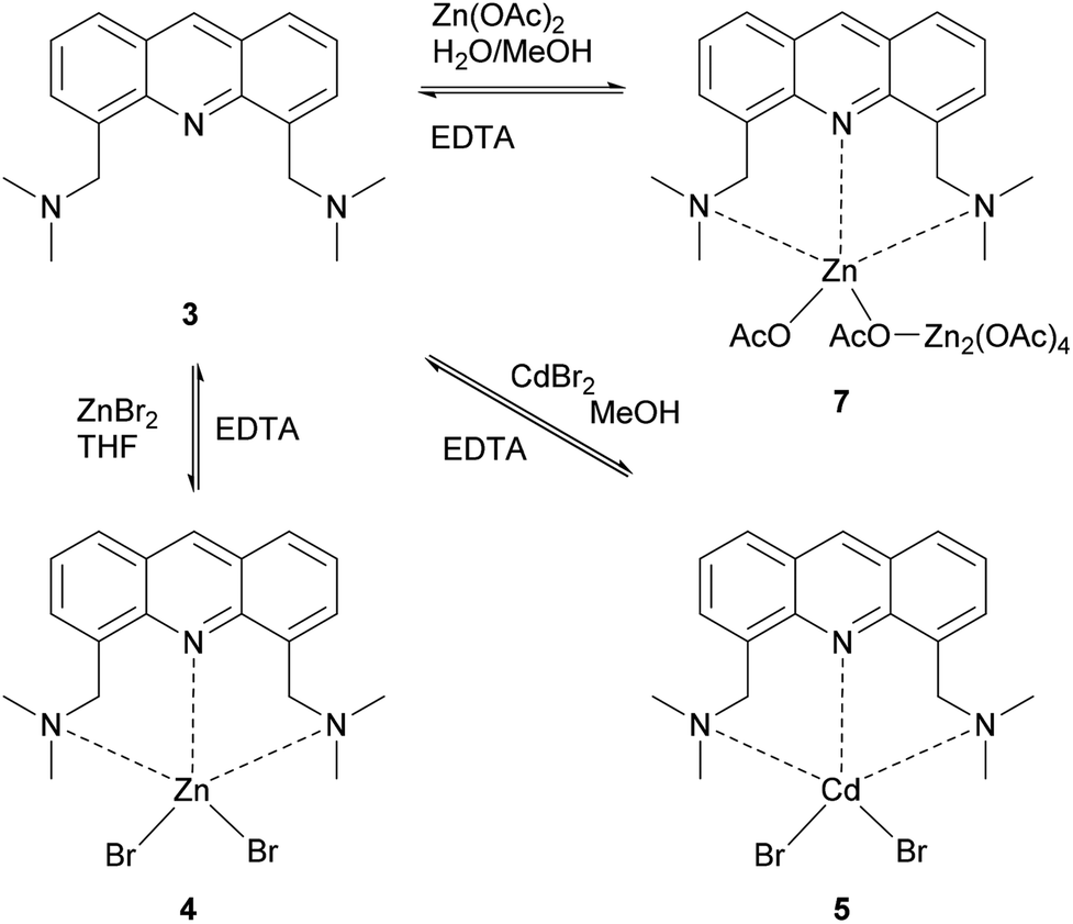 | ||
| Scheme 2 Reaction pathways to the metal ion complexes 4, 5 and 7. | ||
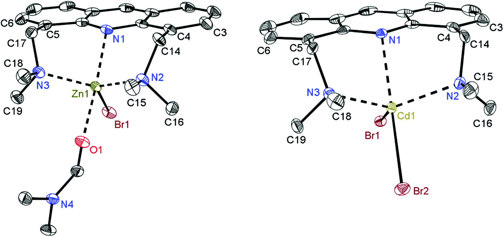 | ||
| Fig. 4 Solid state structures of [(dmf)ZnBr{(Me2NCH2)2Acr}]+ (4a, left) and [CdBr2{(Me2NCH2)2Acr}] (5, right); crystallized from a THF/DMF mixture. The omitted counter anion in the asymmetric unit of the zinc structure is a solvent-separated [(dmf)ZnBr3]− unit. All hydrogen atoms are omitted for clarity. Anisotropic displacement parameters are depicted at the 50% probability level. | ||
The solid state structures indicate the coordination of the Zn2+ and Cd2+ ions by all three present nitrogen atoms. This binding motif where the methylene bridged amines at the 4- and 5-positions and the aromatic nitrogen atom of the acridine are all involved in metal ion coordination is currently not present in the CSD. Only two examples with methylene bridged phosphorus atoms coordinating a ruthenium ion can be found in the database.18 Overall, a structural search concerning coordination of an acridine unit to any metal ion merely results in 56 hits.19
From the nitrogen–metal bond lengths in Table 1, it is obvious that the average distances from the amine nitrogen atoms to the metal ion (208.49(17) for Zn2+ and 232.01(19) pm for Cd2+) are shorter than that from the acridine nitrogen atom (230.39(16) and 254.07(18) pm, respectively). The shorter distances are in perfect agreement with the literature values found in the CSD, compared to general N–Zn2+/Cd2+ bonds. The longer distances can still be found in the database quoted as bonds but less frequently. One reason for this different bonding situation results from the rotational ability of the amines to improve the orbital overlap with the target ions. The C–N–C angles of the sp3-hybridized amines are very close to the ideal tetrahedral angle. The resulting best geometry forces the metal ion underneath the acridine plane which reduces the dative bonding to the ring nitrogen atom. Moreover, the lone pair of this sp2-hybridized nitrogen atom is partly delocalized in the π-system of the aromatic system and has therefore a reduced donor capacity. The N–M bond encloses a relatively acute angle of 44.5°(4a) and 38.5°(5), to the plane of the central acridine ring. A closer look at the bonding situation and the angle values in Table 1 reveals the coordination polyhedron of a trigonal bipyramid at each metal atom. This geometry is depicted in Fig. 6.
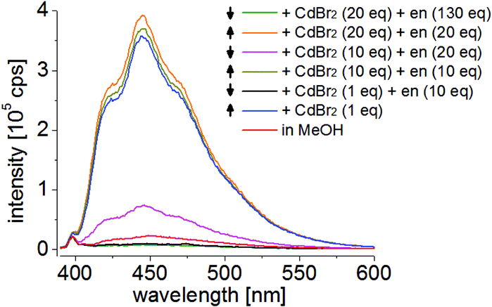 | ||
| Fig. 5 Reversibility of metal complexation of 3 with alternating addition of CdBr2 and ethylenediamine (en); all substances are dissolved in methanol (λexc = 357 nm). | ||
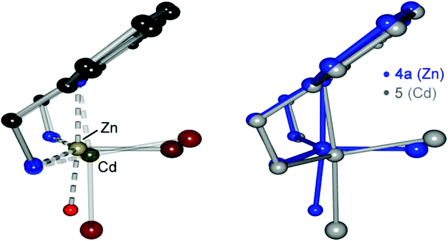 | ||
| Fig. 6 Superpositions of both metal ion structures (4a, 5), showing the trigonal bipyramidal geometry of the coordination motif. The methyl groups at the amines and the main part of the DMF molecule are omitted for clarity. | ||
| Bond [pm] | 4a (Zn) | 5 (Cd) | Angle [°] | 4a (Zn) | 5 (Cd) |
|---|---|---|---|---|---|
| N1–M | 230.39(16) | 254.07(18) | N1–M–N2 | 90.05(6) | 82.91(6) |
| N2–M | 207.89(16) | 232.31(19) | N1–M–N3 | 89.81(6) | 82.81(6) |
| N3–M | 209.09(17) | 231.70(19) | N2–M–N3 | 128.68(6) | 120.87(7) |
| Br1–M | 239.83(4) | 259.13(4) | N1–M–O1/Br2 | 175.31(6) | 174.08(4) |
| O1/Br2–M | 218.82(14) | 266.16(4) | Angular sum of N2 | 325.0(5) | 327.1(6) |
| Angular sum of N3 | 324.0(5) | 327.0(6) | |||
| C3–C4–C14–N2 | −114.8(2) | −105.3(3) | |||
| C6–C5–C17–N3 | 112.7(2) | 105.9(3) |
Although the longer N–M2+ distances can still be found in the CSD, the solid state investigations are not sufficient evidence for the existence of a bonding situation.20 In order to judge on the main binding forces of the metal ion coordination, and to relate the findings to the chemistry in solution, we conducted a series of electronic structure calculations on the Zn2+ and Cd2+ compounds. All structures were optimized at the B3LYP-D3/def2-TZVPP21 level of theory. Included were the complexes 4a and 5, identified in the solid state structures, as well as the hypothetical complex 4. The structures obtained are in good agreement with the crystal data (ESI†). A natural bond orbital (NBO) analysis22 was carried out to gain insight into the coordination of the metals to the acridine derivative 3. The data in Table 3 give the second order perturbation theory energies23 for the interaction between the lone pairs of the different nitrogen atoms and the zinc or cadmium ion. As expected, in these structures the metal has a weaker coordination to N1, compared to the other nitrogen atoms.24 Substitution of one bromide anion by a DMF solvent molecule increases this value from 5.6 to 17.0 kcal mol−1 (4 in comparison with 4a). Another possibility would be that the bulky bromide anion pulls the metal away from the acridine ring nitrogen atom.
| Compound | λ exc [nm] | Intensity [104 cps] | Relative intensity |
|---|---|---|---|
| 3 + ZnBr2 | 357 | 84.15 | 0.12 |
| 3 + CdBr2 | 357 | 105.83 | 0.15 |
| 3 + Zn(OAc)2 | 357 | 678.63 | 0.95 |
| Anthracene | 373 | 711.91 | 1.00 |
| E (PT2) [kcal mol−1] | N1 | N2 | N3 |
|---|---|---|---|
| [(dmf)ZnBr{(Me2NCH2)2Acr}]+ (4a) | 17.0 | 25.4 | 25.1 |
| [ZnBr2{(Me2NCH2)2Acr}] (4) | 5.6 | 24.7 | 25.4 |
| [CdBr2{(Me2NCH2)2Acr}] (5) | 4.7 | 22.8 | 22.7 |
| [(MeOH)2Zn{(Me2NCH2)2Acr}]2+ (in MeOH) | 21.9 | 25.4 | 26.5 |
| [(MeOH)2Cd{(Me2NCH2)2Acr}]2+ (in MeOH) | 15.0 | 32.1 | 31.8 |
The NPA charges for the two compounds are 1.49 (4) and 1.60 (4a), so that the charge difference is not very significant. The second possibility, hence the interaction with a second bromide to weaken the coordination to the acridine seems to be the most likely explanation for this effect. Furthermore, we conducted calculations on the hypothetical complexes formed in solution, [(MeOH)2Zn{(Me2NCH2)2Acr}]2+ and [(MeOH)2Cd{(Me2NCH2)2Acr}]2+. Since we are interested in replicating the conditions in solution as close as possible the COSMO continuum solvation model25 was applied (for standard methanol solution conditions). The NBO results show that the coordination to the acridine nitrogen is strengthened (21.9 and 15.0 kcal mol−1 for Zn2+ and Cd2+, respectively). This is in line with the observations made when comparing 4 and 4a. The complexes formed in the solid state have a weaker binding to the acridine moiety due to the crystallization with bromide anions. Our computed structures show smaller N1–Zn/Cd distances in agreement with this observation. The values are 216 and 237 pm, for Zn2+ and Cd2+ respectively. In methanol solution, sizeable interactions between the metal ion and the N1 nitrogen atom can be confirmed. The interactions with the side arm nitrogens are relatively constant, just slightly enhanced in solution. In Fig. 7, the computed difference electron density map of 4a is illustrated. The electron densities of the individual parts (3, Zn2+, Br−, and DMF) were computed and subtracted from the electron density of 4a; all with the same geometry. This facilitates us to illustrate the regions where the electron density is enhanced due to the interaction of the relevant atoms.
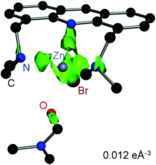 | ||
| Fig. 7 Computed difference electron density map of 4a at an isosurface level of 0.012 e Å−3. | ||
The green cloud represents the accumulated electron density. Here, the interaction of the lone pairs of all three nitrogen atoms with the metal ion is accentuated. When comparing the results of the computational studies with the fluorescence properties, it is apparent from the difference density plot that the metal coordination does not affect the π-density significantly in agreement with the almost unchanged emission wavelength. A possible explanation would be that the coordination does not influence the π-system significantly. Considering the geometry optimisations for the complex in methanol solution, both metal ion interactions with the aromatic nitrogen atom become stronger.
This should also be the case for the titration experiment of the fluorescence measurements. For a deeper insight into the liquid phase, we recorded several 1H NMR spectra depicted in Fig. 8. In comparison with the spectrum of the pure ligand, the complexes show significantly broadened signals. This is often caused by dynamic processes. Consequently, we measured the spectrum of 4 at different temperatures (Fig. 9). The increase of the temperature sharpens the broad signals until one definite set of signals is obtained (353 K). At lower temperatures, the spectrum splits into two different sets of signals with a maximum intensity at around 253 K. At this temperature they exhibit an intensity ratio of around 2![[thin space (1/6-em)]](https://www.rsc.org/images/entities/char_2009.gif) :1 which can be monitored by the singlet of the H-9 proton at 9.57 ppm and 9.17 ppm, respectively. This splitting is most likely to be attributed to the exchange of the bromine atoms with the solvent DMF. To prove this statement, we carried out a pseudo 2D 1H DOSY experiment at low temperatures and added an excess of NaBr to the NMR sample. After the addition, a third set of signals appears whereby the intensity ratio of the other two sets of signals is reversed (ESI Fig. S3.13†). The diffusion coefficient of the rising signals decreases slightly which can be explained by the smaller radius/mass ratio of a bromine atom compared to a DMF molecule.26 Consequently, the latter should represent the dibrominated species (4). The new signals have a diffusion coefficient which lies between the two others and are therefore assigned to the monobrominated compound (4a). Furthermore, the signals at room temperature are no longer broadened after the addition of NaBr. That is why the dynamic processes can be attributed to this solvent/bromide exchange.
:1 which can be monitored by the singlet of the H-9 proton at 9.57 ppm and 9.17 ppm, respectively. This splitting is most likely to be attributed to the exchange of the bromine atoms with the solvent DMF. To prove this statement, we carried out a pseudo 2D 1H DOSY experiment at low temperatures and added an excess of NaBr to the NMR sample. After the addition, a third set of signals appears whereby the intensity ratio of the other two sets of signals is reversed (ESI Fig. S3.13†). The diffusion coefficient of the rising signals decreases slightly which can be explained by the smaller radius/mass ratio of a bromine atom compared to a DMF molecule.26 Consequently, the latter should represent the dibrominated species (4). The new signals have a diffusion coefficient which lies between the two others and are therefore assigned to the monobrominated compound (4a). Furthermore, the signals at room temperature are no longer broadened after the addition of NaBr. That is why the dynamic processes can be attributed to this solvent/bromide exchange.
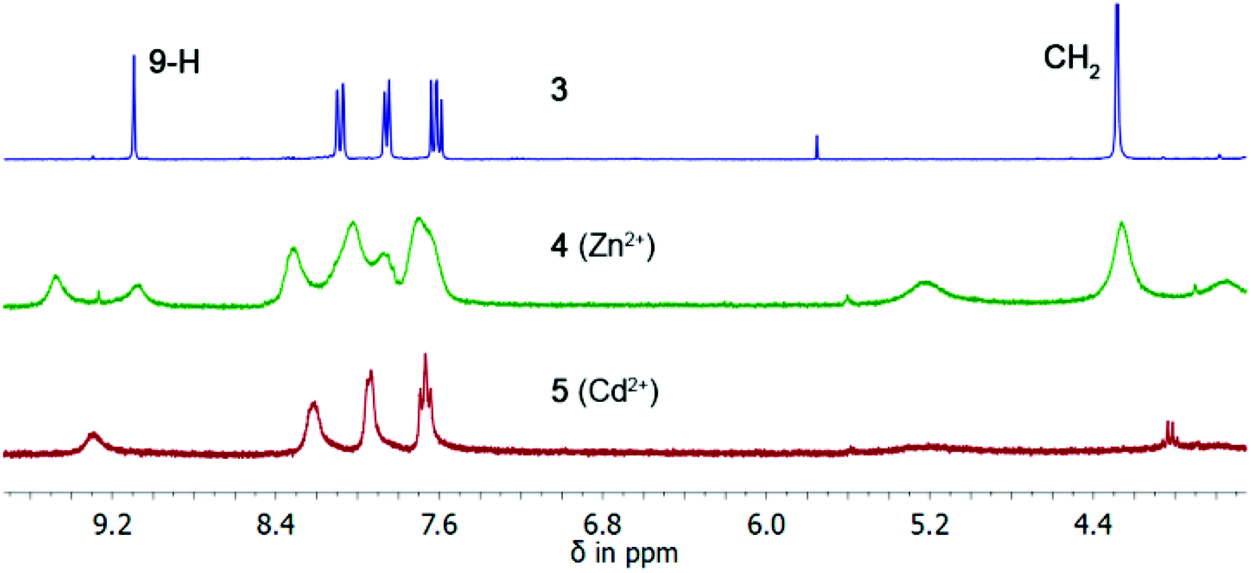 | ||
| Fig. 8 1H NMR spectra of compounds 3–5 in DMSO-d6 at ambient temperature. | ||
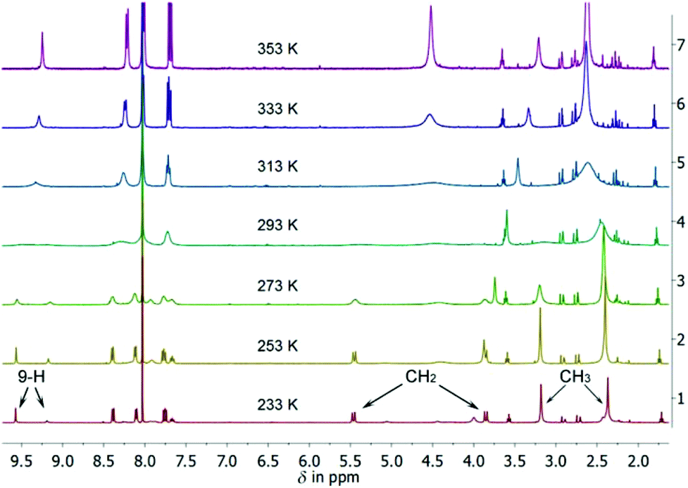 | ||
| Fig. 9 Temperature-dependent 1H NMR spectra of 4 in DMF-d7. The discussed signals are marked at 233 K. | ||
At the coalescence temperature of nearly 303 K, the diastereotopicity of the CH2 protons becomes visible. The signal of the four methyl groups at the amines splits into two signals. The velocity of their hindered rotation caused by the coordination of the zinc ion lies underneath the NMR timescale at lower temperatures and can be monitored. Zinc ions are spectroscopically silent which makes it difficult to detect a ligand–metal interaction by NMR spectroscopy. However, we measured the 15N NMR chemical shifts of the free ligand and of the zinc complex. The comparison of the resulting shifts shows a small but significant deshielding of Δδ = 8.6 ppm.27
In principle, the 1H NMR spectra of 5 show similar dependencies upon temperature variation (ESI Fig. S3.17†). The coalescence temperature in DMF-d7 is around 313 K where the diastereotopicity of the CH2 protons becomes visible as well as the mentioned splitting of the signal of the methyl groups. But in this case, no second set of signals appears and the spectrum is less broadened. The advantage of this complex is the NMR active 113Cd nucleus. Employing a 2D 113Cd, 1H-HMBC experiment at low temperatures, we could identify vicinal couplings of the methyl and methylene protons to the metal ion (ESI Fig. S3.18†). The chemical shift of the cadmium isotope is −345 ppm, referenced to Me2Cd. Interestingly, a coupling with the 113Cd nucleus was only observed for one proton of each methylene group. The coupling with the other proton is likely not to be observed due to an unfavourable angle between the related atoms.28 From the NMR spectroscopy experiments it is clear that the complexes adopt the same contact ion pairs in solution as observed in the solid state. Another analytical method which allows for investigating the transferability of the solid state structure to solution is mass spectrometry. We chose time-of-flight (TOF) spectrometry in combination with the mild electrospray ionisation method because of the poor solubility in most of the common solvents (e.g. THF, MeOH, MeCN). The measurements were performed in pure THF for the zinc complex and in THF/H2O for the cadmium complex due to the even lower solubility. However, both complexes were sufficiently present in solution for TOF spectrometry. Fig. 10 depicts a comparison of the two mass patterns. The black lines represent the experimental data from the zinc complex, whereas the blue lines illustrate the simulated spectrum for the proposed structural motif (in the positive ion mode one bromide anion was omitted for the calculated spectrum). The comparison demonstrates a perfect match of the predicted and observed species. Furthermore, the full mass spectrum is free of any ionic fragments because only signals generated by the acridine derivative, with and without metal ions, and related fragments can be detected. The same is valid for the Cd2+ complex (ESI Fig. S6.5†). Both methods, NMR spectroscopy and mass spectrometry, emphasize that the solid state structure is maintained in solution. As mentioned in the introduction, molecular sensors play a key role in a wide range of areas, especially in medicinal or analytical applications. In this publication, we have mainly shown the possibility of using our sensor molecules in very polar solvents (fluorescence measurements in MeOH; NMR in DMF; ESI-MS in THF). However, using water as a solvent frequently evokes serious challenges. Despite the poor solubility of organic molecules, the self-ionization of water mainly results in the protonation of the amine side-arm receptor units. This dramatically hampers the coordination of analytes. Additionally, the pKa value of acridine rises from 5.45 to 10.7 when becoming excited in the fluorescence spectrometer,29 an even higher value than the aliphatic amine (∼9.7, value of the related N,N-dimethylbenzylamine).30 The formation of the protonated species can be easily monitored by fluorescence and NMR spectroscopy. In the laboratory, the addition of hydrochloric acid to a clear solution of 3 in toluene resulted in a yellow precipitate. The solubility in this solvent is reduced due to the induced charge in the product. After purification, a 1H NMR spectrum in DMSO-d6 shows the attached proton and the related coupling constants. Furthermore, the solid state crystal structure of this salt could be determined (ESI Fig. S3.19 and S7.2†). With 2, the protonation experiment was not feasible. On account of the aniline like structure, the lone pair of the nitrogen atom is partly delocalized in the phenyl ring which results in a lower basicity (+M effect).31
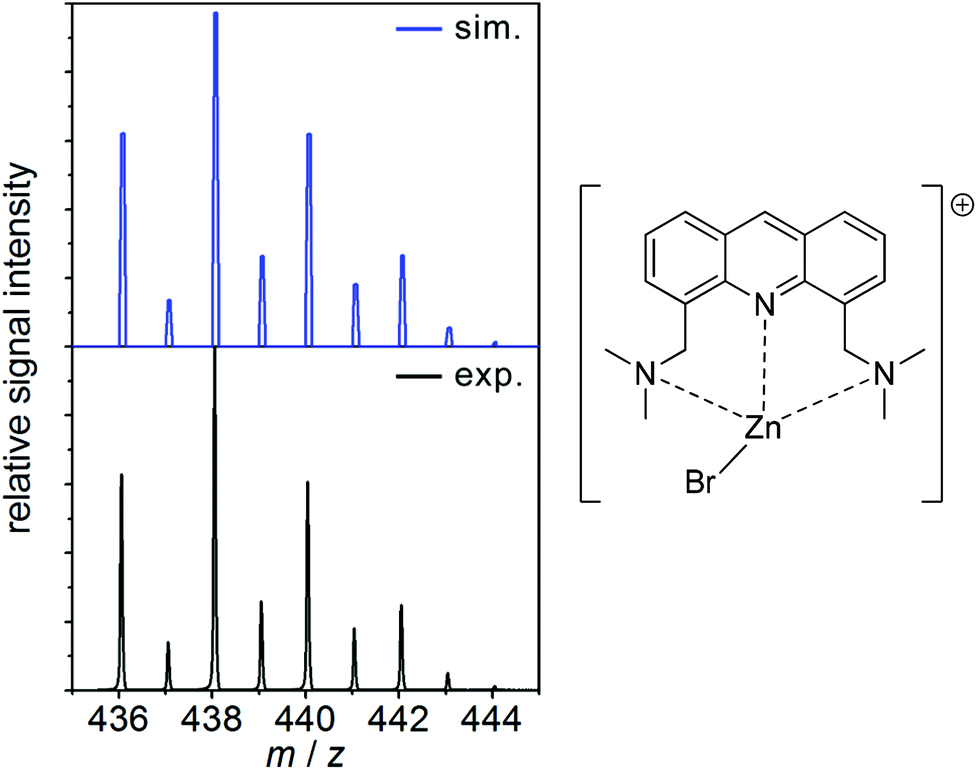 | ||
| Fig. 10 Extract from the mass spectrum of the cation 4, showing the simulated (blue) and the experimental (black) isotope pattern of the positively charged complex. | ||
To achieve an enhancement of the emission intensity, a large excess of hydrochloric acid was needed, whereas for 3, already two equivalents of the acid resulted in a notable change of the spectrum (ESI Fig. S5.3–4†). The latter was expected for this compound since with regard to the lone pair the bonding to a proton is equivalent to the coordination of a metal ion. In both cases, the PET is quenched and the emission of light is facilitated. According to these results, measurements in water were only reasonable with a buffer system in a high pH region (>11). However, we could prevent these difficulties by dissolving the ligand in methanol and the analyte in water. In Fig. 11, the good sensitivity of the sensor system towards Cd2+ ions is still present even in the presence of water.
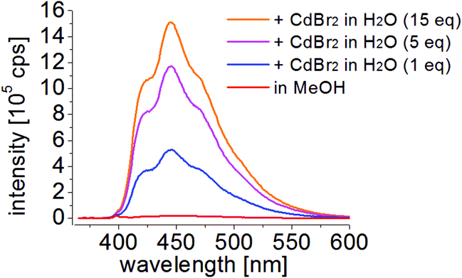 | ||
| Fig. 11 Emission spectra of 3 in methanol for various concentrations of CdBr2 dissolved in purified water (λexc = 357 nm). | ||
Conclusions
The fluorescence spectra of ligand 3 show a remarkable metal ion selectivity and an increase in fluorescence emission upon titration with dissolved ZnBr2 and CdBr2 in methanol/water. We could demonstrate the high sensitivity of the acridine based sensor molecule and the good stability of the systems. The solid state contact ion pairs 4 and 5 determined by X-ray structure analyses were confirmed to be present both in the gas phase by computational chemistry and in solution by NMR spectroscopy and mass spectrometry. This features the 4,5-bis(N,N-dimethylaminemethylene)acridine as a valuable ligand in the medically and environmentally important photochemical detection of zinc and cadmium ions. Compared to the numerous publications concerning zinc sensors, only a very few provide structural evidence of the metal ion coordination, although this is inevitable for the accurate understanding of fluorescence processes.Experimental section
Materials and measurements
All materials were purchased from Sigma-Aldrich, VWR International, and ABCR Chemicals and used without further purification – except water which was purified first through a Millipore water purification system Milli-RO 3 plus and finally with a Millipore ultrapure water system Milli-Q plus 185. The NMR measurements were performed on a Bruker Avance III 300 and Bruker Avance III HD 400 spectrometer. The chemical shifts (δ) are reported in parts per million (ppm) relative to the residual proton signals of the incompletely deuterated solvents. The fluorescence measurements were carried out on a Horiba Jobin-Yvon Fluoromax-4 spectrometer. All emission spectra were recorded with an excitation wavelength of 357 nm. For electrospray ionization mass spectrometry studies, sample solutions of c ≈ 5 mm were continuously administered into the ESI source of a micrOTOF-Q II mass spectrometer (Bruker Daltonik) by using a step motor-driven gas-tight syringe (flow rate: 0.5 ml h−1). This instrument combines a quadrupole mass filter with a time-of-flight analyzer. The simulated isotope patterns were calculated using the COMPASS® software package from Bruker Daltonik.Single-crystal structural analysis
Suitable single crystals for X-ray structural analysis were selected from a Schlenk flask under an argon atmosphere and covered with perfluorinated polyether oil on a microscope slide, which was cooled with a nitrogen gas flow using the X-TEMP2 device.32 An appropriate crystal was selected using a polarizing microscope, mounted on the tip of a MiTeGen MicroMount, fixed to a goniometer head and shock-cooled by using the crystal cooling device. The data for 4a were collected on an INCOATEC Mo microsource33 with mirror optics and Mo-Kα radiation with λ = 71.073 pm. The data for 5 were collected at an INCOATEC Ag microfocus source with INCOATEC Quazar mirror optics and Ag radiation at λ = 56.086 pm. Both diffractometers were equipped with an APEX II detector with a D8 goniometer and a low-temperature device. The data for 4a and 5 were integrated with SAINT34 and an empirical absorption correction (SADABS)35 was applied. The structures were solved by direct methods using SHELXT36 and refined by full-matrix least-squares methods against F2 (SHELXL)37 within the SHELXLE GUI.38 The hydrogen atoms were refined isotropically on calculated positions using a riding model with their Uiso values constrained to equal 1.5 times the Ueq. of their pivot atoms for terminal sp3 carbon atoms and 1.2 times for all the other carbon atoms. The disordered moieties were refined using bond length restraints and anisotropic displacement parameter restraints.39 Crystallographic data for the structures reported in this paper have been deposited with the Cambridge Crystallographic Data Centre. The CCDC numbers, crystal data, and experimental details for the X-ray measurements are listed in Table S7.2 of the ESI.†Computational studies
All geometry optimizations were carried out at the B3LYP-D3/def2-TZVPP21,40 level of theory (including Becke–Jones type damping41 of the dispersion correction21f). In the case of Cd, the Stuttgart/Dresden ECP28MDF42 preudopotential was used. The electronic densities and the corresponding NBO analysis22,23 were computed at the same level. All calculations were carried out with the ORCA43 program package. B3LYP calculations were performed under the RIJCOSX approximation.44:1) mixture. The crystals were formed at rt after two weeks. 1H NMR (400 MHz, DMF-d7, 353 K): δ 9.25 (s, 1 H, H9), 8.22 (d, 3J = 8.3 Hz, 2 H, H1,8), 8.01 (d, 3J = 7.1 Hz, 2 H, H3,6), 7.69 (dd, 3J = 8.3, 7.1 Hz, 2 H, H2,7), 4.52 (4 H, CH2), 2.62 (s, 12 H, CH3). 13C{1H} NMR (100 MHz, DMF-d7, 243 K): δ 148.7 (2 C, C4a,10a), 142.0 (1 C, C9), 135.5 (2 C, C3,6), 130.5 (2 C, C1,8), 127.7 (2 C, C8a,9a), 126.4 (2 C, C2,7), 63.1 (2 C, CH2), 48.9 (2 C, CH3), 46.4 (2 C, CH3). 15N NMR (40.6 MHz, DMF-d7, 243 K): δ −346.7 (NMe2). ESI-TOF: m/z: 813.15 [(C19H23N3)2ZnBr2H]+, 438.05 [(C19H23N3)ZnBr]+, 294.20 [(C19H23N3)H]+. Anal. Calcd for C19H23N3ZnBr2: C, 44.00; H, 4.47; N, 8.10; Br, 30.81. Found: C, 44.00; H, 4.50; N, 8.10; Br, 30.00.
:2) mixture, heated up to the boiling point of THF and then cooled to rt overnight. The crystals thus formed were suitable for X-ray diffraction. 1H NMR (400 MHz, DMF-d7, 353 K): δ 9.32 (s, 1 H, H9), 8.28 (d, 3J = 8.5 Hz, 2 H, H1,8), 8.08–7.99 (m, 2 H, H3,6), 7.71 (dd, 3J = 8.5, 6.8 Hz, 2 H, H2,7), 4.56 (s, 4 H, CH2), 2.74 (s, 12 H, CH3). 113Cd, 1H-HMBC (66.6 MHz, DMF-d7, 243 K): δ −345.5. ESI-TOF: m/z: 861.15 [(C19H23N3)2CdBr2H]+, 486.02 [(C19H23N3)CdBr]+, 294.21 [(C19H23N3)H]+. Anal. Calcd for C19H23N3CdBr2: C, 40.35; H, 4.10; N, 7.43; Cd, 19.87. Found: C, 40.28; H, 4.31; N, 7.20; Cd, 19.00.
Note added in proof
During the production of this paper related aciridine complexes were published: E. V. Solovyeva, G. L. Starova, L. A. Myund, A. S. Denisova, Polyhedron, 2016, 106, 1–9.Acknowledgements
Thanks to the Danish National Research Foundation (DNRF93) funded Center for Materials Crystallography (CMC) for partial support and the Land Niedersachsen for providing a fellowship in the GAUSS PhD program. We also want to thank Timo Schillmöller for the support of some fluorescence measurements.Notes and references
- (a) W. N. Lipscomb and N. Sträter, Chem. Rev., 1996, 96, 2375–2434 CrossRef CAS PubMed; (b) R. McRae, P. Bagchi, S. Sumalekshmy and C. J. Fahrni, Chem. Rev., 2009, 109, 4780–4827 CrossRef CAS PubMed.
- (a) J.-Y. Koh, S. W. Suh, B. J. Gwag, Y. Y. He, C. Y. Hsu and D. W. Choi, Science, 1996, 272, 1013–1016 CrossRef CAS PubMed; (b) A. I. Bush, Curr. Opin. Chem. Biol., 2000, 4, 184–191 CrossRef CAS PubMed.
- (a) M. Cortesi, E. Fridman, A. Volkov, S. S. Shilstein, R. Chechik, A. Breskin, D. Vartsky, N. Kleinman, G. Kogan, E. Moriel, V. Gladysh, M. Huszar, J. Ramon and G. Raviv, Prostate, 2008, 68, 994–1006 CrossRef CAS PubMed; (b) Z. Medarova, S. K. Ghosh, M. Vangel, R. Drake and A. Moore, Am. J. Cancer Res., 2014, 4, 385–393 Search PubMed.
- (a) J. M. Berg and Y. Shi, Science, 1996, 271, 1081–1085 CrossRef CAS PubMed; (b) N. Wellinghausen, M. Martin and L. Rink, Eur. J. Immunol., 1997, 27, 2529–2535 CrossRef CAS PubMed; (c) M. J. Salgueiro, M. Zubillaga, A. Lysionek, M. I. Sarabia, R. Caro, T. De Paoli, A. Hager, R. Weill and J. Boccio, Nutr. Res., 2000, 20, 737–755 CrossRef CAS.
- (a) K. Nogawa, A. Ishizaki and E. Kobayashi, Environ. Res., 1979, 18, 397–409 CrossRef CAS PubMed; (b) C. Tohyama, Z. A. Shaikh, K. Nogawa, E. Kobayashi and R. Honda, Arch. Toxicol., 1982, 50, 159–166 CrossRef CAS.
- Y. Li, T. Liu, H. Liu, M.-Z. Tian and Y. Li, Acc. Chem. Res., 2014, 47, 1186–1198 CrossRef CAS.
- T. Saha, A. Sengupta, P. Hazra and P. Talukdar, Photochem. Photobiol. Sci., 2014, 13, 1427–1433 Search PubMed.
- J. B. Briks, Photophysics of Aromatic Molecules, Wiley - Interscience, London, New York, Sydney, Toronto, 1st edn, 1970 Search PubMed.
- (a) R. A. Bissel, A. P. de Silva, H. Q. N. Gunaratne, P. L. M. Lynch, G. E. M. Maguire, C. P. McCoy and K. R. A. S. Sandanayake, in Photoinduced Electron Transfer V, ed. J. Mattay, Springer, Berlin, Heidelberg, 1993, vol. 168, pp. 229–230 Search PubMed; (b) K. Kubo, in Topics in Fluorescence Spectroscopy, ed. C. D. Geddes and J. R. Lakowicz, Springer, US, 2005, ch. 6, vol. 9, pp. 219–247 Search PubMed; (c) A. P. de Silva, J. Phys. Chem. Lett., 2011, 2, 2865–2871 CrossRef CAS.
- (a) A. P. de Silva, H. Q. N. Gunaratne, T. Gunnlaugsson, A. J. M. Huxley, C. P. McCoy, J. T. Rademacher and T. E. Rice, Chem. Rev., 1997, 97, 1515–1566 CrossRef CAS PubMed; (b) Z. Fei, N. Kocher, C. J. Mohrschladt, H. Ihmels and D. Stalke, Angew. Chem., Int. Ed., 2003, 42, 783–787 ( Angew. Chem. , 2003 , 115 , 807–811 ) CrossRef CAS PubMed; (c) A. Ajayaghosh, P. Carol and S. Sreejith, J. Am. Chem. Soc., 2005, 127, 14962–14963 CrossRef CAS PubMed; (d) K. Ono, J. K. Klosterman, M. Yoshizawa, K. Sekiguchi, T. Tahara and M. Fujita, J. Am. Chem. Soc., 2009, 131, 12526–12527 CrossRef CAS PubMed; (e) Z. Xu, J. Yoon and D. R. Spring, Chem. Soc. Rev., 2010, 39, 1996–2006 RSC; (f) H. Woo, S. Cho, Y. Han, W.-S. Chae, D.-R. Ahn, Y. You and W. Nam, J. Am. Chem. Soc., 2013, 135, 4771–4787 CrossRef CAS PubMed; (g) M. Yamashina, M. M. Sartin, Y. Sei, M. Akita, S. Takeuchi, T. Tahara and M. Yoshizawa, J. Am. Chem. Soc., 2015, 137, 9266–9269 CrossRef CAS PubMed.
- (a) J. Joseph, E. Kuruvilla, A. T. Achuthan, D. Ramaiah and G. B. Schuster, Bioconjugate Chem., 2004, 15, 1230–1235 CrossRef CAS PubMed; (b) Z. Ma, J. R. Choudhury, M. W. Wright, C. S. Day, G. Saluta, G. L. Kucera and U. Bierbach, J. Med. Chem., 2008, 51, 7574–7580 CrossRef CAS PubMed.
- (a) H. N. Lee, H. N. Kim, K. M. K. Swamy, M. S. Park, J. Kim, H. Lee, K.-H. Lee, S. Park and J. Yoon, Tetrahedron Lett., 2008, 49, 1261–1265 CrossRef CAS; (b) J. Kertész, B. Bognár, A. Kormos, I. Móczár, P. Baranyai, M. Kubinyi, T. Kálai, K. Hideg and P. Huszthy, Tetrahedron, 2011, 67, 8860–8864 CrossRef.
- M. S. Park, K. M. K. Swamy, Y. J. Lee, H. N. Lee, Y. J. Jang, Y. H. Moon and J. Yoon, Tetrahedron Lett., 2006, 47, 8129–8132 CrossRef CAS.
- (a) S. Cao, H. Li, T. Chen and J. Chen, J. Solution Chem., 2009, 38, 1520–1527 CrossRef CAS; (b) E. B. Veale and T. Gunnlaugsson, Annu. Rep. Prog. Chem., Sect. B, 2010, 106, 376–406 RSC.
- J. Chiron and J.-P. Galy, Synlett, 2003, 2349–2350 CrossRef CAS.
- M. E. Huston, K. W. Haider and A. W. Czarnik, J. Am. Chem. Soc., 1988, 110, 4460–4462 CrossRef CAS.
- J. F. Mammone, S. K. Sharma and M. Nicol, J. Phys. Chem., 1980, 84, 3130–3134 CrossRef CAS.
- (a) C. Gunanathan, L. J. W. Shimon and D. Milstein, J. Am. Chem. Soc., 2009, 131, 3146–3147 CrossRef CAS PubMed; (b) X. Ye, P. N. Plessow, M. K. Brinks, M. Schelwies, T. Schaub, F. Rominger, R. Paciello, M. Limbach and P. Hofmann, J. Am. Chem. Soc., 2014, 136, 5923–5929 CrossRef CAS PubMed.
- (a) F. H. Allen, Acta Crystallogr., Sect. B: Struct. Sci., 2002, 58, 380–388 CrossRef; (b) Cambridge Structural Database, v5.37 (November 2015), Cambridge, UK, 2015 Search PubMed.
- U. Flierler and D. Stalke, in Structure and Bonding, ed. D. Stalke, Springer, Berlin, New York, 2012, vol. 146, pp. 1–20 Search PubMed.
- (a) A. D. Becke, Phys. Rev. A, 1988, 38, 3098–3100 CrossRef CAS; (b) C. Lee, W. Yang and R. G. Parr, Phys. Rev. B: Condens. Matter, 1988, 37, 785–789 CrossRef CAS; (c) A. D. Becke, J. Chem. Phys., 1993, 98, 5648–5652 CrossRef CAS; (d) P. J. Stephens, F. J. Devlin, C. F. Chabalowski and M. J. Frisch, J. Phys. Chem., 1994, 98, 11623–11627 CrossRef CAS; (e) F. Weigend and R. Ahlrichs, Phys. Chem. Chem. Phys., 2005, 7, 3297–3305 RSC; (f) S. Grimme, J. Antony, S. Ehrlich and H. Krieg, J. Chem. Phys., 2010, 132, 154104–154119 CrossRef PubMed.
- (a) J. P. Foster and F. Weinhold, J. Am. Chem. Soc., 1980, 102, 7211–7218 CrossRef CAS; (b) A. E. Reed, R. B. Weinstock and F. Weinhold, J. Chem. Phys., 1985, 83, 735–746 CrossRef CAS; (c) E. D. Glendening, J. K. Badenhoop, A. E. Reed, J. E. Carpenter, J. A. Bohmann, C. M. Morales and F. Weinhold, GenNBO 5.9, University of Wisconsin, Madison, 2009 Search PubMed.
- A. E. Reed, L. A. Curtiss and F. Weinhold, Chem. Rev., 1988, 88, 899–926 CrossRef CAS.
- C. Maaß, D. M. Andrada, R. A. Mata, R. Herbst-Irmer and D. Stalke, Inorg. Chem., 2013, 52, 9539–9548 CrossRef PubMed.
- A. Klamt and G. Schüürmann, J. Chem. Soc., Perkin Trans. 2, 1993, 799–805 RSC.
- (a) A. Bondi, J. Phys. Chem., 1964, 68, 441–451 CrossRef CAS; (b) R. Neufeld and D. Stalke, Chem. Sci., 2015, 6, 3354–3364 RSC.
- (a) H. Gornitzka and D. Stalke, Eur. J. Inorg. Chem., 1998, 311–317 CrossRef CAS; (b) T. E. Wood, B. Berno, C. S. Beshara and A. Thompson, J. Org. Chem., 2006, 71, 2964–2971 CrossRef CAS PubMed.
- M. Karplus, J. Am. Chem. Soc., 1963, 85, 2870–2871 CrossRef CAS.
- J. R. Lakowicz and A. Balter, Biophys. Chem., 1982, 16, 117–132 CrossRef CAS PubMed.
- U. Knips and F. Huber, Z. Naturforsch., B: Anorg. Chem. Org. Chem., 1983, 38b, 434–436 CAS.
- J. N. Murrell, Proc. Phys. Soc., London, Sect. A, 1955, 68, 969–975 CrossRef.
- (a) T. Kottke and D. Stalke, J. Appl. Crystallogr., 1993, 26, 615–619 CrossRef; (b) T. Kottke, R. J. Lagow and D. Stalke, J. Appl. Crystallogr., 1996, 29, 465–468 CrossRef CAS; (c) D. Stalke, Chem. Soc. Rev., 1998, 27, 171–178 RSC.
- T. Schulz, K. Meindl, D. Leusser, D. Stern, J. Graf, C. Michaelsen, M. Ruf, G. M. Sheldrick and D. Stalke, J. Appl. Crystallogr., 2009, 42, 885–891 CrossRef CAS.
- SAINT v8.30C, Bruker AXS Inc., Madison, WI, USA, 2013 Search PubMed.
- L. Krause, R. Herbst-Irmer, G. M. Sheldrick and D. Stalke, J. Appl. Crystallogr., 2015, 48, 3–10 CrossRef CAS PubMed.
- G. M. Sheldrick, Acta Crystallogr., Sect. A: Fundam. Crystallogr., 2015, 71, 3–8 CrossRef PubMed.
- G. M. Sheldrick, SHELXL in SHELXTL v2014/7, Madison, WI, USA, 2014 Search PubMed.
- C. B. Huebschle, G. M. Sheldrick and B. Dittrich, J. Appl. Crystallogr., 2011, 44, 1281–1284 CrossRef CAS PubMed.
- P. Müller, R. Herbst-Irmer, A. L. Spek, T. R. Schneider and M. R. Sawaya, Crystal structure refinement - A crystallographer's guide to SHELXL, Oxford University Press, Oxford, 8th edn, 2006 Search PubMed.
- K. Eichkorn, F. Weigend, O. Treutler and R. Ahlrichs, Theor. Chem. Acc., 1997, 97, 119–124 Search PubMed.
- (a) E. R. Johnson and A. D. Becke, J. Chem. Phys., 2005, 123, 024101 CrossRef PubMed; (b) A. D. Becke and E. R. Johnson, J. Chem. Phys., 2005, 123, 154101 CrossRef PubMed; (c) E. R. Johnson and A. D. Becke, J. Chem. Phys., 2006, 124, 174104 CrossRef PubMed.
- D. Andrae, U. Häußermann, M. Dolg, H. Stoll and H. Preuß, Theor. Chim. Acta, 1990, 77, 123–141 CrossRef CAS.
- F. Neese, Wily Interdiscip. Rev.: Comput. Mol. Sci., 2012, 2, 73–78 CAS.
- (a) E. J. Baerends, D. E. Ellis and P. Ros, Chem. Phys., 1973, 2, 41–51 CrossRef CAS; (b) J. L. Whitten, J. Chem. Phys., 1973, 58, 4496–4501 CrossRef CAS; (c) F. Neese, F. Wennmohs, A. Hansen and U. Becker, Chem. Phys., 2009, 356, 98–109 CrossRef CAS.
Footnote |
| † Electronic supplementary information (ESI) available: Further spectroscopic data like NMR, UV/vis, fluorescence, and mass spectra as well as crystallographic and computational tables. CCDC 1061423, 1061424, 1423218 and 1423219. For ESI and crystallographic data in CIF or other electronic format see DOI: 10.1039/c6dt00557h |
| This journal is © The Royal Society of Chemistry 2016 |