A multidisciplinary approach to the study of the brightening effects of white chalk ground layers in 15th and 16thcentury paintings
Vanessa
Antunes
*ab,
António
Candeias
cd,
João
Coroado
e,
Vitor
Serrão
a,
Mário
Cachão
f and
Maria L.
Carvalho
b
aARTIS-Instituto História da Arte da Faculdade de Letras da Universidade de Lisboa (ARTIS-FLUL), Alameda da Universidade, 1600-214 Lisboa, Portugal. E-mail: vanessahantunes@gmail.com
bLIBPhys-UNL, Laboratório de Instrumentação, Engenharia Biomédica e Física da Radiação, Departamento de Física, Faculdade de Ciências e Tecnologia, Universidade Nova de Lisboa, 2829-516, Caparica, Portugal
cLaboratório José de Figueiredo / Direcção-Geral do Património Cultural (LJF-DGPC), Rua das Janelas Verdes 37, 1249-018 Lisboa, Portugal
dLaboratório HERCULES, Escola de Ciências e Tecnologia, Universidade de Évora, Largo Marquês de Universidade de Évora, Largo Marquês de Marialva 8, 7000-676 Évora, Portugal
eInstituto Politécnico de Tomar (IPT)/GeoBioTec ID&T unit, 2300-313 Tomar, Portugal
fInstituto Dom Luiz, Faculdade de Ciências, Universidade de Lisboa, Campo Grande, 1749-016 Lisboa, Portugal
First published on 4th May 2016
Abstract
This paper employs various techniques to analyze the mixture of chalk and binder materials used, by Portuguese and Flemish painters in the 15th and 16th centuries, to enhance the reflection of light in paintings. The cases studied show evidence of the search by painters for light effects created when combining specific fillers and binders to obtain absorbent or non-absorbent ground layers in order to reflect superficial or deep light in paintings. These brightening effects are largely provided by microscopic coccoliths and calcispheres – the main constituents of chalk. The composition, size and slightly concave-convex shield-like shape of calcareous nannofossils (micrometrical dimensions) also facilitate application, thereby increasing the speed of handling. These calcareous nannofossils are crucial proof that chalk was used in the ground layers of Portuguese paintings. They have proved to be important in defining the various stages of Portuguese painting workshops, such as the Viseu Workshop (1501–1569), which used powdered chalk in the first phase and powdered limestone in the second phase in the production of a ground layer. A two-layer structure has been verified in some Flemish paintings of the period, but the use of different binders to provide different levels of light absorption and reflection in these artworks had not been previously identified. The results showing this two-layer ground structure are significant in making the connection between Flemish and Portuguese art in the context of a specific painting technique. The use of calcium carbonate ground layers was verified by SEM-EDS and confirmed by μ-XRD, and μ-Raman, while binders were analyzed by μ-FTIR and optical microscopy, using staining tests.
1. Introduction
Investigation of painting techniques demands a significant understanding of the types of material used as well as the possible combinations of these in order to understand an artist's reason for choosing such materials. This research, using analytical practices, alongside several protocols found in the literature concerning the study of painting materials, compares similar ground layers in various paintings from the 15th and 16th centuries. These paintings have been deeply studied in order to identify different techniques used by painters during this period.1–6 When dealing with materials which are undetectable to the naked eye, as it is the case of the painting ground layers, analytical procedures require a detailed and exhaustive research plan to understand this “invisible matter”.7 In Portugal, the study of ground layers in paintings from the 15th and the 16th centuries has proved that two different types of material exist in these layers: calcium sulphate and calcium carbonate.1–4,7–9 The existence of calcium carbonate in the ground layers of Flemish paintings in Portugal and in Portuguese paintings has been proved by earlier studies8,9 but the possible reasons that led to Portuguese painters selecting this particular material is a little studied issue7 and this paper aims to bring some new data on this subject. In some cases, the limestone was powdered and used as a ground layer while in other cases a special kind of limestone, i.e. pure chalk, was used. Chalk is a soft limestone composed mainly of small biogenic calcium carbonate particles (biomicrite). Typically it dates from the Cretaceous period and was usually taken from the sea cliffs of England and France, but can also be found in Paleogene (Danian) deposits (North Sea, Denmark, Maastricht). Its genesis is associated with deep marine deposits and is characterized by the presence of calcareous nannofossils of unicellular marine phytoplankton organisms, the coccolithophores. Coccoliths are microscopic calcium carbonate shells of various shapes, the most common morphotype being the discoidal placoliths. These are produced by the coccolithophores, unicellular organisms where the cell is enclosed in an exoskeleton of calcite, the coccosphere.10Today, chalk is extracted from several regions and its primary characteristics include fineness, softness and whiteness.
A two-layer glue/oil structure has been verified in some Flemish paintings of the period,11 indicating that this technique might have been taught in artist workshops at the time. However, the use of different binders to provide different levels of light absorption and reflection in these art works had not previously been discussed. This two-layered ground technique was proved, during a study of Rembrandt's paintings, to have been used in the 17th century. Rembrandt had worked on panels since c. 1624 and had used ground layers that fundamentally followed the Flemish technique. Treatises such as the de Mayerne Manuscript, of 1620–1646, refer to the use of glue and chalk ground layers,12 but chalk in oil as a ground layer is reported very uncommonly, being found in a German 19th century treatise.13,14 Some panel preparations used by Rembrandt, and other Flemish, French and Italian painters, had a two-layered ground.12 In Rembrandt's panel paintings the first ground layer might consist of glue and chalk (with or without pigments) in order to seal the wood's porosity; a second oil-containing layer could be added, obtaining a smooth, yellowish (or other color surface) on which to paint. This second layer might contain chalk mixed with other pigments.12
The present study reveals that the two-layer structure used in the ground, and the phenomenon of using chalk material, provided largely by calcareous nannofossils (mainly coccoliths and calcispheres), to reflect light, could have been a practice that had been taught in painting workshops since the 15th century. Calcareous nannofossils are calcite skeletons of very small dimensions (typically between 2 and 20 μm), including coccoliths, nannoliths and calcispheres. Its excellent workability and lubricating qualities come from physical and mechanical properties such as fineness and softness, as well as the rock being naturally pulverulent.7,15,16 The composition and size of the slightly concave-convex shield-shaped microfossiliferous limestone, facilitate the speed of application of the substances. The quick application of the chalk as a painting ground layer allows its homogenization before drying, and therefore facilitates the aggregation of particles and their subsequent flattening.
The unique properties of chalk allow for the identification of different phases of work in a painting workshop. This was observed in the first decades of the so-called Viseu Workshop, led by the painter Vasco Fernandes (Grão-Vasco) (c. 1475–1542) in the north of Portugal. This painter was the precursor to the Portuguese Renaissance, being distinguished among other painters due to his technique of polishing the painting ground layer with oil, to facilitate a smoother finish with the paintbrush, as is referenced in the 17th century Portuguese treatise Breve tratado de iluminaçam.17
In addition to its fineness and smoothness, chalk has a special feature which was sought after and exploited by the artists who used it – a particular ability to reflect light. From this perspective, it is possible to identify a distinctive ground layer stratigraphy in these paintings. This ground layer is thought to contain a proteinaceous binder in the lower layer and in the upper layer an oily binder, as can be seen in the historical case studies of Flemish and Portuguese paintings.7,18,19 The mixture of chalk and animal glue has characteristics of “superficial light”, since chalk elements are kept in suspension by adhesive bonds, allowing the light to penetrate through the layers and cause a reflection mainly at the surface of the layer.
When mixed with oil, chalk provides the phenomenon of “deep light”, with the decrease in the coating power of the mixture and the slow-drying oil oxidation and polymerization that encapsulate the particles. This factor allows light to penetrate deeply at the same time as being partially absorbed by the ground layer.20,21 However, the absorption of the oil by the pigment or filler (material added to a substance to modify its physical properties) depends on the quality of both materials, requiring differing amounts according to this inherent characteristic. An excess of oil can cause yellowing of the ground. According to the oil absorption index required for each pigment or filler, a certain amount of oil is required to obtain a homogeneous and efficient mixture. These values have been studied and tabulated. It takes approximately 18.9 g of oil to 100 g of chalk, stirring white chalk into the oil until it turns yellow-gray in color and eventually becomes a hardened paste.21
Thus, the ideal percentage of oil for each pigment or filler is closely related to the proper protection of its particles and with the accurate degree of plasticity required for the mixture. When the amount of oil is too high this causes the yellowing of the paintings, their hardening, and consequently, the detachment of the layers. Complex reactions between the pigments and the binder affect the drying of the mixture, its consistency, drying speed, extent of oxidation and the flexibility, hardness, durability and color stability of the resulting paint film.21 To adjust the degree of drying in the mixture artists might mix the linseed oil with other materials such as poppy oil which has a slower drying rate.21
As evidenced by the panels studied in this work, the reflection of light was caused by the presence of the minute calcite crystals of the coccoliths in the chalk/binder mix.
This combination of materials was influenced by Northern European tradition and was selected for the earlier works of this renaissance workshop due to the long-lasting nature of the mixture. The results showing this two-layer ground structure (chalk/binder mix) are significant in making the connection between Flemish and Portuguese art in the context of a specific painting technique.
2. Experimental
The studied ground layers were analysed by complementary techniques, in order to confirm and fulfil the different results:Optical microscopy (OM) was carried out using a Leitz Wetzlar optical microscope coupled with digital camera Leica DC 50 equipped with visible light, dark and light field. Samples were observed by OM with visible and ultraviolet light. In UV, samples were observed with an emission filter of 490 nm after a staining test had been performed. This test was achieved with a highly sensitive fluorescent staining protein (Orange Sypro©) observable between 300 and 472 nm (excitation) and 490–570 nm (emission) following established protocols.22–24
SEM imaging (secondary electrons (SE) and back-scattered electrons (BSE) modes) were executed using a Hitachi S-3700N scanning electron microscope coupled with a Bruker XFlash 5010 SDD detector. This technique confirmed the exact location of the ground layers between the support and the painting layers.
SEM-EDS identified elemental composition of inorganic calcium carbonate compounds in the ground layers. In some cases samples were covered with a conductive film of carbon to improve measurement of calcium carbonate.
Micro X-ray diffraction (μ-XRD), performed on a Bruker AXS D8 Discovery diffractometer with Cu Kα radiation and Gadds detector, permitted the detection of crystalline phases through the EVA software. This procedure established the presence of calcium carbonate mineral calcite rich ground layers.
Micro-confocal Raman spectroscopy (μ-Raman) was performed using an Xplora (Horiba) spectrometer equipped with a 785 nm laser diode, a 100× objective and a 1200 l mm−1 optical grating with a spectral resolution of about 4 cm−1. The incident laser power on the samples was 5 mW and the compounds were detected comparing to literature and databases (RRUFF, Crystal Sleuth, and Spectral ID).25
Micro-Fourier transform infrared spectroscopy (μ-FTIR) was analysed in transmission mode after the sample was compressed in a Spectra-Tech (Sample Plan micro-compression diamond cell). A Thermo Nicolet Nexus spectrometer coupled to a Nicolet Continuum microscope, equipped with a Nicolet mercury–cadmium–telluride (MCT-A) detector (working range: 4000–650 cm−1) was used. For each spectrum 256 scans were recorded with a spectral resolution of 4 cm−1. Samples were mounted in an epoxy polymeric resin and cross-sectioned with a Leica microtome in a thickness of 20 μm in order to remove micro-samples of the upper and lower areas of each ground layer.
Ground layer reconstructions were set according to the recommendations of treatises, oral tradition and other documentation referring to chalk ground layers.20,26–28 Reconstructions were made taking into account the maximum thickness found in the studied historical samples (c. 300 μm). On the wooden oak support a sizing layer of animal glue was first applied (from the brand Manuel Riesgo, Spain). It was followed by a layer consisting of glue/chalk or oil/chalk or both, overlaying this last to first. This superposition was made with chalk that had been more finely powdered, mixed with oil and applied after the first layer was dry and polished with silicon carbide, the thickness of each layer being c. 150 μm in this case. In some of these last reconstructions a sizing between both layers was applied. The chalk (Belgian chalk powdered and exported by Omya company, average size of the particles 2.6 μm) was mixed with a warmed solution of animal glue (with previous 10% dilution in H2O, animal glue) in 1![[thin space (1/6-em)]](https://www.rsc.org/images/entities/char_2009.gif) :
:![[thin space (1/6-em)]](https://www.rsc.org/images/entities/char_2009.gif) 1 and 3
1 and 3![[thin space (1/6-em)]](https://www.rsc.org/images/entities/char_2009.gif) :
:![[thin space (1/6-em)]](https://www.rsc.org/images/entities/char_2009.gif) 1 v/v proportions, respectively. The same proportions were used in the mixture of oil/chalk (Windsor&Newton drying linseed oil). These reconstructions had a curing time of 3 months at room temperature. Specimens were characterized by their brightness and color index using the CIE systems, from the initials of the Commission Internationale de l'Eclairage, the following parameters: CIE L* a* b* (L* represents the lightness, a* the redness-greenness, b* the yellowness-blueness) and CIELCH (L*C*h° – L* specifies lightness, C* denotes chroma and h° defines hue angle, with angular measurement).29
1 v/v proportions, respectively. The same proportions were used in the mixture of oil/chalk (Windsor&Newton drying linseed oil). These reconstructions had a curing time of 3 months at room temperature. Specimens were characterized by their brightness and color index using the CIE systems, from the initials of the Commission Internationale de l'Eclairage, the following parameters: CIE L* a* b* (L* represents the lightness, a* the redness-greenness, b* the yellowness-blueness) and CIELCH (L*C*h° – L* specifies lightness, C* denotes chroma and h° defines hue angle, with angular measurement).29
The spectrophotometer used was a Datacolor CHECK® II-Plus with the apertures Large Area View (LAV), measured values at 11 mm aperture and Ultra-Small Area View (USAV), measured values at 2.5 mm aperture, scanning from 60 to 700 nm at 10 nm intervals for LAV and USAV, Lab at D65/10°.White tile calibration was used with the label in the upright position, centered over the port opening before start measuring. The illuminant/observer conditions were kept in order to collect comparable data from the reconstructions.
3. Results and discussion
3.1. Chalk-raw material characterization
White chalk is composed mostly of calcite (from 90 to 98% CaCO3), less than 1% of minerals such as micas, quartz and heavy minerals which have been derived mainly from the erosion of metamorphic and igneous rocks, and a small percentage of clay minerals (circa 1%).30 Its content is a volume of between 20% and 50%, composed of the inorganic skeletal remains of marine phytoplankton cells with micrometrical dimensions (less than 62 μm) and overall denominated calcareous nannofossils.30 These microfossils occurred in past marine environments such as ocean sediment and are important lithogenetic contributors in that they are abundant and geographically widely distributed. They are biostratigraphic indicators dating from the Mesozoic and Cenozoic rocks since they first appeared in the Triassic period 220 million years ago.31The fact that individualized calcareous nannofossils are of small dimensions, typically between 5 μm and 20 μm, offers the advantage of their shells being well preserved and in large numbers even within a small sample size.31 The finer the chalk, the greater number of calcareous nannofossils which can be found.30 Despite the workability and processing capacity of this material, these microfossil rocks have good mechanical strength against being powdered.31
The natural function of these coccolith shells is to protect the coccolithophore, as well as playing a role in the regulation of the light by acting as reflectors.31 This fact is of utmost importance when dealing with the theme of chalk ground layers in painting. A major reflection of light was one of the maximum objectives in the execution of a ground layer for the studied epochs.
Calcareous nannofossils found in the studied paintings have different types, corresponding to different levels of reflection existing in the ground layers. Its presence in the ground layers suggests that the material was used in its natural form, without the artificial synthesizing of calcium carbonate by calcination or recarbonation. The species of these calcareous nannofossils, usually identifiable by OM and SEM techniques, are difficult to recognize in ground layers, since some are partly macerated and immersed in protein or oily binders which have been used as a mixture for the application of the ground layer. The OM analysis usually requires the disintegration of the chalk sample in order to individualize and thus identify the coccoliths. Therefore, it was decided to identify them using the SEM analytical technique in order to preserve the historical samples, assuming the impossibility of classifying many of the coccoliths. Calcareous nannofossils mostly found in these ground layers are divided into two distinct groups: coccoliths and calcispheres. In coarser granulometry ground layers also contain larger microfossils with morphology compatible with foraminifera, along with other indeterminate bioclast fragments with possible connections to bivalve remains. Foraminifera are unicellular marine protists that typically produce chambered shells. These shells also exist in the chalk, and although they are larger than calcareous nannofossils they are still microfossil remains of marine organisms. Bivalves (marine shelled molluscs), serpulids (tube-building annelid shelled worms), bryozoans (aquatic invertebrate animals that may produce mineralized exoskeletons), and equinoderms can also be found.
Coccoliths found in the painting of São Pedro of Tarouca Monastery church, c. 1530–1535, Viseu, Portugal and attributed to Vasco Fernandes and Gaspar Vaz (1515–1568) who were both painters of the Viseu Workshop, resemble a typical morphotype of Watznaueria britannica, the dominant coccolith of Jurassic and Cretaceous periods, although rarer in the lower Cretaceous32 (Fig. 1).
This species is found in various ancient deposits of calcareous nannofossils, such as the formation of the Upper Jurassic Kimmeridge Clay of Wessex in southern England, and it is considered a good indicator of paleoproductivity, promoting low diversity of the species.33 Some of the specimens of W. britannica32 studied in the upper Jurassic deposits of southern Germany, show that this coccolith is abundant in the stratigraphic Balingen-Tieringen section and varies in size, morphology and degree of mineralization of the species, according to the progressive changes of paleoenvironmental factors.34 Abundance and diversity of these calcareous nannofossil species have been used to characterizes oil stratigraphic sections, based on the geological sequence.33 It occurs abundantly in deposits in southern England and southern Germany, despite its presence in other outcrops during these periods. The presence of calcareous nannofossils of the placolith type in the ground layer of the painting of São Pedro of Tarouca Monastery church shows that the concentration of these coccoliths is high in the chalk that was used. This might suggest that the chalk for this painting was sourced from southern England or southern Germany. Fig. 1 shows SEM images (SE mode) of coccoliths found in the cross-section of sample 1a of the painting of São Pedro of the Tarouca Monastery church, illustrating coccoliths and a coccosphere comprising a morphology compatible with W. britannica.
The clear rings of the coccoliths were also disclosed in the altarpiece of Viseu Cathedral (1501–1506), and attributed to Vasco Fernandes and the Flemish painter Francisco Henriques (Flanders, ?-Portugal, 1518). Identifiable in the SEM image (BSE mode) of Fig. 2, which shows the cross-section of sample 2a of the Apresentação no Templo painting from the altarpiece of Viseu Cathedral, are the rounded forms of calcispheres in the ground layer; cross-sections from the samples 3a and 3b of the Adoração dos Magos painting from the same altarpiece show unrated coccoliths. These nannofossils were also found in the altarpiece of the main chapel of Lamego Cathedral, contracted by Vasco Fernandes and constructed between 1506 and 1511, (nowadays the remaining paintings are in Lamego Museum (ML)) and in the Assunção da Virgem painting (National Museum of Ancient Art (MNAA), Lisbon),attributed to Vasco Fernandes between 1511 and 1515. These were also found in Flemish paintings such as the old Flemish polyptych in Évora Cathedral and the Esporão Chapel altarpiece of the same cathedral, both in Museu de Évora (ME) and attributed to the circle of Gerard David (after 1495).
Calcispheres occur most frequently in the lower levels of the ground layers studied, as proved by the SEM image (BSE mode) of the cross-section from sample 1b of the painting of São Pedro of Tarouca Monastery church (Fig. 3a). They were produced using calcareous dinoflagellates and constitute the exoskeleton of these unicellular microorganisms of marine phytoplankton. They may occur in limestone from the Devonian to the Cretaceous. In this case, most are probably from the Cretaceous, considering that they were found in the chalk and are the second most abundant group of microfossils in rocks of the Upper Cretaceous.35 With the shape of a hollow sphere, their walls consist of calcite crystals, bodies with a diameter of 400 μm or less. These microorganisms often form two circular calcareous shells, one that protects the body and another that grows in parallel, bonded thereto. These specimens were found in the same pictorial groups that disclosed coccoliths.
In the ground layers studied, fragments of foraminifera are also present with macrofossils of possible bivalves or other marine organisms (serpulids, bryozoans). These bioclasts present various shapes and dimensions although typically less than 1 mm in size.30
Such bioclasts were also found in the ground layers of paintings from the Viseu workshop, such as the painting of São Pedro of Tarouca Monastery church, the Viseu Cathedral altarpiece, the old Flemish polyptych of the Évora Cathedral and the Esporão Chapel altarpiece of the same cathedral. Fig. 3b SEM image (BSE mode) of the cross-section from sample 1a of the painting of São Pedro of Tarouca Monastery church, shows the remnants of macrofossil, and a detailed image (SE mode) at a higher magnification. Fig. 3c also presents a diffractogram of sample 1c from the same painting, highlighting the increased intensity of calcite (CaCO3) peaks, the main component of the chalk calcium carbonate ground layer.
The SEM image (BSE mode) of the cross-section from sample 4a of the Cristo perante Pilatos painting from the Esporão Chapel altarpiece of Évora Cathedral (ME) demonstrates the presence of macrofossil remnants; the SEM image (BSE mode) of the cross-section from sample 3b of the Adoração dos Magos painting, belonging to the altarpiece of Viseu Cathedral also shows macrofossil remnants in the ground layer; the Raman spectrum obtained for the last sample compound exhibits the increasing intensity of calcite peaks (1088 cm−1, 715 cm−1, and 281 cm−1), constituent of the chalk calcium carbonate ground layer (Fig. 4).
3.2. Chalk-processing technology
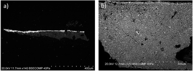
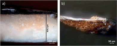
A feature of major importance in the context of our study is that the higher the content of these calcareous nannofossils, the coarser the chalk.30 This aspect suggests the need for grinding, and thereby partly destroying the shells of macrofossils in order to apply a smoother and thinner material to the upper layer of the ground. The studied cases suggest that chalk needs to be processed by grinding before being mixed with a binder thereby enabling a thinner ground layer technology, a different method from it being used directly after mining and washing, as some authors state.36Fig. 5 illustrates the SEM image (BSE mode) of the cross-section from sample 2a of the Apresentação no Templo painting from the altarpiece of Viseu Cathedral showing a thin ground layer with coarser granulometry in the lower part, and thinner in the upper area; the SEM image (BSE mode) of the cross-section from sample 1b of the painting of São Pedro of Tarouca Monastery church shows an increased amount of calcareous nannofossils having the same granulometry throughout the ground layer. The analysis made of the chalk from the altarpiece of Viseu Cathedral, when compared to the one from the painting of São Pedro of Tarouca Monastery church, deserves further interpretation. It presents a finely ground aspect (sample 5a from the Circuncisão painting from the Viseu Cathedral altarpiece (MGV), measuring about 45 μm thick) and apparently with a lower quantity of fossil in the upper layer while the one of São Pedro of Tarouca Monastery church, presents a greater thickness (sample 1b has 624.2 μm thick) and a coarser chalk in the ground layer (Fig. 6).Chalk material is also an identifier of the technique used in a specific epoch of certain Portuguese painting workshops, as in the case of the Viseu Workshop. After a primary phase of working with chalk material and using a ground layer technology with coarser grains in the lower part and finer powder in the upper area of the ground layer, the second phase of this workshop technique used calcium carbonate ground layers. The absence of coccoliths or other microfossils in some of the cases studied, however, led to the conclusion that powdered limestone was used instead of chalk, as in the case of the altarpiece of the church of Freixo-de-Espada-à-Cinta, Bragança (about 1530, a possible partnership between Vasco Fernandes and António Vaz (act. 1537–1569)).37 This altarpiece is made of chestnut oak and prepared with powdered limestone, coarser in the first layer and finer in the second layer. Fig. 7 shows an OM image of the cross-section of sample 6a of the Ecce Homo painting, presenting a saturated darker tone in the upper area of the ground layer; the SEM image (BSE mode) of the cross-section from the same sample presents coarser calcium carbonate (bigger grains of CaCO3) in the first layer and finer powder in the upper area. The altarpiece was recently restored due to its extreme state of degradation.7,38–40
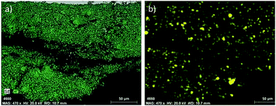
Despite limestone being an important natural resource in Portugal, this powdered material was only found in two of the groups studied. This move, made by the Viseu Workshop, into using chalk could be a consequence of the difficulty in getting chalk overseas, since it does not exist as a natural resource in Portugal, but also in an attempt to explore natural resources within the geographical proximity, such as the Ançã stone, Coimbra. This oolitic limestone, much used by the 15th and 16th century sculptors, contains silica (SiO2), since the oolites are formed around quartz grains.7 The maps of elemental analysis by SEM-EDS from powdered calcite in the ground layer of the sample 6a of the Ecce Homo painting from the altarpiece of the church of Freixo-de-Espada-à-Cinta shows the detected Ca and Si elements (Fig. 8).
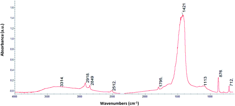
The later work of the workshop evidenced a move towards the use of gesso sottile in the ground layers of their subsequent paintings, as in the case of the São Pedro palla made for Viseu Cathedral (MGV) or the two paintings of saints São Vicente and Santo António, c. 1550, from Cavernães Church, Viseu, and attributed to António Vaz, follower of Vasco Fernandes. In Fig. 9 both the OM images of cross-section from sample 7a and the SEM image (BSE mode) of the Santo António painting in Cavernães Church show an homogeneous mixture of filler and binder ground layer; the detailed SEM image (BSE mode) of the same sample shows acicular particles characteristic of gesso sottile. The semi-quantitative measurements of μ-XRD conducted in the areas of gypsum and anhydrite peaks revealed the presence of 67% to 91% of gypsum and 9% to 33% of anhydrite. This analysis also detected the presence of hydrocerussite, certainly corresponding to Pb found by the SEM-EDS analysis. The presence of Pb may be linked with the addition of lead carbonate to the ground layer. This Pb is associated with aluminosilicates Mg, K and Fe. Iron, besides being associated with aluminosilicates, also coexists in grains whose major constituent is Fe but which also contain a large amount of Pb, with traces of Mg, Al, S, P, S, K, Ca and Cu; these grains correspond to red ocher, as already verified by OM,41 being the main factor responsible for the color of the ground layer. In Fig. 10 the spectrum obtained by μ-FTIR of the coarser lower part of the ground layer of sample 7a is largely dominated by bands related to the presence of dehydrated gypsum 3539 cm−1 and 3405 cm−1 correspond to an elongation νOH of water molecules present in the gypsum structural network; 1621 cm−1 is due to deformation of the δOH group of water molecules of the structural network; the bands at 1139 cm−1 match an antisymmetric group νSO4 elongations of the band to 1008 cm−1 corresponds to a symmetrical elongation νSO4 group, and the band at 670 cm−1 is attributable to deformation of the sulfate group, δSO4. The proteins with a weak band at 1448 cm−1 and two minor bands at 1546 cm−1 and 1683 cm−1, are covered by the coexistence of a typical gypsum band in the spectra; these two bands relating to N–H deformation and elongation C–N (amide II) and elongation amides of C![[double bond, length as m-dash]](https://www.rsc.org/images/entities/char_e001.gif) O (amide I), respectively, bands associated with the presence of protein. The presence of alkyl group bands, are evident in a wide range of organic compounds, suggesting in this case the occurrence of oil, material stated in Portuguese treatises as a polisher of gypsum ground layers;17 the oil is identifiable in the bands at 2926 cm−1 and 2857 cm−1 corresponding to an antisymmetric and symmetric elongation, respectively νCH alkyl groups linked, the band related to the elongation νC
O (amide I), respectively, bands associated with the presence of protein. The presence of alkyl group bands, are evident in a wide range of organic compounds, suggesting in this case the occurrence of oil, material stated in Portuguese treatises as a polisher of gypsum ground layers;17 the oil is identifiable in the bands at 2926 cm−1 and 2857 cm−1 corresponding to an antisymmetric and symmetric elongation, respectively νCH alkyl groups linked, the band related to the elongation νC![[double bond, length as m-dash]](https://www.rsc.org/images/entities/char_e001.gif) O ≈ 1740 cm−1 is less detectable, and is only visible with a small shoulder. These paintings show a homogeneous mixture of gypsum and binder in the ground layer.
O ≈ 1740 cm−1 is less detectable, and is only visible with a small shoulder. These paintings show a homogeneous mixture of gypsum and binder in the ground layer.
3.3. Binders-absorbent and non-absorbent systems
The difference in coarseness of the powder from the lower part to the upper part of the ground layer, while having the mechanical aim of sustaining the panel oscillations, also has aesthetic intentions with specific absorbency qualities.The upper area of the chalk ground layer in painting has been noted in recent studies.19,42 The increased transparency in this upper level when compared to the lower part of the ground layer is an optical quality which can be attributed to the penetration of the oil used as medium in the painting layers. This oil can be absorbed through the ground layer, due to less porosity in the upper area when compared to the lower part of the ground.42 The upper area is also responsible for slow drying, in order to obtain the singular transparency seen in Flemish paintings.7,19
Previous studies show that chalk has a lower refractive index (n = 1.50–1.64) than the remaining pigments. Its coating power is activated when it is mixed in an aqueous solution of glue, which in a concentration of about 10% has a refractive index of around n = 1.35 (Table 1).20
| Type | Material | Refractive index (n) |
|---|---|---|
| Pigment/filler | Chalk | 1.50–1.64 |
| Aqueous binder | Animal glue (diluted to 10%) | 1.348 |
| Oil binder | Linseed oil | 1.484 |
When chalk is placed in a mixed solution of linseed oil with a refractive index n = 1.48, its coating power decreases due to the close refractive index of both materials (of about 0.07).43 On the other hand, if chalk is mixed with animal glue, with a greater difference in the refractive index, the mixture improves its coating power, even while wet. Fig. 11 exemplifies the reconstruction of mixture of chalk with animal glue showing the drying of the first ground layer, even while wet, with an improved coating power. The SEM images (BSE mode) of the reconstruction of Portuguese powdered calcareous rock and of the cross-section of the sample 6a of the Ecce Homo painting from the altarpiece of the church of Freixo-de-Espada-à-Cinta puts has evidence of powdered calcite on its angular faces. The coating power of the mixture continues to improve upon drying of the surface since the chalk remains primarily surrounded by air.20
The color parameters were registered in both apertures LAV and USAV for ground layer reconstructions of glue/chalk, oil/chalk or both, overlaying this last to first, in order to evaluate color differences between the samples and to investigate the reflectance percentage. In Table 2 we verify the calculated values so that: L* > 0, the sample became more luminous; L* < 0, the sample darkened; a* > 0, the sample became redder; a* < 0, the sample became greener; b* > 0, the sample became more yellow; b* < 0, the sample became more blue,29C* > 0, the sample became more chromatic and h° denotes hue angle. The results presented in Table 2 show that samples that contain oil are less luminous, having lower reflectance percentage, than animal glue samples. The colorimetric results showed that the total change in color is significant when mixing chalk with oil or with glue. They confirmed the decreasing refractive index of the grayish mixture oil/chalk (lower values of a* when comparing to b*) in contrast with the whitish mixture of glue/chalk (lower values of a*and b* when comparing to oil/chalk mixture). Lightness has higher values in the chalk/animal glue reconstructions but color/chroma parameters (a* b* and C*) are higher in chalk/oil reconstructions, giving the ground layers a grayish half-tone for painting.
| Ingredients | Amounts | Sample | CIE Lab Ch D65/10Deg | ||||
|---|---|---|---|---|---|---|---|
| CIE L | CIE a | CIE b | CIE C | CIE h | |||
| Chalk/animal glue | 1![[thin space (1/6-em)]](https://www.rsc.org/images/entities/char_2009.gif) : :![[thin space (1/6-em)]](https://www.rsc.org/images/entities/char_2009.gif) 1 (v/v) 1 (v/v) |
1 | 83.31 | 1.54 | 11.88 | 11.98 | 82.62 |
| Chalk/animal glue | 3![[thin space (1/6-em)]](https://www.rsc.org/images/entities/char_2009.gif) : :![[thin space (1/6-em)]](https://www.rsc.org/images/entities/char_2009.gif) 1 (v/v) 1 (v/v) |
2 | 90.55 | 0.96 | 6.91 | 6.98 | 82.07 |
| Chalk/oil over chalk/animal glue with sizing layer between them | 1![[thin space (1/6-em)]](https://www.rsc.org/images/entities/char_2009.gif) : :![[thin space (1/6-em)]](https://www.rsc.org/images/entities/char_2009.gif) 1 (v/v) 1 (v/v) |
3 | 72.13 | 3.79 | 21.31 | 21.64 | 79.91 |
| Chalk/oil over chalk/animal glue | 1![[thin space (1/6-em)]](https://www.rsc.org/images/entities/char_2009.gif) : :![[thin space (1/6-em)]](https://www.rsc.org/images/entities/char_2009.gif) 1 (v/v) 1 (v/v) |
4 | 71.11 | 3.8 | 21.41 | 21.75 | 79.93 |
| Chalk/oil over chalk/animal glue with sizing layer between them | 3![[thin space (1/6-em)]](https://www.rsc.org/images/entities/char_2009.gif) : :![[thin space (1/6-em)]](https://www.rsc.org/images/entities/char_2009.gif) 1 (v/v) 1 (v/v) |
5 | 71.65 | 4.59 | 19.72 | 20.25 | 76.91 |
| Chalk/oil over chalk/animal glue | 3![[thin space (1/6-em)]](https://www.rsc.org/images/entities/char_2009.gif) : :![[thin space (1/6-em)]](https://www.rsc.org/images/entities/char_2009.gif) 1 (v/v) 1 (v/v) |
6 | 71.96 | 4.63 | 19.64 | 20.18 | 76.72 |
| Chalk/oil over chalk/animal glue with sizing layer between them | 25 gr c + 4.72 gr o; 1c:1/2g (v/v) | 7 | 73.2 | 4.75 | 19.04 | 19.63 | 75.98 |
| Chalk/oil over chalk/animal glue | 25 gr c + 4.72 gr o; 1c:1/2g (v/v) | 8 | 75.09 | 4.36 | 19.09 | 19.58 | 77.14 |
| Chalk/animal glue | 1![[thin space (1/6-em)]](https://www.rsc.org/images/entities/char_2009.gif) : :![[thin space (1/6-em)]](https://www.rsc.org/images/entities/char_2009.gif) 1/2 (v/v) 1/2 (v/v) |
9 | 89.7 | 1.09 | 7.34 | 7.43 | 81.52 |
| Chalk/oil | 25 gr c + 4.72 gr o | 10 | 69.98 | 4.48 | 19.1 | 19.62 | 76.79 |
| Chalk/oil | 1![[thin space (1/6-em)]](https://www.rsc.org/images/entities/char_2009.gif) : :![[thin space (1/6-em)]](https://www.rsc.org/images/entities/char_2009.gif) 1 (v/v) 1 (v/v) |
11 | 69.22 | 4.19 | 20.9 | 21.32 | 78.66 |
| Chalk/oil | 3![[thin space (1/6-em)]](https://www.rsc.org/images/entities/char_2009.gif) : :![[thin space (1/6-em)]](https://www.rsc.org/images/entities/char_2009.gif) 1 (v/v) 1 (v/v) |
12 | 71.86 | 4.05 | 19.94 | 20.35 | 78.52 |
| Chalk/strong animal glue mixture | — | 13 clear surface | 77.27 | 2.18 | 23.66 | 23.76 | 84.75 |
| Chalk/rabbit glue | — | 14 clear surface | 78.71 | 1.28 | 24.68 | 24.71 | 87.02 |
| Chalk/strong animal glue mixture | — | 13 dark surface | 58.93 | 9.38 | 24.53 | 26.26 | 69.07 |
| Chalk/rabbit glue | — | 14 dark surface | 58.22 | 9.41 | 25.16 | 26.86 | 69.5 |
Animal chalk/glue historical reconstructions made in 1973 by Reis Santos in Laboratório José de Figueiredo (samples 13 and 14) when measured and compared to the above mentioned reconstructions made by us show that the lightness of the mixture decreases, probably due to the yellowing of the binder, also visible by naked eye. These samples are heterogeneous in color when comparing to the reconstructions made by us. Results show that darker surfaces of the mixture are less luminous than clearer areas.
In the reconstructions with overlapping layers, glue/chalk on the base and oil/chalk on the top, it is possible to achieve an average slight improvement of lightness when compared to the oil/chalk single layer reconstruction, proofing the utility of this 2-layer structure. Results confirm also data brought by historical texts referring to the prominence of the white ground layers with the aging of the paintings.11,44 With aging, shadows and half-shadows disappear (probably due to the aging of the binders in the ground layer, as shown by the results of decreasing lightness of the historical reconstructions analyzed), and as such, it was probably required by the painters the search of a grayish half-tone ground, preventing these aging problems.11,44 These results corroborate the need for a two-layered glue/chalk and oil/chalk ground structure.
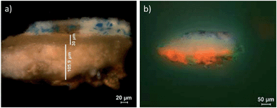
The first ground layer applied over the panel, after being sealed with a sizing layer, is a mixture of chalk and animal glue, promoting the adhesion of the ground layer to the support.3,4,7,19 Although the distinction between these two layers is sometimes difficult to recognize in OM, there are cases where this difference is evident, as in sample 6a of the Ecce Homo painting from the altarpiece of the church of Freixo-de-Espada-à-Cinta (powdered limestone) (Fig. 7) or sample 4a of the Cristo perante Pilatos painting from the Flemish altarpiece of the Esporão Chapel (ME) (chalk). Fig. 12 contains a diagram which has been adapted from a pictorial layer structure.21 The diagram shows the reflection of “superficial light” on a white ground layer, thus bringing a greater level of brightness and luminosity to the pictorial layer, which is equivalent to a lower layer of chalk and glue (Fig. 12a); the reflection on the “deep light” translucent background reduces the brightness of the color layer (Fig. 12b); the cross-section of sample 4a of the Cristo perante Pilatos painting from the altarpiece of the Esporão Chapel (ME) shows an historical case of the reflection scheme of “superficial light” from the bottom proteinaceous ground layer and “deep light” from the oil binder upper layer of the chalk ground. The example shows the distinction between binders in the ground layer, oil for the upper layer and animal glue for the level below, reproducing the search for a different refractive index of the materials to cover the surface of the support and to obtain distinct reflecting light effects. In this sample, which is considered to be complete (due to the wood fiber remains of the support) the thickness of the ground layer mixed with animal glue is about 105.9 μm, with the thickness of the upper part mixed with oil being about 30 μm (Fig. 13a). This technical aspect is understandable, given the fact that particle size, in conjunction with the concentration, influences the refractive index of the covering capacity of the material. In general, the finer the particles the greater the coating power.20 By applying a first layer of coarser chalk bonded with animal glue, the artist achieved a ground with hydrophilic characteristics i.e. having a strong affinity for water. At the second stage, to give the necessary transparency and facilitate the drawing and painting of the future composition, a powder layer was applied with an oil binder, providing a compatible surface to optimize the integration between the painting and the ground layer. This non-absorbent ground allows for the painting of the following layers maintaining the hydrophobic effect and promoting the union between films, contributing to the cohesion of the painting.
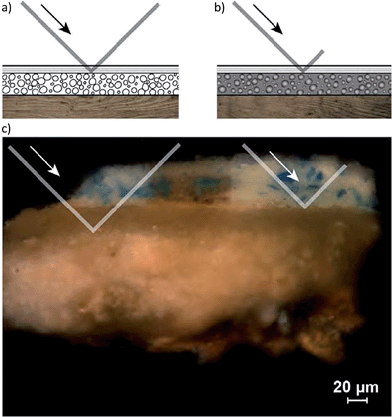 | ||
| Fig. 12 (a) Diagram of the reflection of “superficial light” on white background, giving brightness and luminosity to the pictorial layer, equivalent to the bottom of chalk and glue, adapted diagram from;21 (b) diagram of the reflection on the “deep light” translucent background, reducing the amount of reflection and the brightness of the color layer, adapted diagram from;21 (c) cross-section of sample 4a of painting “Cristo perante Pilatos”, Altarpiece of the Esporão Chapel (ME) showing the reflection scheme of “superficial light” of the bottom proteic ground layer and “deep light” in the oil binder top area of the chalk ground. | ||
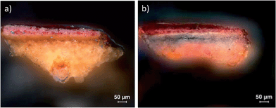
When observed by OM with ultraviolet light and an emission filter of 490 nm, sample 4a highlights a medium-rich layer of higher density in the upper area of the chalk ground, marking the area of the corresponding ground layer with animal glue proteins in a fluorescent orange color. This test was performed using a highly sensitive fluorescent protein dye observable between 300/472 nm (excitation) and 490/570 nm (emission) (Orange Sypro©).22,23Fig. 13b and 14b show, via OM image, historical cross-sections from sample 4a of the Cristo perante Pilatos painting from the altarpiece of the Esporão Chapel (ME) and sample 8a of the Morte da Virgem painting from the old Flemish polyptych in Évora Cathedral, under ultraviolet light and stained in fluorescent orange (Orange Sypro©). These figures focus on the proteinaceous area of the ground layer, thus distinguishing the thicker proteinaceous bottom from the thinner oil binder of theupperareas of the ground layers.
This non-absorbent surface area of the chalk ground layer, when saturated, has the ability to control the penetration of the descendant oil coming from the painting layers, maintaining a homogeneous and regular distribution of the deep light effect. In the examples studied we observe that paintings with different granulometry in the ground layer, using oil in the upper area, retain the binder homogeneously. Painting with a thicker proteinaceous ground layer using the same granulometry has a heterogeneity in the absorption of the binder coming from painting, priming or polishing layers, as is the case with the altarpiece of Viseu Cathedral, when compared with the painting São Pedro of Tarouca Monastery church (Fig. 6a).
Mixing the pigments with the binder is essential since it disintegrates the agglomerates, surrounding each particle of the binder and thus obtaining a homogeneous mixture, creating greater plasticity and stability.21
A highly accurate technique of ground layer execution was required, based on the expertise and traditional knowledge of Flemish painters and passed to Portuguese masters, as in the case of the Viseu Workshop.
4. Conclusions
From this work we can conclude that one of the main objectives of the ground layer is to create the best possible reflection of light, using thin transparent layers to enhance the vibrancy of the colors painted upon them. This technique was used by Flemish painters and seems to have been adapted by Portuguese painters during the early stages of the Viseu Workshop to achieve translucent and thin layers with an aesthetic appearance.Furthermore, it was observed that coarser calcium carbonate (chalk or powdered limestone) and glue emulsion are the first phase of a ground layer to be applied, after sizing the panel. A second, thinly powdered ground layer of the same filler material was then applied with oil to achieve specific light effects. This granular difference between the upper area and the lower area is essential to increase resistance to mechanical movements of the panel and to control light effects of the ground layer. In addition, using a thinner ground layer technology facilitates the ease of application, thereby increasing the speed of handling.
In addition, it was verified that the aim of this ground layer technique, using oil and proteinaceous binders separately, was to create a saturated system of a non-absorbent ground layer with a reflective capacity of superficial and deep light. Provided by the intentional use of oil as a binder, the upper ground layer was received by an aqueous system of the absorbent ground layer (using glue as a binder) and permitting a superficial light reflection. Both systems (absorbent/non-absorbent) led to the reflection of superficial and deep light and might have been used intentionally by the artists to obtain the thinness and translucency of the grounds. Therefore, it is consistent that the oily upper layer was finely powdered, and upon being mixed with oil, its coating power was reduced and this made possible the reflection of the underlying coarser protein ground layer which was thick and white, taking advantage of “deep light” and “superficial light” brought by both binders. The results of the reconstructions corroborate the need for a two-layered glue/chalk and oil/chalk ground structure, as found for historical samples of the studied paintings.
The reconstructions prove the less luminous (deep light) grayish tone achieved when combining oil and chalk when compared to the glue/chalk mixture (superficial light). By overlapping both layers, with oil/chalk on the top, it is possible to achieve a slight improvement of lightness when compared to the oil/chalk single layer, proofing the utility of this 2-layer structure (deep and superficial light). A grayish half-tone of ground layers combining superficial and deep light for painting could have been an achievement of the painters of the 15th and 16th centuries.
Finally, to accomplish the durability of the layers, and to conserve its light, careful grinding combined with specific quality and compatibility between materials was required.
This constructive technology has enabled these panel paintings to preserve their quality until today: a Baltic oak panel, isolated by an aggregating sizing layer and chalk ground layers, coarser in the base and finely powdered in the upper area to achieve mechanical resistance and translucent light effects.
We can also conclude that different granulometry of the powdered limestone medium forming a ground layer did not bring about the desired results of superficial and deep light required by the artists since it was only used at one point in time. Also the use of gesso sottile suggests characteristics of smoothness and capture of the light closer to chalk provided by its shield-shaped particles, being the next most used ground material at the Viseu Workshop, after chalk.
The characterization of inorganic and organic materials existing in ground layers, confirmed by the new analytical results obtained, have led to novel conclusions about the techniques applied to Flemish and Portuguese paintings of the 15th and 16th centuries. This ground layer technique could have been the origin of the canvas priming technique, used in the 16th and 17th centuries and thereafter, where the use of oil overlies the use of proteinaceous ground layers.
Acknowledgements
The authors acknowledge Fundação para a Ciência e Tecnologia for financial support (Post-doc grant SFRH/BPD/103315/2014) through program QREN-POPH-typology 4.1., co-participated by the Social European Fund (FSE) and MCTES National Fund. Also wish to acknowledge Dr Irina Sandu for staining protocols, Dr José Mirão for the text revision, Dr Maria José Oliveira for the assistance at the μ-XRD, Dr Luís Dias for the assistance at the SEM-EDS, Dr Stéphane Longelin for the assistance at the μ-Raman, Dr Catarina Miguel and Dr Ana Cardoso for the assistance at the μ-FTIR.References
- V. Antunes, A. Candeias, M. L. Carvalho, M. J. Oliveira, M. Manso, A. I. Seruya, J. Coroado, L. Dias, J. Mirão, S. Longelin and V. Serrão, J. Instrum., 2014, 9, C05006 CrossRef.
- V. Antunes, A. Candeias, M. L. Carvalho, M. J. Oliveira, A. I. Seruya, J. Coroado, L. Dias, J. Mirão, S. Longelin and V. Serrão, J. Raman Spectrosc., 2014, 1026–1033 CrossRef CAS.
- V. Antunes, M. J. Oliveira, H. Vargas, A. Candeias, A. Seruya, L. Dias, V. Serrão and J. Coroado, Microsc. Microanal., 2014, 20, 66–71 CrossRef CAS PubMed.
- V. Antunes, M. J. Oliveira, H. Vargas, V. Serrão, A. Candeias, M. L. Carvalho, J. Coroado, J. Mirão, L. Dias, S. Longelin and A. I. Seruya, Anal. Methods, 2014, 6, 710–717 RSC.
- O. Syta, K. Rozum, M. Choińska, D. Zielińska, G. Z. Żukowska, A. Kijowska and B. Wagner, Spectrochim. Acta, Part B, 2014, 101, 140–148 CrossRef CAS.
- V. Antunes, A. Candeias, M. J. Oliveira, M. Lorena, A. I. Seruya, M. L. Carvalho, M. Gil, J. Mirão, J. Coroado, V. Gomes and V. Serrão, Microchem. J., 2016, 125, 290–298 CrossRef CAS.
- V. Antunes, Tese doutoral, Faculdade de Letras da Universidade de Lisboa, 2014.
- J. Campelo, A. Pais and N. Escobar, Cadernos de conservação e restauro: O retábulo Flamengo de Évora, Instituto dos Museus e da Conservação, Lisboa, 2008/2009 Search PubMed.
- Instituto José de Figueiredo Boletim Informativo do Instituto José de Figueiredo, 1987–1988, Secretaria de Estado da Cultura, Instituto Português do Património Cultural, Lisboa, Minerva do Comércio edn, 1989.
- M. Cachão and T. Moita, Mar. Micropaleontol., 2000, 39(1/4), 131–155 CrossRef.
- M. Stols-Witlox, in Oud Holland, ed. Q. f. D. A. History, 2015, vol. 128, pp. 171–186 Search PubMed.
- K. Groen, in A Corpus of Rembrandt Paintings IV: Self-Portraits,Volume 4 of Rembrandt Research Project Foundation, ed. E. v. d. Wetering, Springer, Netherlands, 2007, vol. IV, pp. 318–334 Search PubMed.
- M. Stols-Witlox, PhD thesis, Amsterdam School for Culture and History (ASCH), 2014.
- L. Simis, Grondig onderwijs in de schilder- en verwkunst: bevattende eene duidelijke onderrigting in den aard, den oorsprong, sterkte en zwakte der verwen, olijeteiten, enz., het bereiden en behoorlijk of kwalijk gebruik derzelven, ook over het behandelen der werken, huizen en rijtuigen, gladhout en marmer schilderen, vergulden, verzilveren en metalen, alsmede over het verlakken en witwerken, het maken en gebruiken van lakken en vernissen, het verwen van wollen, saijetten, zijden, enz., over de craijon-pennen en waterverwen, schilderen van papier en het glassnijden, doormengd met vele andere en soortgelijke wetenschappen en kunstgeheimen: alles niet alleenlijk onontbeerlijk voor den leerling, maar ook hoogstnuttig voor den meer ervaren schilder: na eene zes en dertig jarige ondervinding zamengebragt, Erven H. Gartman, 2 edn, 1829.
- M. I. F. P. Cardoso, Tese doutoral, University College London, 2010.
- M. I. F. P. Cardoso, Infocus, 2006, 4(41), 64–86 Search PubMed.
- P. Monteiro and A. J. Cruz, in The Materials of the Image. As Matérias da Imagem, ed. L. U. Afonso, Cátedra de Estudos Sefarditas Alberto Benveniste da Universidade de Lisboa, Lisboa, 2010, pp. 237–286 Search PubMed.
- M. Lorena, in Colóquio Internacional sobre As Preparações na Pintura Portuguesa dos Séculos XV e XVI, Auditório do Museu Nacional de Arte Antiga, 28 e 29 de Junho de 2013, ed. V. Serrão, V. Antunes and A. I. Seruya, Faculdade de Letras da Universidade de Lisboa, Lisboa, Overprint- graphic prodution management/Torreana edn, 2013, pp. 133–142 Search PubMed.
- M. Lorena, Tese doutoral, Universidade de Évora, 2013.
- K. Nicolaus, Manual de restauración de cuadros, Konemann, Köln, 1999 Search PubMed.
- R. Mayer, Manual do artista de técnicas e materiais, Martins Fontes, São Paulo, 1999 Search PubMed.
- I. C. A. Sandu, S. Schäfer, D. Magrini, S. Bracci and C. A. Roque, Microsc. Microanal., 2011, 18, 860–875 CrossRef PubMed.
- I. C. A. Sandu, A. C. A. Roque, P. Matteini, S. Schäfer, G. Agati, C. R. Correia and J. F. F. P. Viana, Microsc. Res. Tech., 2011, 75, 316–324 CrossRef.
- S. Kuckova, I. C. A. Sandu, M. Crhova, R. Hynek, I. Fogas, V. S. Muralha and A. V. Sandu, Microchem. J., 2013, 110, 538–544 CrossRef CAS.
- B. Lafuente, R. T. Downs, H. Yang and N. Stone, in Highlights in Mineralogical Crystallography, ed. T. Armbruster and R. M. Danisi, W. De Gruyter, Berlin, Germany, 2015, pp. 1–30 Search PubMed.
- S. T. T. d. Mayerne, Pictoria, sculptoria et quae subalternarum artium (the 'Mayerne manuscript'), B.L. Sloane 2052, British Library, 1620–1646 Search PubMed.
- L. Carlyle, J. J. Boon, R. Haswell and M. Stols-Witlox, in Preparation for Paintings: the Artist's Choice and its Consequences, ed. J. H. Townsend, T. Doherty, G. Heydenreich and J. Ridge, Archetype Books, 2008, pp. 110–122 Search PubMed.
- F. L. T. d. C. d. Silva, Pintura Simples, Lisboa, 1898 Search PubMed.
- R. L. Feller, Accelerated Aging; Photochemical and Thermal Aspects, Getty Conservation Institute, Marina del Rey, California, 1990 Search PubMed.
- J. Dercourt and J. Paquet, Geology: Principles & Methods, Gulf Pub. Co., Book Pub. Division, 1985 Search PubMed.
- University College London and JISC, Calcareous Nannofossils, MIRACLE, microfossil image recovery and circulation for learning and education, http://www.ucl.ac.uk/GeolSci/micropal/calcnanno.html, accessed 9/5/2014.
- Nannotax3 Website, ed. J. R. Young, P. R. Bown and J. A. Lees, International Nannoplankton Association, 2014 Search PubMed.
- R. W. Jones, Applications of Palaeontology: Techniques and Case Studies, Cambridge University Press, 2011 Search PubMed.
- F. Giraud, B. Pittet, E. Mattioli and V. Audouin, Mar Micropaleontol, 2006, 60, 205–225 CrossRef.
- J. E. Wendler, I. Wendler, B. T. Huber and K. G. Macleod, GSA Denver Annual Meeting 31 October 3 November 2010, Geological Society of America, 2010, vol. 42, p. 131 Search PubMed.
- M. Stols-Witlox, in Conservation of Easel Paintings-routledge Series in Conservation and Museology, ed. J. H. Stoner and R. Rushfield, Taylor & Francis, 2012, pp. 161–185 Search PubMed.
- L. Reis-Santos, Vasco Fernandes e os pintores de Viseu do século XVI, Edição do autor, Lisboa, 1946 Search PubMed.
- B. C. Maia, J. C. Frade, A. Calvo and A. Candeias, Santiago de Compostela, Universidade de Santiago de Compostela, 2012 Search PubMed.
- B. C. Maia, J. C. Frade, A. Calvo and A. Candeias, Porto, Escola das Artes da Universidade Católica Portuguesa (Centro Regional do Porto), 2012 Search PubMed.
- A. Calvo, C. Nodal, J. Carballo, E. Parra and L. Bravo, Estudo técnico-científico de 16 pinturas sobre madeira da igreja matriz de Freixo de Espada-à-Cinta Centro de Conservação e Restauro da Escola das Artes da Universidade Católica Portuguesa, Porto, 2004 Search PubMed.
- I. Rochinha, Relatório n° 41-92, análise material da pintura “Santo António” da Igreja paroquial de Cavernães,Viseu, Laboratório de Conservação e Restauro José de Figueiredo, Instituto de José de Figueiredo, Laboratório Central do Instituto Português do Património Cultural, Lisboa, 20 de Outubro de 1992 Search PubMed.
- E. S. B. Ferreira, R. Morrison and J. J. Boon, in Preparation for Paintings: The Artist's Choice and Its Consequences, ed. J. H. Townsend, T. Doherty, G. Heydenreich and J. Ridge, Archetype Books, 2008, pp. 50–58 Search PubMed.
- F. W. Tegethoff, J. Rohleder and E. Kroker, Calcium Carbonate: From the Cretaceous Period Into the 21st Century, Springer, 2001 Search PubMed.
- J.-B. Oudry, in Le Cabinet de l'Amateur, E. Piot, 1861-1862, pp. 107–117 Search PubMed.
| This journal is © The Royal Society of Chemistry 2016 |


