Relationship between negative thermal expansion and lattice dynamics in a tetragonal PbTiO3–Bi(Mg1/2Ti1/2)O3 perovskite single crystal
Kai Jiangab,
Peng Zhanga,
Jinzhong Zhanga,
Guisheng Xuc,
Wenwu Lia,
Zhigao Hu*a and
Junhao Chua
aDepartment of Electronic Engineering, East China Normal University, Shanghai 200241, China. E-mail: zghu@ee.ecnu.edu.cn; Fax: +86-21-54345119; Tel: +86-21-54345150
bNational Laboratory for Infrared Physics, Shanghai Institute of Technical Physics, Chinese Academy of Science, Shanghai 200083, China
cKey Laboratory of Transparent Opto-Functional Advanced Inorganic Materials, Shanghai Institute of Ceramics, Chinese Academy of Sciences, Shanghai 201899, China
First published on 15th December 2015
Abstract
A range of compelling information on the thermal expansion behavior and lattice dynamics of a novel ferroelectric perovskite-type 0.62PbTiO3–0.38Bi(Mg1/2Ti1/2)O3 single crystal has been revealed by means of temperature-dependent X-ray diffraction and polarized Raman scattering. X-ray diffraction analysis suggests a strong ferroelectricity and structural change (tetragonal to cubic) upon heating. The A1(1TO) soft mode, associated with the order parameter spontaneous polarization displacement, abnormally shifts downward and vanished, indicating the change in polar nanoregions and phase transition. Both the temperature dependent unit cell volume and frequency of the soft mode confirm the correlation between spontaneous polarization and negative thermal expansion. The experimental evidence of spontaneous volume ferroelectrostriction will further help in understanding the relationship between the structure and optical properties of perovskite-type ferroelectric oxides.
1 Introduction
In recent years, complex mixed-ion ferroelectric materials have been extensively investigated in order to achieve optimum properties as well as to understand the underlying factors for property tweaking.1–4 PbTiO3 (PT)-based perovskite compounds are important multifunctional materials, which have been investigated in the last half century due to their controllable physical properties, such as ferroelectric, piezoelectric, optical, and electric properties.5–8 Most recently, the research hotspot of PbTiO3–BiMeO3 ferroelectrics, where Me can be a single cation of valency +3 (e.g. Sc3+, In3+ and Fe3+)9–14 or a mixture of cations with an average valence of +3 (e.g. Zn1/2Ti1/2, Mg1/2Ti1/2, and Ni1/2Ti1/2),6,15–18 have stimulated much interest. Their excellent optoelectronic/piezoelectric properties and high Curie temperature (TC) are the key parameters in piezoelectric devices for applications with a stable structure and performance under specific temperature conditions.A significant alternative system of PbTiO3–BiMeO3 is involved in PbTiO3–Bi(Mg1/2Ti1/2)O3 (PT–BMT), which has being considerably studied due to its good performances such as high TC, low cost, low thermal expansion, and other good properties.3,13,19–21 In particular, the morphotropic phase boundary (MPB) of xPT–(1 − x)BMT ceramics is in the range of 0.36 ≤ x ≤ 0.38, with a relatively high TC of about 700 K and piezoelectric coefficient d33 above 200 pC N−1.3 In order to avoid the redundant phase transition and improve the stability in a wide temperature range, we have systematically investigated the piezoelectric properties of tetragonal phase 0.62PT–0.38BMT single crystals, which can be considered as novel piezoelectric materials for high-performance actuators and transducers.21 According to the lattice dynamical theory and experiments, the A1(1TO) soft mode in PT-based solid solutions is considered to be proportional to the parameter spontaneous polarization PS.13 Especially in a PT-based ferroelectric system, where polarization is determined by the degree of local distortions, it is crucial to understand the structural behavior at the atomic level.22 Therefore, further investigation is necessary in order to illustrate the physical mechanism between the lattice dynamics and negative thermal expansion (NTE) of a 0.62PT–0.38BMT crystal. Up to now, a small amount of information could be found on the vibration behavior of a PT–BMT system, which is attributed to the difficult growth technology of pure perovskite BMT. Fortunately, the 0.62PT–0.38BMT crystals were successfully grown using a high-temperature solution method.21
In this work, we present the results from temperature dependent polarized Raman scattering experiments complemented by X-ray diffraction data, which depict the features regarding the local structure of the 0.62PT–0.38BMT single crystal. The anomalies were discussed according to the results of the unit cell volume and A1(1TO) soft mode as a function of temperature. The results provide evidence of a consistent correlation between thermal expansion and PS displacement.
2 Experimental details
The 0.62PT–0.38BMT powder and single crystals were grown using the flux-Bridgman method. Details about the crystal growth, microstructure and characterization can be found elsewhere.21 Structural analysis was undertaken using temperature-dependent X-ray diffraction (XRD), which was carried out using a Bruker D8 Discover High Resolution XRD in the temperature range from 300 to 873 K (TCU100 Temperature Control Unit, LNC Nitrogen Suction Equipment, Anton Paar). The temperature dependent Raman scattering was recorded on heating using a Jobin-Yvon LabRAM HR 800 micro-Raman spectrometer with the frequency range of 10–1000 cm−1 and a THMSE 600 heating/cooling stage (Linkam Scientific Instruments) in the temperature range from 80 to 850 K. The crystals were excited using a 632.8 nm He–Ne laser and recorded in back-scattering geometry in parallel 〈x|zx|y〉 (VH) and perpendicular 〈x|zz|y〉 (VV) polarization configurations. The laser beam was focused through a 50× microscope with a working distance of 18 mm. An air-cooled charge coupled device (CCD) (−70 °C) with a 1024 × 256 pixels front illuminated chip was used to collect the scattered signal dispersed on 1800 grooves per mm grating with a resolution better than 1 cm−1. The measured spectra have been treated with the Bose–Einstein phonon occupation factor and fitted with Lorentzian functions to determine the scattering information.23–253 Results and discussion
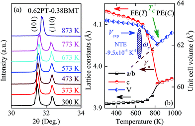
Room temperature (RT) XRD patterns of the 0.62PT–0.38BMT powder ground from the single crystals are shown in Fig. 1a and b. The single perovskite phase can be obtained according to the diffraction peaks, which is in good agreement with PDF #06-0452 and other literature.6,13,16 The typical tetragonal symmetry can be characterized by the splitting of the (001) and (002) peaks at 22° and 44°, respectively. No impurity phases, such as Bi12TiO20 and Bi4Ti3O12 phases, are observed in the composition presented here. The slight splitting of the (001) and (100) peaks from the 0.62PT–0.38BMT crystal can be found in Fig. 1b, indicating that the habitual faces of the grown crystals are {001}. The c/a ratio of the 0.62PT–0.38BMT single crystal is 1.061, which is much larger than that of PT–BMT in the MPB regions because the composition largely deviates from its MPB.21 Note that the c/a ratio of a single crystal is much larger than that of ceramics, suggesting a higher ferroelectricity and PS.6High temperature XRD measurements were performed to identify the phase transition and the change of lattice parameters. Fig. 2(a) shows a typical transition of the tetragonal phase 0.62PT–0.38BMT crystal. The large splitting of the two diffraction peaks (101) and (110), representing the tetragonal phase, reduces with increasing temperature. When the temperature approaches above TC (about 800 K), the two peaks merge into one, signifying that the cubic phase is dominant. Fig. 2(b) shows the lattice parameters [a(b) axis and c axis] and unit cell volume (abc) of the 0.62PT–0.38BMT system as a function of temperature. In the temperature range of 300–800 K, the c axis continually decreases while the a(b) axes slightly increase with increasing temperature. The unit cell volume, which is highly correlated with the change in ferroelectricity, contracts and reaches a minimum at TC. Therefore, the temperature dependence of the XRD results demonstrates that the ferroelectric to paraelectric phase transition (FE–PE) of the 0.62PT–0.38BMT crystal is located at ∼800 K. Furthermore, it is interesting to find that temperature strongly affects the c axis of the 0.62PT–0.38BMT crystal when compared with the a(b) lattice parameters. Therefore, the unit cell volume is dominated by the c axis, which is also a common character for PT-based compounds.6
According to the first-principle calculations and experimental results,4 the relatively small temperature dependence of the a(b) lattice parameters is related to the fact that the B-site Ti/Mg cation is more strongly covalent with four neighboring O2 oxygens in the ab plane than the two O1 oxygens (O1–Ti–O1) along the c axis. The stiff Ti–O2 bonds make compression and elongation in the ab plane energetically unfavorable, while the bonds of the O1–Ti–O1 unit along the c axis (the polar direction) are soft.4,8 As shown in Fig. 2(b), 0.62PT–0.38BMT exhibits a very weakened NTE in the tetragonal phase. The volume coefficient of thermal expansion (CTE) αV can be defined by the equation:  . Where VTC and VRT are the unit cell volume at TC and RT, respectively. In a lower temperature range (RT to 673 K), the unit cell volume shows little dependence on temperature with an average CTE of αV = −7.3 × 10⁻⁴ K−1. However, it contracts dramatically at elevated temperature with αV = −3.5 × 10⁻³ K−1 (673 to 800 K). The results demonstrate that the unit cell volume shrinks in the tetragonal phase but with a different slope as a function of temperature, which is similar to the results of the polar c axis length. The NTE in PT can be attributed to the thermal fluctuation of the cubic configurations in the tetragonal matrix.26 The average CTE in the temperature range from RT to 800 K is αV = −9.5 × 10⁻⁴ K−1. Moreover, a high positive thermal expansion (PTE, αV = −3.7 × 10⁻³ K−1) could be found beyond the temperature of 800 K, indicating a structural phase transition.
It is known that two factors contribute to the unit cell volume of the ferroelectric phase: anharmonic lattice phonon vibration and ferroelectric order.6 With decreasing temperature, the contribution from the factor of anharmonic lattice phonon vibration decreases. Thus the nominal unit cell volume (Vn) should normally contract, which can be described using the Debye–Grüneisen model.6,27 As shown in Fig. 2(b), Vn below TC, which is only determined by anharmonic lattice phonon vibrations, should be the extrapolation from the high-temperature paraelectric phase to the low-temperature ferroelectric one. However, the experimental unit cell volume (Vexp) of PT–BMT actually shows an abnormal increase with decreasing temperature below TC, signifying that the contribution from the ferroelectric order is simultaneously enhanced. The deviation of the unit cell volume from Vn to Vexp may be ascribed to the ferroelectric volume effect, which can be quantitatively described by spontaneous volume ferroelectrostriction (SVFS) using the equation: ωs = (Vexp − Vn)/Vn × 100%.28 As shown in Fig. 2(b), the value of SVFS for 0.62PT–0.38BMT is ωs = 8.0% at RT, which gradually decreases with increasing temperature, and reaches zero at TC (800 K). The mechanism of SVFS can be used to explain the phenomenon of NTE in the 0.62PT–0.38BMT crystals. It is known that the large ferroelectric polarization of PT-based compounds originates from the unique strong hybridization of the Pb 6s and O 2p orbitals, which stabilizes the large tetragonal distortion. This can be attributed to a stronger ferroelectric activity of A-site ions of Pb and Bi with a higher PS displacement value of 0.45 Å and 0.80 Å, respectively. Therefore, there is strong coupling between PS and NTE, which can be preliminarily characterized by the lattice dynamics results as follows.
Raman spectroscopy is a sensitive technique for investigating structure modifications and phonon modes, which can give information on the changes of lattice vibrations and ferroelectricity.25,29 For the tetragonal P4mm structure of 0.62PT–0.38BMT,16 a total of 8 optical lattice modes are predicted: ΓP4mm = 3A1 + 4E + B1.30,31 The simultaneous infrared and Raman activity produces additional splitting of each A1 and E mode into longitudinal-optical and transverse-optical components, so that, up to 15 modes should be visible for the P4mm unit cells. However, the scattering efficiency from some of these modes may be weak, and, generally in PT-based materials, scattering contributes only by increasing the width of neighboring modes with a higher intensity. For the above reasons, all the Raman modes in PT-based materials are convoluted into three main observable peaks, and a complete assignment of the underlying phonon modes has never been reported.
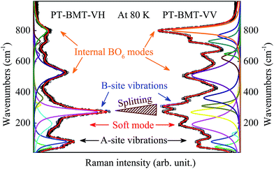
Fig. 3 shows the polarized Raman spectra of the 0.62PT–0.38BMT single crystal at a representative temperature of 80 K. Spectral deconvolution was performed according to 13 Lorentzian peak functions by means of a best-fitting algorithm. The spectrum is consistent with previous reports of ABO3-type ferroelectric materials, and can be linked to a tetragonal crystal structure. The three main regions can be distinguished in the spectrum; each of them has a relationship with different kinds of vibration in the lattice: (i) low frequency below 150 cm−1, which can be associated with the vibrations of the perovskite A-site, involving Pb and Bi ions. According to the assignment of a PT single crystal,31 the Raman phonon modes at about 80 and 131 cm−1 can be assigned to the E(1TO) and E(1LO) phonon modes, respectively. The presence of such distinct modes in the frequency range suggests possible cation ordering at the A-site. (ii) The frequency range of 150–450 cm−1, which is related to the Ti–O/Mg–O stretching vibrations. In particular, the mode with the highest intensity in the VH geometry, at about 276 cm−1, has been assigned as the B1 + E mode, closely related to the strength of the Ti/Mg–O bond. The splitting of the Ti/Mg–O modes is found in the VV geometry, which is believed to correspond to a typical ferroelectric material.32 The A1(1TO) soft mode at about 190 cm−1 consists of displacements of the BO6 octahedron relative to the Pb/Bi ions parallel to the PS direction (c-axis). The ferroelectric soft mode is highly associated with the behavior dependence on temperature and will be discussed later in the present work. (iii) High frequency bands above 450 cm−1 have all been associated with BO6 vibrations, namely the breathing and stretching modes of the oxygen octahedra. It should be emphasized that the Pb-based ferroelectrics with aliovalent chemistry on the B site such as 0.62PT–0.38BMT exhibit relatively strong Raman scattering near 680–850 cm−1, in contrast to the A-site complex with homovalent chemistry on the B site, such as (Na0.5Bi0.5)TiO3–7%BaTiO3 and (K0.5Na0.5)NbO3–5%LiNbO3.33,34 The Raman scattering near 680–850 cm−1 has been proven to arise from the symmetrical BO6 stretching in a double-perovskite structure, which generates the A1 mode in the cubic Fm![[3 with combining macron]](https://www.rsc.org/images/entities/char_0033_0304.gif) m structure. The local-scale doubling of the perovskite unit cell is attributed to the existence of a local-scale NaCl-type chemical B-site order even in compounds with no long-range B-site chemical order. The Raman spectra suggest a certain degree of local B-site chemical order for the 0.62PT–0.38BMT single crystal with simultaneous A-site and B-site doping.
m structure. The local-scale doubling of the perovskite unit cell is attributed to the existence of a local-scale NaCl-type chemical B-site order even in compounds with no long-range B-site chemical order. The Raman spectra suggest a certain degree of local B-site chemical order for the 0.62PT–0.38BMT single crystal with simultaneous A-site and B-site doping.
To further understand the relationship between lattice dynamics and NTE behavior for the 0.62PT–0.38BMT crystal, Fig. 4 presents the temperature dependence of the polarized Raman spectra in the temperature range from 80 to 850 K. With increasing temperature, the peak position and intensity of the polarized Raman phonon modes present a decreasing trend, while the broadening of the phonon modes is observed in the whole temperature range, which is similar to other ferroelectric materials.29 It should be pointed out that the lowest intensity for all the phonon modes occurs at the temperature range of 600–700 K (marked by the dashed rectangles). A similar anomaly range can be found from the XRD results (different CTE values). Note that the change of the polarized Raman spectra above 700 K demonstrates that there are some minor structural changes in the tetragonal phase structure of the PT–BMT crystal. This phenomenon can be ascribed to the influence of the dynamics of the polar nanoregions (PNRs) and SVFS mechanism.
 | ||
| Fig. 4 Temperature evolution of Raman intensities for the 0.62PT–0.38BMT single crystal. The dashed rectangles mark the temperature ranges of the most pronounced changes. | ||
The temperature dependence of the Raman phonon modes from the VV geometry is shown in Fig. 5. In the absence of any phase transition, one would expect steady phonon softening upon heating. However, in the case of the 0.62PT–0.38BMT crystal, one clear anomaly is visible at about 670 K. It should be emphasized that the reason for the anomaly is quite different from the phase transition temperature at about 800 K as determined using the XRD pattern and dielectric permittivity.21 The anomaly is an abrupt change in the wavenumber (softening of the phonon modes) that takes place in the A-site-related modes at about 670 K. The A1(1TO) soft mode varies linearly as a function of temperature, which can be described using a harmonic model:  . Where ω is the vibration frequency, k is the force constant related to the Pb/Bi–BO6 bond strengths, and m* is the reduced mass of the soft mode. Furthermore, temperature dependent polarization can also be indicated by the variation of four short Pb/Bi–O2 bonds, since the Pb atom forms covalent bonds only with the four nearest-neighbor oxygens.8,13 The Pb/Bi–O2 bonds elongate as a function of temperature for PT-based ferroelectric materials, suggesting a weakened Pb/Bi–O2 interaction followed by decreasing PS displacement. The decreased PS displacement of Pb/Bi introduces the centroids of Pb/Bi and BO6 further, resulting in the decreased force constant k. On the other hand, with increasing temperature, the reduced PS displacement makes the anharmonic potential energy shallow. As a result, the transition energy between excited states is reduced, and accordingly the vibration frequency ω of the soft mode is weakened. The tetragonality (c/a) in the PT–BMT single crystal could indicate NTE, since c/a is correlated with the lattice distortion. With increasing temperature up to TC, the larger decrease of c/a for PT–BMT results in strong NTE, which is demonstrated in Fig. 2(b). The temperature dependence of c/a is strongly coupled with the Raman-active soft mode (i.e. NTE and PS displacement) as shown by comparison of Fig. 2(b) and 5(b). Thus, it is apparent that there is a strong coupling interaction between PS and NTE as determined using the soft mode and XRD patterns.
It is well-known that the PNRs are believed to play an important role in lead-based ferroelectric materials. Note that below the so-called Burns temperature (TB), the temperature at which PNRs appear, the refractive index n, lattice parameters, unit cell volume V, or reciprocal dielectric permittivity 1/ε, begin to deviate from the linear changes of high temperatures.35 The frequency of the soft mode actually shows an abnormal increase with decreasing temperature (at about 670 K), as shown in Fig. 5(b), indicating some structural changes. In the whole temperature range, two different slopes of lines are observed, which can be attributed to the variation of the soft mode frequency induced by the local PS inside the PNRs. The dA1(1TO)/dT above and below TB is about −0.011 and −0.030 cm−1 K−1, respectively. Similar abnormal results were found by Zhang et al.17 and Bokov et al.35 using thermal expansion and dielectric measurements. The varying tendency of the A1(1TO) soft mode frequency with temperature agrees well with the PNRs in Pb-based ferroelectrics, in which the unit cell volume deviates from the linear thermal expansion at TB according to the XRD results in Fig. 2(b). Furthermore, as the temperature cools down from TB, the effect of SVFS is evident due to the frozen PNRs.28 Therefore, the NTE behavior of ferroelectrics is sensitive to spontaneous polarization displacement, which could be reflected by the A1(1TO) soft mode. The present lattice dynamics observations further support the results demonstrated by the temperature dependent XRD measurements.
4 Conclusions
To summarize, with the detailed analysis of temperature dependent XRD and polarized Raman scattering results for the tetragonal 0.62PT–0.38BMT single crystal, we present clear experimental evidence for the mechanisms of NTE and PS in structural phase transition driven by temperature. The TC is found to be at about 800 K, which is suitable for high temperature piezoelectric devices. Furthermore, the major finding is that the lattice parameter, unit cell volume and frequency of the soft mode behavior in the tetragonal phase have abnormal shifts at about 670 K, which can be linked to the appearance of polar nanoregions and TB. The present results show that the negative thermal expansion of the PT–BMT single crystal is correlated to its ferroelectricity and spontaneous polarization. The current study presents a novel method to characterize the relationship between the NTE and PS of perovskite ferroelectric materials.Acknowledgements
This work was financially supported by the Major State Basic Research Development Program of China (Grant No. 2013CB922300 and 2011CB922200), Natural Science Foundation of China (Grant No. 11374097 and 61376129), Projects of Science and Technology Commission of Shanghai Municipality (Grant No. 15JC1401600, 14XD1401500, 13JC1402100, and 13JC1404200), and the Program for Professor of Special Appointment (Eastern Scholar) at Shanghai Institutions of Higher Learning.References
- I. Grinberg, V. R. Cooper and A. M. Rappe, Nature, 2002, 419, 909 CrossRef CAS PubMed.
- D. D. Khalyavin, A. N. Salak, N. P. Vyshatko, A. B. Lopes, N. M. Olekhnovich, A. V. Pushkarev, I. I. Maroz and Y. V. Radyush, Chem. Mater., 2006, 18, 5104 CrossRef CAS.
- C. A. Randall, R. Eitel, B. Jones, T. R. Shrout, D. I. Woodward and I. M. Reaney, J. Appl. Phys., 2004, 95, 3633 CrossRef CAS.
- R. E. Cohen, Nature, 1992, 358, 136 CrossRef CAS.
- D. W. Wang, M. S. Cao and S. J. Zhang, J. Am. Ceram. Soc., 2012, 95, 3220 CrossRef CAS.
- J. Chen, L. Hu, J. X. Deng and X. R. Xing, Chem. Soc. Rev., 2015, 44, 3522 RSC.
- X. B. Li, X. Y. Zhao, B. Ren, H. S. Luo, W. W. Ge, Z. Jiang and S. Zhang, Scr. Mater., 2013, 69, 377 CrossRef CAS.
- Y. Kuroiwa, S. Aoyagi, A. Sawada, J. Harada, E. Nishibori, M. Takata and M. Sakata, Phys. Rev. Lett., 2001, 87, 217601 CrossRef CAS PubMed.
- L. Zhang, Z. Xu, L. H. Cao and X. Yao, Mater. Lett., 2007, 61, 1130 CrossRef CAS.
- S. P. Zhang, X. H. Wang, J. L. Zhu, C. Q. Jin, H. L. Gong and L. T. Li, Scr. Mater., 2014, 82, 45 CrossRef CAS.
- F. F. Wang, L. L. Fan, Y. Ren, J. Chen and X. R. Xing, Appl. Phys. Lett., 2014, 104, 252901 CrossRef.
- L. Zhang, Z. Xu, Z. R. Li, S. Xia and X. Yao, J. Electroceram., 2008, 21, 605 CrossRef CAS.
- J. Chen, K. Nittala, J. S. Forrester, J. L. Jones, J. Deng, R. B. Yu and X. R. Xing, J. Am. Chem. Soc., 2011, 133, 11114 CrossRef CAS PubMed.
- V. F. Freitas, O. A. Protzek, L. A. Montoro, A. M. Goncalves, D. Garcia, J. A. Eiras, R. Guo, A. S. Bhalla, L. F. Cótica and I. A. Santos, J. Mater. Chem. C, 2014, 2, 364 RSC.
- T. Sebastian, I. Sterianou, D. Sinclair, A. Bell, D. Hall and I. Reaney, J. Electroceram., 2010, 25, 130 CrossRef CAS.
- A. Upadhyay and A. K. Singh, J. Appl. Phys., 2015, 117, 144102 CrossRef.
- Q. Zhang, Z. R. Li and Z. Xu, Mater. Lett., 2011, 65, 3143 CrossRef CAS.
- L. B. Kong, T. S. Zhang, J. Ma and F. Boey, Prog. Mater. Sci., 2008, 53, 207 CrossRef CAS.
- P. H. Hu, J. Chen, X. Y. Sun, J. X. Deng, X. Chen, R. B. Yu, L. J. Qiao and X. R. Xing, J. Mater. Chem., 2009, 19, 1648 RSC.
- T. Leist, J. Chen, W. Jo, E. Aulbach, J. Suffner and J. Rödel, J. Am. Ceram. Soc., 2012, 95, 711 CrossRef CAS.
- J. F. Liu, X. X. Chen, G. S. Xu, D. F. Yang, Y. F. Tian and X. Zhu, CrystEngComm, 2015, 17, 5605 RSC.
- C. M. Foster, M. Grimsditch, Z. Li and V. G. Karpov, Phys. Rev. Lett., 1993, 71, 1258 CrossRef CAS PubMed.
- A. Dubroka, J. Humlíček, M. V. Abrashev, Z. V. Popović, F. Sapiña and A. Cantarero, Phys. Rev. B: Condens. Matter Mater. Phys., 2006, 73, 224401 CrossRef.
- K. Datta, A. Richter, M. Göbbels, R. B. Neder and B. Mihailova, Phys. Rev. B: Condens. Matter Mater. Phys., 2015, 92, 024107 CrossRef.
- K. Jiang, W. W. Li, X. G. Chen, Z. N. Zhan, L. Sun, Z. G. Hu and J. H. Chu, J. Raman Spectrosc., 2012, 43, 583 CrossRef CAS.
- H. Z. Fang, Y. Wang, S. L. Shang and Z.-K. Liu, Phys. Rev. B: Condens. Matter Mater. Phys., 2015, 91, 024104 CrossRef.
- F. Sayetat, P. Fertey and M. Kessler, J. Appl. Crystallogr., 1998, 31, 121 CrossRef.
- J. Chen, F. F. Wang, Q. Z. Huang, L. Hu, X. P. Song, J. X. Deng, R. B. Yu and X. R. Xing, Sci. Rep., 2013, 3, 02458 Search PubMed.
- J. Z. Zhang, W. Y. Tong, J. J. Zhu, J. Y. Xu, Z. H. Duan, L. P. Xu, Z. G. Hu, C. G. Duan, X. J. Meng, Z. Q. Zhu and J. H. Chu, Phys. Rev. B: Condens. Matter Mater. Phys., 2015, 91, 085201 CrossRef.
- M. D. Fontana, H. Idrissi, G. E. Kugel and K. Wojcik, J. Phys.: Condens. Matter, 1991, 3, 8695 CrossRef CAS.
- G. Burns and B. A. Scott, Phys. Rev. B: Solid State, 1973, 7, 3088 CrossRef CAS.
- J. Fu and R. Zuo, Acta Mater., 2013, 61, 3687 CrossRef CAS.
- T. Huang, S. Guo, L. P. Xu, C. Chen, Z. G. Hu, H. S. Luo and J. H. Chu, J. Appl. Phys., 2015, 117, 224103 CrossRef.
- L. P. Xu, K. Jiang, J. Z. Zhang, G. S. Xu, Z. G. Hu and J. H. Chu, Appl. Phys. Lett., 2015, 106, 122901 CrossRef.
- A. A. Bokov and Z. G. Ye, J. Mater. Sci., 2006, 41, 31 CrossRef CAS.
| This journal is © The Royal Society of Chemistry 2016 |


