Wenwen Zhaoab,
Qian Ye*a,
Haiyuan Hua,
Xiaolong Wanga and
Feng Zhou*a
aState Key Laboratory of Solid Lubrication, Lanzhou Institute of Chemical Physics, Chinese Academy of Sciences, Lanzhou 730000, China. E-mail: yeqian213@gmail.com; zhouf@licp.cas.cn; Fax: +86-931-4968163; Tel: +86-931-4968466
bUniversity of Chinese Academy of Sciences, Beijing 100049, China
First published on 10th November 2015
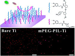
The rational design of an effective antifouling component is challenging but important for many fundamental and applied applications. Herein, we report the synergistic antifouling effects of nonionic, cationic and anionic compounds in combination with a poly(ionic liquid) toward suppressing marine fouling. Firstly, nonionic, cationic and anionic compounds were respectively grafted onto substrate surfaces by a light-induced click reaction of mPEG-SH, bromohexadecyl nicotinate (1-sulfydryl) ethyl ester and sodium 2-mercaptoethane sulfonate. Subsequently the poly(ionic liquid) brushes were grafted onto as-prepared surfaces via surface-initiated ring-opening metathesis polymerizations to obtain binary components modified surfaces. The antifouling properties of the sole component and binary components modified surfaces were evaluated by an algae adhesion assay using Navicula spores and Dunaliella tertiolecta. The antifouling results show that the binary compound modified substrate surfaces exhibit better anti-adsorption performance than the sole component, which is owing to the synergistic antifouling effect of the binary components.
Recently, polymer brushes have often been used to alter surface properties due to their higher mechanical and chemical robustness, and higher long-term stability.5 Polymer brushes with large exclusion volumes, to inhibit protein and bacterial adhesion, or bactericidal functional groups, are of great importance in antifouling fields.6 So polymeric materials were investigated extensively as environmentally benign anti-biofouling replacements.7 Therefore, various types of polymers have been explored as anti-biofouling coatings, including PEGylated polymers,2 hydrophobic polymers,8,9 hydrophilic polymers,10 amphiphilic polymers,11 zwitterionic polymer brushes,12 and bioinspired polymers,13 which are suited for the preparation of bio-inert interfaces to prevent protein adsorption, cell adhesion, and bacterial attachment. Poly(ethylene glycol) (PEG) and its derivatives acted as commonly used antifouling materials and exhibit good antifouling attributed to the steric barrier, osmotic repulsion, excluded-volume effects, and the mobility or flexibility of the highly hydrophilic layer.4,14,15 A polymer consisting of methoxy-terminated poly(ethylene glycol) (mPEG) conjugated to L-3,4-dihydroxyphenylalanine (DOPA) exhibited a substantial decrease in attachment of both cells of Navicula perminuta and zoospores of U. linza, as well as the highest detachment of attached cells under flow.16 Recently, poly(ionic liquid)s (PILs) consisting of ammonium, pyridinium, phosphonium, imidazolium etc. were found to play an important role in the fields of polymer chemistry and materials science due to their particular properties, such as high ion conductivity, inherent conductivity, high thermal stability, excellent mechanical properties and biocompatibility.17,18 Attaching ionic liquid moieties to polymer chains is developing novel effective antimicrobial agents and anti-biofouling materials.19,20 Tiller et al. designed derivatizing poly(vinyl-N-hexylpyridinium bromide)-modified surfaces of the synthetic polymers high-density polyethylene, low-density polyethylene, polypropylene, nylon 6/6, and poly(ethylene terephthalate), which exhibited significant antibacterial properties against S. aureus and E. coli cells.21 In our previous work, high-density poly(ionic liquid) brushes were grafted onto surfaces via surface-initiated ring-opening metathesis polymerizations (ROMP) and the as-prepared poly(ionic liquid) possessed excellent anti-bacterial and anti-biofouling properties.22
Furthermore, cationic antimicrobials are also well-known for the development of self-sterilizing surfaces and are widely used on hospital surfaces, surgical equipment, protective clothes in hospitals, food packaging materials, and everyday consumer products.23 With respect to cationic antimicrobials, quaternary ammonium salts (QAS), which can cause the death of cells by destroying the membrane integrity, show great promise in the field of antimicrobial coatings.24,25 Surfaces modified with QAS-containing polymers are effective at inhibiting microbial biofilm formation and killing a wide range of microorganisms.26,27 For example, pH responsive quaternary ammonium modified chitosan was prepared by Composto’s group, which shows the weakest bacterial adhesion compared to the silicon oxide surface and chitosan brush layer.28 Anionic components that display a negative charge are another important functional antifouling agent, such as 3-sulfopropyl methacrylate (SPMA), 4-styrenesulfonic acid sodium (NaSS) and glycidyl methacrylate (GMA), which display excellent performance for preventing the adhesion of bacteria and microorganisms.29 Terada et al. have reported that biofilms formed on negatively charged surfaces were easily removed by liquid high shear washing.30
Although various polymers possess antibiofouling characteristics, there remains a drawback to using single component systems as many biofoulers release glycoprotein adhesives, therefore the amphiphilic polymer with hydrophilic domains and hydrophobic domains has been demonstrated to have more effective antibiofouling properties than only a single polymeric component.31,32 Surfaces modified with the polymer brushes 2-hydroxyethyl methacrylate (HEMA) and (dimethylamino)ethyl methacrylate (DMAEMA) exhibited an excellent resistance to bacterial fouling compared to the single-component.33 Herein, we present a novel strategy combining a light induced-click reaction with surface-initiated ROMP to prepare binary component antifouling materials based on a poly(ionic liquid). mPEG-SH, sodium 2-mercaptoethanesulfonate (MESNA) and bromohexadecyl nicotinate (1-sulfydryl) ethyl ester (SH-Py16) were grafted on the surface respectively via a light-induced click reaction, then poly(ionic liquid) brushes were grafted via a surface-initiated ROMP. The synergistic antifouling properties of the binary component antifouling materials based on poly(ionic liquid) have been evaluated. This work hopefully provides a promising component motif for the design of new effective antifouling surfaces (Scheme 1).
1H-NMR (400 MHz, CDCl3) δ 9.95 (d, J = 6.0 Hz, 1H, H-1), 9.71 (s, 1H, H-5), 8.99 (d, J = 8.0 Hz, 1H, H-3), 8.34 (m, 1H, H-2), 5.17 (t, J = 7.2 Hz, 2H, H-10), 4.56 (t, J = 6.4 Hz, 2H, H-7), 3.26 (s, 1H, H-9), 2.97 (s, 2H, H-8), 2.06 (s, 2H, H-11), 1.22–1.39 (m, 26H, H-(12–24)), 0.87 (t, J = 7.2 Hz, 3H, H-25).
13C-NMR (100 MHz, CDCl3) 160.9 (C-6), 149.0 (C-1), 145.5 (C-3), 145.2 (C-5), 130.3 (C-4), 128.8 (C-2), 68.4 (C-10), 62.8 (C-7), 32.0 (C-23), 31.9 (C-11), 29.7–29.0 (C13-22), 26.1 (C-12), 23.1(C-8), 22.7 (C-24), 14.1(C-25).
HRMS (ESI+): for C24H42NO2S M+: calculated: 408.2931, found: 408.2887.
1H-NMR and 13C-NMR spectra were recorded on a 400 MHz spectrometer (Bruker AM-400) using CDCl3 as solvent. The chemical composition information of the samples was obtained by X-ray photoelectron spectroscopy (XPS); the measurement was carried out on a PHI-5702 multi-functional spectrometer using Al Ka radiation (29.35 eV) and the binding energies were referenced to the C 1s line at 284.8 eV from adventitious carbon. FT-IR spectra were recorded on a TENSOR 27 instrument (BRUCKER). The morphologies were investigated by transmission electron microscopy (TEM) (Hitachi Model JEM-2010). Atomic force microscopy (AFM) measurements were performed on an Agilent Technologies 5500 AFM using a MacMode Pico SPM magnetically driven dynamic force microscope. The zeta potential was measured using a Nano-ZS 90 Nanosizer (Zetasizer Nano ZS, Malvern Instruments Ltd., UK). Scanning electron microscopy (SEM) images were carried out on a field-emission scanning electron microscope (JSM-6701F, JEOL Inc., Japan). Optical micrographs were taken on OLYMPUS BX51.
Prior to the antifouling investigation, the successful modification of the binary components was ascertained by XPS, IR and TEM. XPS measurements were used to monitor the surface chemical composition change of each reaction step. Fig. 1A displays the XPS full survey spectra of bare TiO2 nanowires, initiator-modified TiO2 nanowires, mPEG-TiO2 nanowires and mPEG-PIL-TiO2 nanowires. The successful anchoring of the initiator onto TiO2 nanowires was ascertained by XPS.22 After 0.5 h of click reaction, the C 1s high-resolution spectrum of mPEG-TiO2 nanowires shows the peak components at binding energies of about 286.4 eV and 284.8 eV, attributed to the C–O bonds and the C–C bonds, as shown in Fig. 1C. As shown in Fig. 1B, the N 1s peak of the initiator-modified TiO2 and mPEG-TiO2 nanowires display one peak with a binding energy at 398.3 eV. After 2 h SI-ROMP of the monomer NM-MIm-PF6, the grafting of the poly(ionic liquid) brushes was confirmed by a sharp increase in the N signal at 400.6 eV, attributable to ammonium nitrogen (N+), and the presence of strong F 1s (686.4 eV) and P 2p (136.5 eV) signals. In Fig. S2B and S2E,† MESNA and SH-Py16 modified TiO2 nanowires were confirmed by the appearance of Na and Br signals at 1071.6 and 62.5 eV, respectively. The N1s spectrum of the Py16-TiO2 nanowires shows a broad peak at binding energies of about 399.8 eV and 401.8 eV, attributed to the OC–N bonds in the catecholic initiator and ammonium nitrogen (N+) in Fig. S3.† As shown in Fig. S2C and F,† MESNA-PIL-TiO2 and Py16-PIL-TiO2 nanowires also display a sharp increase of the N signal, attributable to the ammonium nitrogen (N+) at 401.8 eV and F 1s at 686.4 eV after 2 h SI-ROMP of NM-MIm-PF6.
The successful grafting of mPEG-SH and PIL onto TiO2 nanowires was also confirmed by FT-IR spectroscopy (Fig. 2). Blank TiO2 shows a featureless spectrum, except for the wide absorption peaks of absorbed water above 3000 cm−1.22 Fig. 2 demonstrates the FT-IR spectrum evolution from blank TiO2 nanowires, initiator modified TiO2 nanowires, and mPEG-TiO2 nanowires, to mPEG-PIL-TiO2 nanowires (2 h). FT-IR analysis of the mPEG-modified surfaces (Fig. 2-c) reveals characteristic adsorption bands at 1238, 1398 and 1650 cm−1, which can be attributed to the C–O stretching vibration, C–H vibration mode and CO stretching vibration of the alkyl chains, which indicate the attachment of mPEG-SH onto TiO2 nanowires. After click reaction with MESNA, the S
O stretching bands of the sulfonate group have been observed in the region of 1183 to 1293 cm−1 and the symmetric S
O stretching mode appears near 1156 cm−1 in Fig. S4-a.†35 As shown in Fig. S4-c,† after the modification of Py16-SH, the characteristic adsorption bands at 1130 and 1301 cm−1 appeared, which can be attributed to the C
O stretching vibration, and the adsorption bands at 2855 and 2924 cm−1 are owed to the C–H stretching vibration mode of the alkyl chains. After grafting the second component PIL, the strong absorption peak at 841 cm−1 is attributed to the absorbance of P–F stretching modes of PF6−, and the characteristic vibration bands at 1571, 1460 and 1159 cm−1 correspond to the imidazolium ring cation,22 indicating that PIL has been successfully grafted onto the TiO2 nanowires.
![]() | ||
Fig. 2 FT-IR spectra of (a) TiO2 nanowires, (b) initiator-modified TiO2 nanowires, (c) mPEG-TiO2 nanowires and (d) mPEG-PIL-TiO2 nanowires. |
The zeta potentials (ζ) of TiO2 nanowires before and after modification with mPEG and mPEG-PIL were investigated by a Nano-ZS 90 Nanosizer in ethanol media. The zeta potential of bare TiO2 nanowires is −5.71 ± 0.9 mV, after modification with mPEG, the zeta potential becomes positive (−1.2 ± 0.3 mV). Then, after grafting the poly(ionic liquid) brushes on mPEG-TiO2 nanowires, the zeta potential becomes more positive (31.9 ± 3.4 mV) due to the imidazolium cation (Table S1†). The zeta potentials of MESNA-TiO2 nanowires and Py16-TiO2 nanowires are −29.9 ± 0.4 and 22.0 ± 1.2 mV, respectively; after grafting PIL, the zeta potentials of MESNA-PIL nanowires and Py16-PIL nanowires also became more positive, 21.5 ± 1.8 and 31.1 ± 3.4 mV, respectively.
The presence of the mPEG-SH and PIL layer was directly observed by TEM characterization. Fig. 3 shows transmission electron microscopy (TEM) images of the surface morphology of mPEG-SH and mPEG-PIL coated TiO2 nanowires. From Fig. 3A, the TiO2 nanowire surfaces were coated with a very uniform mPEG-SH coating layer and the interface boundary is very clear; the thickness of mPEG-SH is close to 3 nm. After modification with PIL (Fig. 3B), the thickness of the polymer layer increased to 15 nm and it has a lighter color than the bulk TiO2 nanowires grafted with PIL. From the above results, we can conclude that the TiO2 nanowires had been successfully modified with mPEG-SH and PIL.
To further support the results, atomic force microscopy (AFM) was used to investigate the surface morphology of the mPEG-SH and mPEG-PIL modified Ti surfaces. Fig. 4 shows the 3 μm × 3 μm tapping-mode AFM phase images of Ti, mPEG-Ti and mPEG-PIL-Ti. Before modification (Fig. 4A), small particles are uniformly distributed over the whole Ti surface. After modification with mPEG-SH on the Ti surface (Fig. 4B), the roughness of the mPEG-Ti surface becomes low (RMS decreased from 0.874 nm to 0.505 nm), which shows that mPEG-SH has masked the Ti surface. The roughness of MESNA-Ti and Py16-Ti also decreases (RMS decreases to 0.606 nm and 0.693 nm respectively, Fig. S5†). After modification with the second component PIL, small grains are uniformly distributed over the whole surface, and the rms roughness of mPEG-PIL-Ti increases to 0.720 nm (Fig. 4C). After 2 h ROMP of the monomer NM-MIm-PF6, the roughness of the MESNA-PIL and Py16-PIL modified Ti surfaces have the same varying trend (RMS increases to 0.91 nm and 0.791 nm respectively, Fig. S5†), indicating that PIL has been successfully grafted onto the Ti surfaces.
Fouling results from the settlement and growth of marine organisms on the surfaces of a submerged object. The first stage of biofouling involves the formation of biofilms which provides the condition of microfouling. However, marine organisms may settle at the same time, such as zoospores of the alga Ulva linza and cyprids of the barnacle Amphibalanus amphitrite (A. amphitrite).34 Therefore, preventing the settlement of microalgae is the important objective for an effective antifouling coating.
In order to investigate the anti-biofouling effect of different components, mPEG-Ti, mPEG-PIL-Ti, MESNA-Ti, MESNA-PIL-Ti, Py16-Ti and Py16-PIL-Ti were prepared and the modified substrates were thereafter tested in biological assays with two ubiquitous biofouling organisms: organic Navicula and Dunaliella spores under artificial seawater conditions for 24 h. The results of the settlement assays of Navicula. sp and Dunaliella tertiolecta on different surfaces are shown in Fig. 5, and we could observe significant differences in the settlement behavior of Navicula. sp and Dunaliella tertiolecta. As shown in Fig. 5A, the average spore density of mPEG-SH, MESNA and SH-Py16 modified surfaces was 1088 ± 49, 1248 ± 91, and 1160 ± 145 spores mm−2 after 24 h in the Navicula spores culture media. Meanwhile, a similar trend was observed for the settlement of Dunaliella on the as-prepared surfaces (Fig. 5B). The average Dunaliella spores density of mPEG-SH, MESNA and SH-Py16 modified surfaces was 2573 ± 243, 2802 ± 145, and 2601 ± 141 spores mm−2, respectively.
![]() | ||
Fig. 5 Organic (A) Navicula and (B) Dunaliella spore settlement data on (a) bare Ti, (b) mPEG-Ti, (c) mPEG-PIL-Ti, (d) MESNA-Ti, (e) MESNA-PIL-Ti, (f) Py16-Ti, and (g) Py16-PIL-Ti. |
It was observed that mPEG-SH modified surfaces exhibited a lower settlement than that of the MESNA and SH-Py16 modified surfaces, which could be attributed to the highly hydrated PEG chains in water.14 In comparison to the uncoated Ti surface, the fractions of settlement on the anionic MESNA-Ti surface exhibited the lowest resistance to microalgae, with a decrease in Navicula and Dunaliella adhesion of 13.3% and 8.6%, which might be due to electrostatic interaction between the anionic surface and the positively charged lysine of microalgae.8 As seen in Fig. 5A-f and 5B-f, the Py16-Ti surface exhibited a higher efficiency in deterring microalgae than the MESNA-Ti surface, with settlement fractions of 19.4% and 15.2%, respectively. In Fig. 5A, after introducing the second antifouling component PIL, mPEG-SH, MESNA and SH-Py16 with PIL resulted in a reduction of approximately 38%, 4.2% and 46.3% for Navicula adhesion, respectively. As in the case of Dunaliella in Fig. 5B, a dramatic reduction in the settlement on mPEG-SH and SH-Py16 modified surfaces was observed (47.7% and 49.5%, respectively). So, the PIL can enhance the antifouling performance of the mPEG-SH and SH-Py16 based surface. The MESNA-PIL-Ti shows a poor inhibition effect for the spore, because the electronegativity of MESNA has decreased the catalytic activity of the Grubbs catalyst.36 The results indicate that the binary compound based poly(ionic liquid) grafting surfaces exhibited better anti-adsorption performance than the sole component, owing to the synergistic effect.
The settled Navicula and Dunaliella spores on different surfaces were fixed in 2.5% glutaraldehyde in phosphate buffer pH 7.4 for 3 h and processed in the conventional way for SEM (dehydration, critical point drying and sputter-coating with gold) before viewing. The adhesive form of Navicula and Dunaliella spores on different surfaces was not identical. Fig. 6 and 7 show the conformation of Dunaliella and Navicula spores on bare Ti, mPEG-Ti, mPEG-PIL-Ti, MESNA-Ti, MESNA-PIL-Ti, Py16-Ti, and Py16-PIL-Ti surfaces respectively. Spores can adhere to the surfaces via a secreted adhesive glycoprotein.37 In Fig. 6, it is clear that there is a circular blot around the Dunaliella spores. After modification with PIL, the circular blot became small. Fig. 7 shows the adhesion of Navicula spores on different surfaces. After immersion in the culture suspension for 3 h, it can be seen that there is a blot around the spore in Fig. 7A, B, D and F. Meanwhile, there are few blots around the spore adhesion on surfaces after grafting PIL. This phenomenon proves that binary components are very effective against microalgae.
![]() | ||
Fig. 6 SEM images of Dunaliella tertiolecta settlement on (A) bare Ti, (B) mPEG-Ti, (C) mPEG-PIL-Ti, (D) MESNA-Ti, (E) MESNA-PIL-Ti, (F) Py16-Ti, and (G) Py16-PIL-Ti. |
![]() | ||
Fig. 7 SEM images of Navicula spores settlement on (A) bare Ti, (B) mPEG-Ti, (C) mPEG-PIL-Ti, (D) MESNA-Ti, (E) MESNA-PIL-Ti, (F) Py16-Ti, and (G) Py16-PIL-Ti. |
Footnote |
† Electronic supplementary information (ESI) available. See DOI: 10.1039/c5ra23391g |
This journal is © The Royal Society of Chemistry 2015 |