The potential cytotoxicity and mechanism of VO2 thin films for intelligent thermochromic windows

Huaijuan Zhou ab, Jinhua Liab, Shanhu Baoa, Donghui Wangab, Xuanyong Liu*a and Ping Jin*ac
aState Key Laboratory of High Performance Ceramics and Superfine Microstructure, Shanghai Institute of Ceramics, Chinese Academy of Sciences, Shanghai 200050, China. E-mail: p-jin@mail.sic.ac.cn; xyliu@mail.sic.ac.cn; Fax: +86 21 6990 6221; Fax: +86 21 52412409; Tel: +86 21 6990 6208 Tel: +86 21 52412409
bUniversity of Chinese Academy of Sciences, Beijing 100049, China
cMaterials Research Institute for Sustainable Development, National Institute of Advanced Industrial Science and Technology, Nagoya 463-8560, Japan

Received 13th October 2015 , Accepted 2nd December 2015

First published on 7th December 2015


Abstract

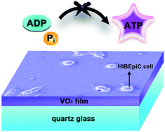
A thermochromic vanadium dioxide (VO2) film holds great promise for intelligent windows owing to marvelous semiconductor-metal transitions (SMT), an active response to external temperature stimuli and near-infrared irradiation. To date, however, its potential biological effect on human cells has not been well characterized. In this work, homogeneous high quality VO2 films with different thicknesses (30, 80, 120 nm) were prepared on a quartz glass substrate. Afterwards, for the first time, we demonstrate the time-dependent and dose-dependent cytotoxicity of the VO2 film to human cells. Speciation analysis by 51V NMR spectra and surface zeta potentials revealed the formation of vanadate(+5) and ADPV, an analogue of ATP. On the basis of energy metabolism or bioenergetics, a plausible hypothesis, i.e., “ATP dyssynthesis”, is proposed here to elucidate the potential toxicity mechanism of the VO2 nanomaterial via vanadate-phosphate antagonism including two steps: (i) vanadate speciation from VO2 surface chemistry; (ii) vanadate disturbing ATP synthesis as a phosphate analogue. ATP stores energy to carry out various life processes; once its synthesis is hindered, vital movement will be impaired. From the perspective of surface modification and bioactivation, some practical methods are recommended to compensate for the potential cytotoxicity of the VO2 material. We expect this work can stimulate scientific interest to search for more versatile material design strategies to balance the energy-saving efficiency and environmental safety of VO2 intelligent window coatings.


1. Introduction

Nowadays, energy conservation has become a global trend for sustainable development achieved by increasing energy efficiency and reducing energy consumption.1 From this perspective, more and more attention has been paid to intelligent windows that own switchable light transmittance capability and hold great promise for daily applications.2,3 Particularly, intelligent thermochromic windows, which are based on thermochromism materials like VO2 films, have attracted growing interest due to their active response to environmental temperature stimuli that makes a reversible structural change from an infrared-transparent semiconductive state to an infrared-reflective metallic state.4–6 VO2 undergoes a semiconductor-to-metal transition (SMT) at a critical temperature Tc (68 °C for bulk VO2),7 which has attracted growing attention as a thermochromic material and holds great promise for applications in intelligent windows.8,9 Since the NIR light radiation carries a large amount of solar energy, nowadays, increasing efforts have been made to exploit the marvelous SMT property of VO2 to intelligently control NIR radiation for energy-saving purpose.

By so far, to achieve the daily application of VO2 film in intelligent windows, the main attention has focused on the optimization of preparation methods and enhancement of energy-saving efficiency. However, a metal oxide nanomaterial may hold toxic potential to human health and ecology environment.10 With regard to nanomaterials, a question must be asked, that is, how those man-made nanostructures could potentially interact with or influence biological systems. Exposure to nanomaterials may cause toxicity to biological systems. Although toxicity data for widely used nanomaterials like TiO2 are available,11 but for those newly developed nanomaterials like graphene,12 fullerene,13 VO2, very little is known on their biological actions and interactions with living cells.

In fact, exposure of vanadium compounds can produce toxicity to mammalian respiratory system,14,15 liver16,17 and also cause adverse physiological effects on various microbes.18–20 Toxicity of vanadium depends upon its physicochemical states, especially upon the valence states and solubility.21,22 Vanadium possesses six oxidation states and the most commonly found in the nature are V(+3), V(+4), and V(+5). Vanadium toxicity increases as the oxidation state ascends. However, to our best knowledge, current reports mainly focused on the toxicity of V2O3 and V2O5 particles, and very few studies were reported on the toxicity of VO2, let alone its film. In terms of thermochromic VO2 film for intelligent window coating, its exposure to human bodies is inevitable in the processes of film production, daily application and refuse disposal.

Herein, homogeneous high quality VO2 films were fabricated on quartz glass with the aim to study the potential toxicity of immobilized VO2 nanomaterial to human intrahepatic biliary epithelial cells. Zeta potential and pH measurements were performed to analyze the surface chemistry of VO2 film. ICP-OES was used to study the release behaviors of vanadium species. NMR spectra were recorded to analyze the vanadium speciation. Meanwhile, AlamarBlue assay, live/dead cell staining, SEM, ATP assay and protein assay were combined to evaluate the cytotoxicity.

2. Materials and methods

2.1. Fabrication of specimens

The high quality VO2 films were prepared by means of magnetron sputtering apparatus (ULVAC Corp., Model ACS-4000-C4) and the deposition details can be found in ESI. Since float glass contains some elements such as Na, K, Ca, Mg, etc., which may interfere in the research results, fused quartz (composed of Si and O elements) was used as the substrate for VO2 film deposition. The film thickness was measured by F20 thin-film analyzer (FILMSTRICS corp.). VO2 films with different thicknesses (including 30 nm, 80 nm, 120 nm) were obtained and denoted as MS-30, MS-80 and MS-120, respectively. The quartz glass acted as control group (MS-0).

2.2. Characterization of specimens

The film crystalline structure was studied by X-ray diffractometer (XRD, Rigaku Ultima IV) with Cu Kα radiation (λ = 0.15406 nm) at voltage of 40 kV, current of 40 mA, glancing incidence angle of 1° and scanning rate of 2° min−1. The surface morphology was examined by field-emission scanning electron microscopy (FE-SEM, FEI Magellan 400, US). XPS characterization was carried out on the surfaces of films after soaking them in PBS for 2 weeks to analyze the chemical valence state of V element. The 51V NMR measurements were performed on Agilent DD2 600 MHz spectrometer operating at 25 °C with the fixed testing frequency of 157.7 MHz. The 51V NMR chemical shifts were relative to VOCl3 solutions. The free induction decay (FID) signals were exponentially line broadened (20 Hz for 51V) prior to Fourier transformation.

2.3. Zeta potential measurements

The surface zeta potentials of MS-0, MS-30, MS-80 and MS-120 samples were measured by SurPASS Electrokinetic Analyzer (EKA; Anton Paar GmbH, Graz, Austria) equipped with a special cell for membrane/film. Samples (size 20 mm × 10 mm × 1 mm) were fixed on holders with double-sided adhesive tape (Scheme S1). 0.9% NaCl electrolyte solution was used as medium and pH value was adjusted by adding HCl and NaOH solutions.

2.4. Vanadium release measurements

The release characteristic of vanadium ions from VO2 films soaked in PBS was detected by inductively coupled plasma optical emission spectrometry (ICP-OES, Varian Liberty 150, US). The pH of PBS was adjusted to 6.8 and 7.4 by adding HCl solution. The samples were placed in sterile microcentrifuge tubes (15 ml), immersed in new medium of 10 ml, and accordingly incubated at 37 °C for 1, 4, 7 and 14 days in static condition. At the end of each incubation period, all the leachates were collected for detection and supplemented with fresh media for further incubation.

2.5. pH measurements

To determine the pH changes of solution when soaking VO2 films, samples MS-0, MS-30, MS-80 and MS-120 were immersed in 6 ml NaOH aqueous solution with an initial pH = 10.40 in sterile microcentrifuge tubes and accordingly incubated at 37 °C for 1, 2, 3, 4, 5, 6 and 7 days in static condition. After the incubation of each period, the pH was measured by pH meter (FE20-FiveEasy™, METTLER TOLEDO).

2.6. Cell proliferation and viability

Human intrahepatic biliary epithelial cells (HIBEpiC) were purchased from ScienCell (San Diego, California, US). Cell proliferation and viability were evaluated by AlamarBlue assay (AbD Serotec Ltd., UK). Cells were seeded on samples with density of 5 × 10⁴ cells per well and five samples per group were tested for each culture period (1, 4 and 7 days). After each period, the culture medium was removed and 500 μl fresh medium with 10% AlamarBlue was added to each well, followed by another incubation of 4 h. Cell proliferation and viability were calculated according to the manufacturer's instruction.

2.7. Live/dead cell staining

Live/Dead Cell Staining Kit (BioVision, US) was used following the manufacturer's instruction. Briefly, cells were seeded on samples (5 × 10⁴ cells per well) and cultured for 4 days. Red fluorescent dye, propidium iodide (PI), and green fluorescent dye, calcein-AM were used to stain dead and live cells with final concentrations of 5 μM and 2 μM in PBS, respectively. Gently washed in PBS thrice, 100 μl of mixed solution was added to each well, then incubated at 37 °C for 15 min. A confocal laser scanning microscopy (CLSM, Leica SP8, Germany) was used to visualize the live and dead cells.

2.8. Cell morphology observation

SEM was utilized to further investigate cell morphology and cell–material interactions. After each culture period, the samples were transferred to a new 24-well plate and rinsed gently with PBS thrice and then fixed with 2.5% glutaraldehyde solution for overnight. Prior to SEM observation, the samples were dehydrated in a series of ethanol solutions (30, 50, 75, 90, 95, and 100 v/v%) for 10 min each sequentially, along with the final dehydration conducted in absolute ethanol (twice) followed by drying in hexamethyldisilizane/ethanol solution series.

2.9. Intracellular ATP level

The ATP level inside cells was measured by the luciferin-luciferase method following the protocol of ATP assay kit (Beyotime, China). HIBEpiC were seeded on samples MS-0, MS-30, MS-80 and MS-120 in 24-well plate (1 × 10⁵ cells per well). Five samples were used for each group. After culture of 4 days, cells were washed twice with PBS and then resolved in ATP lysis buffer on ice. ATP in cell lysate was measured using Synergy H4 Hybrid Microplate Reader (BioTek, US), and the ATP concentration was normalized to the standard curve. Protein contents were measured using BCA Protein Assay kit (Beyotime, China) to correct the different samples for comparison of ATP concentration.

2.10. Statistical analysis

Statistically significant differences (P) between various groups were measured using one-way analysis of variance and Tukey's multiple comparison tests on a GraphPad Prism 5 statistical software package. All the data were expressed as means ± standard deviation (SD). A value of P < 0.05 was considered to be statistically significant and denoted as “*”, P < 0.01 was “**”, and P < 0.001 was “***”.

3. Results and discussion

3.1. Sample characterization

Fig. 1a–c reveals thickness-dependent morphology of VO2 films on quartz, with homogeneous compact nanostructures. One can distinguish clear size polydispersity in addition to the stochastic shape of VO2 crystallites. The nanoparticles were worm-like for MS-30 and became much larger for MS-80. For MS-120, however, the particles turned out to be cobblestone-like structures of uneven size. That increasing deposition time produced particle size growing was attributed to the Volmer-Weber growth mode of VO2 nanofilms. XRD θ–2θ scan profiles were measured on as-deposited films at ambient temperature, as shown in Fig. 1d. Only XRD peaks corresponding to the signal of monoclinic VO2 phase were observed. All the positions of peaks were concordant with the standard reference data (JCPDS no. 72-0514), indicating the exclusive formation of single-phase VO2 polycrystalline films. The raise of VO2 film thickness brought about the much stronger diffraction peaks, accounting for better crystallinity. Besides, the value of full width at half maxima (FWHM) of the predominant orientation (011) was declined with the increment of film thickness. According to Scherrer formula, the average grain size of the films had a positive correlation with the reciprocal of FWHM, which provided another evidence for the growing particle size. Herein, we also demonstrate that the SMT property and energy-saving efficiency of VO2 film can be tailored and optimized in a film thickness dependent manner (Fig. S1).
image file: c5ra22582e-f1.tif
Fig. 1 (a–c) FE-SEM images and (d) grazing incident XRD patterns of the various VO2 films with different thicknesses (30 nm for sample MS-30, 80 nm for sample MS-80, 120 nm for sample MS-120).

3.2. Cytotoxicity of VO2 nanofilm

Fig. 2 shows the proliferation and morphology of HIBEpiC cultured on various samples. At early culture of 1 day, HIBEpiC can well adhere on the VO2 films, showing no significant difference between VO2 and control (Fig. 2a). However, after culturing for 4 days, HIBEpiC proliferation was sharply inhibited on VO2 films, while they can maintain a steady growth on control (Fig. 2b), indicating that the presence of VO2 film on quartz glass caused severe cytotoxicity toward HIBEpiC. Prolonging the culture to 7 days, as shown in Fig. 2c, the growth retardation of HIBEpiC continued and implied the ongoing cell death. SEM was used to analyze the cell morphology development and cell–material interaction of HIBEpiC cultured on various surfaces for 4 days. As seen in Fig. 2d, the surface of quartz glass was thoroughly covered by the HIBEpiC and the cells connected well with each other. On the contrary, markedly reduced cells were observed on the VO2 films and most of them still maintained a spherical or spindle morphology (Fig. 2f, h and j). Therefore, the cell morphology was quite consistent with the proliferation trend. At high magnification, the HIBEpiC on quartz glass spread well, exhibiting a multipolar spindle morphology (Fig. 2e). Focusing on the cell membrane, as indicated by the white arrow, visible particulate matter was prevalent, showing the exuberant extracellular matrix mineralization (ECM). Moreover, abundant lamellipodia and filopodia extensions were found (red arrow), indicating the pervasive intercellular communications and cell–materials interactions. With regard to VO2 films, however, spherical cell morphology, and hardly seen lamellipodia/filopodia (indicated by blue arrow in Fig. 2g, i and k) revealed the poor cell viability and stagnant cell fate toward cell death. To visualize and distinguish between live and dead cells, live/dead cell staining was further performed and the results were given in Fig. 3 (green for live cells, red for dead cells). Nearly no dead cells were observed under the confocal laser scanning microscopy and the HIBEpiC grew well on MS-0. However, prevailing cell death were present on VO2 films and even some dying cells were also observed. Overall, the live/dead staining results were quite consistent with the cell proliferation and SEM results. Considering the presence of VO2, it is inferred that the observed cytotoxicity may be caused by the toxic vanadium element.
image file: c5ra22582e-f2.tif
Fig. 2 (a–c) Proliferation results of HIBEpiC after culture of 1, 4 and 7 days on samples MS-0, MS-30, MS-80 and MS-120, accompanied by the SEM morphology of HIBEpiC after culture of 4 days on surfaces of MS-0 (d and e), MS-30 (f and g), MS-80 (h and i) and MS-120 (j and k). Note: white arrow, showing the extracellular matrix mineralization; red arrow, showing the lamellipodia and filopodia extensions; blue arrow, showing the hardly seen lamellipodia/filopodia.

image file: c5ra22582e-f3.tif
Fig. 3 Live/dead staining results of HIBEpiC viability after culture of 4 days on surfaces of samples MS-0, MS-30, MS-80 and MS-120 that were observed on a confocal laser scanning microscopy. Note: green color for live cells, red color for dead cells.

3.3. Plausible cytotoxicity mechanism

In fact, vanadium toxicity has been of a great concern although it is one of the essential trace elements in human body,23 and so, substantial efforts have been made to clarify the mechanisms of vanadium metabolism and toxicology.24–26 To figure out the reason for observed cytotoxicity, it is quite essential here to analyze the surface aqueous chemistry of vanadium dioxide and speciation of vanadium element. The surface zeta potentials versus pH for each group are shown in Fig. 4a and all the curves exhibit descending trends with the ascending pH. It is well known that a strong electric field exists at an aqueous solution/oxide interface.27 This interfacial electric field is produced by the surface charge of oxide, which can interact with the dipole moment of water molecules and induce ordering of up to several layers at the interface.28,29 The strength and direction of the electric field at an aqueous solution/oxide interface are a function of the pH of solution and the IEPS of substrate. The IEPS, namely, isoelectric point of surface, is the pH value of the aqueous solution in which the total surface charge density is zero. The surface of a substrate is negatively (positively) charged when the pH of the solution is greater (less) than the IEPS of the substrate.30–33 According to Fig. 4a, all the surfaces of VO2 films were negatively charged since the present pH range was above the IEPS of VO2. At prevalent oxic conditions near neutral to slightly alkaline pH, soluble vanadate(+5) anion, i.e., H2VO4, is the predominant specie, in which the pentavalent oxidation state is the most stable form among its various oxidation states.23,34 Therefore, an initial chemical redox reaction might occur at the VO2/solution interface:
 
4VO2 + 4OH + O2 + 2H2O = 4H2VO4 (1)

image file: c5ra22582e-f4.tif
Fig. 4 (a) Results of surface zeta potentials (ζ) of samples MS-0, MS-30, MS-80 and MS-120. (b) Changes of pH values when immersing MS-0, MS-30, MS-80 and MS-120 in PBS with initial pH = 7.40. (c and d) Release features of vanadium ions from MS-0, MS-30, MS-80 and MS-120 surfaces. (e) 51V NMR spectra of various extraction solutions after immersing MS-30, MS-80 and MS-120 in PBS (pH = 7.40) for 14 days, accompanied by the corresponding 51V NMR spectra of the above extraction solutions with adding of ADP (50 mM).

To support this, the pKa values for vanadate were given below:35

 
H3VO4 + H2O ↔ H2VO4 + H3O+, pKa = 3.5 (2)
 
H2VO4 + H2O ↔ HVO42− + H3O+, pKa = 7.8 (3)
 
HVO42− + OH ↔ VO43− + H2O, pKa = 12.5 (4)

From the above, H2VO4 formation is reasonable in the present experimental conditions and negatively charged surface can be formed on VO2 in a wide pH range. In fact, only by dissolving in non-oxidizing strong acids can VO2 produce the vanadyl cations (VO2+) with positively charged surface (VO2 + 2H+ = VO2+ + H2O).36 Meanwhile, the surface zeta potentials from various VO2 films showed a thickness-dependent difference (Fig. 4a) that is quite reasonable. This difference showed to diminish gradually with pH increase, especially at pH = 7.4, indicating a chemical equilibrium was established in these aqueous systems. In reality, at the physiological pH and ionic strength, vanadate(+5) is almost exclusively present in the H2VO4 form.23 Besides, for quartz substrate (MS-0), a hydration process occurred at the IEPS of ∼3.7 (SiO2 + H2O = H2SiO3).37,38 At the IEPS, the total charge density of the hydrated surface on quartz was zero. Upon departure from the IEPS, the corresponding chemical reactions occur at the solution/quartz interface (H2SiO3 + OH → HSiO3 + H2O, > IEPS; H2SiO3 + H+ → H3SiO3+, < IEPS). It should be noted that MS-0 had a significantly different characteristic of surface zeta potential from that of VO2 films.

Furthermore, continuous pH measurements were conducted on NaOH aqueous solutions (initial pH = 10.40) for samples immersion within 7 days. It can be seen from Fig. 4b that, for MS-0 sample, namely, quartz, the pH of solution gradually dropped from 10.40 to 9.12 within the 7 days of immersion in a slow manner. In comparison, all the VO2 films samples suffered from an evident drop of solution's pH throughout the immersion period; the pH decreased from 10.40 to 8.45, 7.92 and 7.68 for MS-30, MS-80 and MS-120, respectively. As can be seen in Fig. 4b, an obvious gap existed between the VO2 samples and the quartz. On the basis of the results, it was deliberately concluded that the significant pH change was caused by the OH depletion.39,40 Therefore, the pH changes had an accordant correlation with the surface zeta potentials in Fig. 4a. Specially, eqn (1) depleted OH ions to lower the pH and produced H2VO4 to form a negatively charged surface at physiological pH. It is also worth mentioning that each pH measurement can refresh the air in the sealed microcentrifuge tubes to supply dissolved oxygen at the same time, which was essential for the formation of H2VO4 since its formation depleted O2 molecules as eqn (1). In fact, the oxygen supply is also essential for cell culture. The release feature of vanadium ions from VO2 films was shown in Fig. 4c–d. From this figure, the vanadium ions were released from VO2 films in a time-dependent manner. To demonstrate this, 51V NMR spectra were measured on extraction solution of VO2 films in PBS, as shown in Fig. 4e. The chemical shift (δ) at around −560 ppm corresponded to mononuclear tetrahedral vanadate (T1). The appearance of δ at about −580 ppm belonged to the divanadate (T2).41–43 The transformation of T1 to T2 can be expressed as follow:44,45

 
2T1T2, [T2] = K1[T1]2 (5)

To further demonstrate the formation of V(+5), XPS technique was used to investigate the chemical valence of surface V atoms. The results are shown in Fig. 5. For pure VO2 phase, the peaks assigned to V2p1/2 and V2p3/2 are located at 523.5 eV and 516.3 eV, respectively.6 After immersing the VO2 films in PBS for 2 weeks, their surfaces were gradually oxidized to be the pentavalent (+5) forms, as evidenced by the V2p1/2 peak at 524.7 eV and V2p3/2 peak at 517.4 eV, which belong to the characteristic peaks of V2p in V2O5 phase.46


image file: c5ra22582e-f5.tif
Fig. 5 XPS analysis results on the surfaces of VO2 films ((a) for MS-30, (b) for MS-80, (c) for MS-120) after immersing them in PBS for 2 weeks.

In biological systems, +5 valent H2VO4 is the most common intracellular form and can be actively transported into cells.47–50 Since among subcellular organelle, mitochondria has been proposed as an important target for vanadium accumulation and toxicity,34,51,52 we thus hypothesize that the observed cytotoxicity to HIBEpiC was caused by the vanadate(+5) uptake into cells and accumulation in mitochondria, affecting the normal functions of mitochondria. Mitochondria, the powerhouse of the cell, produce most of the cellular energy supply of ATP to support various vital movements including cell spreading, growth and differentiation, etc.53,54 ATP synthesis from ADP and phosphate (Pi) is catalyzed by ATP synthase (ADP + Pi → ATP) as illustrated in Fig. 6a. With regard to the phosphate(+5) group, its protonation reactions in aqueous solution are given below:35

 
H3PO4 + H2O ↔ H2PO4 + H3O+, pKa = 2.1 (6)
 
H2PO4 + H2O ↔ HPO42− + H3O+, pKa = 7.2 (7)
 
HPO42− + OH ↔ PO43− + H2O, pKa = 12.7 (8)


image file: c5ra22582e-f6.tif
Fig. 6 (a) Electron transport chain in the inner mitochondrial membrane. A sequence of electron carrier molecules (membrane proteins) shuttle electrons down a series of redox reactions that release energy used to produce ATP. (b) Schematic geometrical construction of inorganic phosphate and vanadate ions.

From the similar pKa values for vanadate and phosphate, it can be seen that an analogy may exist in their electronic properties. On the other hand, according to the structure parameters of inorganic vanadate and phosphate: r(O2−) = 1.36 Å, d(V–O) = 1.72 Å, d(P–O) = 1.54 Å; V–O and P–O distances of four-coordination tetrahedral geometry where r(V+5) = 0.36 Å and r(P+5) = 0.17 Å, vanadate and phosphate are structural analogues since the geometrical diameters are 6.2 Å and 5.8 Å, and the volumes of circumscribing spheres are 125 Å3 and 102 Å3, respectively (Fig. 6b).55,56 From a geometrical perspective, vanadate and phosphate are essentially indistinguishable, making vanadate an efficient competitor for phosphate in binding sites commonly targeted by phosphate. Thus, it is reasonable to infer that vanadate should behave as an analogue of phosphate in a biological system due to their electronic and structural analogy.57,58

In view of the structural and electronic analogy between vanadate and phosphate, during ATP synthesis, the formation of phosphate anhydride and ester bonds may be antagonized by the occurrence of vanadate anhydride and ester bonds:56,59

 
ROH + H2VO4 ↔ ROVO3H + H2O (9)
 
ROPO3H + H2VO4 ↔ ROP(O)2OV(O)2OH2− + H2O (10)

The above reactions could produce vanadate ester and vanadate-phosphate anhydride as the analogues of phosphate ester and anhydride, which may cause adverse effects on ATP synthesis through the vanadate-phosphate antagonism. Some ATP analogues, such as ADPV, AMPV, AMV, have been previously reported to exist in the vanadate-adenosine/AMP/ADP/ATP system.42,44,60 Table 1 lists some equilibrium constants for vanadate-AMP and vanadate-phosphate systems at physiological pH. To verify that possibility of ADPV formation, ADP was added to the extraction solution of VO2 in PBS, followed by 51V NMR measurements, as shown in Fig. 4e. The chemical shift δ at around −540 ppm was assigned to Vγ–Pβ anhydride bond in ADPV, an analogue of ATP.41–43 This coordination reaction is given below:44,45

 
T1 + L ↔ TL, [TL] = K2[T1][L] (11)
where L means the ligand ADP, TL is the product ADPV. According to eqn (11), the formation of ADPV will decrease the concentration of mononuclear vanadate (T1), which can make the chemical equilibrium of eqn (5) shift left and thus reduce the concentration of divanadate (T2). As a result, their resonance signals disappeared.

Table 1 Some equilibrium constants for the vanadate/AMP, vanadate/phosphate systems at a physiological pHa
Species Equilibrium constant Reference
a Note: AMP, adenosine 5′-monophosphate; AMPV, adenosine 5′-monophosphate vanadate; AMPV2, adenosine 5′-monophosphate divanadate; Vi-Pi anhydride, vanadate-phosphate anhydride.
AMPV 9.2 M−1 44
AMPV2 12 M−1 44
Vi-Pi anhydride 11.7 M−1 68


Since the ATP synthesis can be interrupted by vanadate, the level of ATP concentration inside a cell will reflect it accordingly. To verify that, intracellular ATP levels were determined on HIBEpiC, as shown in Fig. 7. From the data, the ATP level inside an individual cell was sharply reduced after culturing HIBEpiC on VO2 for 4 days. Meanwhile, combined with the corresponding proliferation results in Fig. 2b, the interference of vanadate on ATP synthesis functioned in a dose-dependent manner. As a result, a cell cannot metabolize normally to produce ATP to support spreading and proliferation, and gradually dies out (Fig. 2 and 3). To detoxify vanadate(+5), a cell will strain every nerve to bioreduce vanadate to vanadyl (VO2+, +4) through reduced glutathione (GSH), flavoenzymes or nicotinamide adenine dinucleotide (FADH2/NADH), nicotinamide adenine dinucleotide phosphate (NADPH), which causes inhibition of mitochondrial respiratory chain as side effect and seems to quench a thirst with poison.34,47,60 In addition, other potential mechanisms of VO2 material may also exist behind the observed cytotoxicity. On the one hand, the vanadyl (VO2+) cation formed by bioreduction of vanadate anion has the potential to induce the production of reactive oxygen species (ROS) in a Fenton-like reaction, for instance, the formation of ˙OH hydroxyl radicals (VO2+ + H2O2 + H+ → VO3+ + H2O + O˙H).34,61 On the other hand, the VO2 material shows a semiconductor state at low temperature. The conduction band, valence band and band gap are −4.85 eV (Ec), −5.45 eV (Ev) and 0.6 eV (Eg), respectively.6 As a result, the Ec value (−4.85 eV) is lower than the cellular redox potential ranging from −4.12 eV to −4.84 eV.62 Therefore, direct electron transfer may occur from biomolecular redox couples, which maintain the cellular redox potential and cellular homeostasis, to the conduction band of VO2, thus causing oxidative stress induced cytotoxicity.63–67


image file: c5ra22582e-f7.tif
Fig. 7 (a) Intracellular ATP levels of HIBEpiC after culture of 4 days on surfaces of MS-0, MS-30, MS-80 and MS-120 samples, accompanied by the corresponding standard curves of ATP concentration (b) and protein level (c). Notes: ***P < 0.001 versus MS-0; ##P < 0.01 versus MS-30; &&P < 0.01 versus MS-80.

Besides, on the basis of the Endosymbiotic theory, a bacterium is analogous to the mitochondrion of a mammalian cell. Therefore, the respiration is similar between bacterium and mitochondrion. As a result, it is reasonable to infer that VO2 film can also exert potential toxic effect on bacteria just as it does to mitochondria, exhibiting antibacterial ability that would be beneficial for application in self-cleaning windows. In this context, further investigation work need to be done to explore the possible antimicrobial properties in future.

As a marvelous thermochromic nanomaterial, VO2 holds great promise for applications in smart windows and nanodevices owing to its unique semiconductor-metal transition, an active response to external temperature stimuli and near-infrared irradiation. The present study pointed out the potential cytotoxicity of VO2 nanofilm for the first time, and thus was expected to be able to stimulate wide scientific interest to search for more versatile material design strategies to enhance the environmental safety of thermochromic VO2 material in the meantime of improving the energy-saving efficiency for smart window applications. From the perspective of surface coating and modification, some oxide materials, such as TiO2, ZnO, etc., hold great promise for compensating the potential cytotoxicity of VO2 film owing to their excellent bioactivity and biocompatibility. By depositing on VO2 film to form spatial barrier, these oxide overlayers can exert their beneficial biological effects and inhibit VO2 oxidation and vanadium release. This feasible surface coating strategy may also endow the composite film with other desired properties, such as enhanced antireflective and self-cleaning functions contributed by their optical effect and photocatalytic activity, which is extremely attractive for intelligent thermochromic windows.

4. Conclusions

In this study, homogeneous thermochromic VO2 nanofilms with high quality (30 nm, 80 nm, 120 nm in thickness) were successfully deposited on quartz glass by reactive magnetron sputtering method. Subsequently, the potential cytotoxicity of VO2 nanofilm to human cells was demonstrated for the first time in a time-dependent and dose-dependent manner. Surface chemistry and speciation analysis results confirmed the formation of vanadate(+5) and ADPV, an analogue of ATP. On the basis of data analysis and energy metabolism, a plausible hypothesis “ATP dyssynthesis” is proposed here to elucidate the potential cytotoxicity mechanism of VO2 nanomaterial through a vanadate-phosphate antagonism including two steps: (i) vanadate speciation from VO2 surface chemistry; (ii) vanadate disturbing ATP synthesis as phosphate analogue. We give some feasible advice for compensating the potential cytotoxicity of VO2 film from the perspective of surface coating and modification, and hope this work can stimulate wide scientific interest to search for more versatile material design strategies to balance the enhancement of energy-saving efficiency and the improvement of environmental safety of thermochromic VO2 nanofilms for intelligent windows. This work is expected to provide new insights for VO2-based versatile material design and practical applications.

Acknowledgements

The authors are grateful to the financial support by the high-tech project of MOST (2014AA032802) and the Science and Technology Commission of Shanghai Municipality (STCSM, No.: 14DZ2261203).

References

  1. O. I. Asensio and M. A. Delmas, Proc. Natl. Acad. Sci. U. S. A., 2015, 112, E510–E515 CrossRef CAS PubMed.
  2. C. M. Lampert, Mater. Today, 2004, 7, 28–35 CrossRef CAS.
  3. R. Baetens, B. P. Jelle and A. Gustavsen, Sol. Energy Mater. Sol. Cells, 2010, 94, 87–105 CrossRef CAS.
  4. W. Ning, H. Yizhong, S. Magdassi, D. Mandler, L. Hai and L. Yi, RSC Adv., 2013, 3, 7124–7128 RSC.
  5. N. Wang, S. Liu, X. T. Zeng, S. Magdassi and Y. Long, J. Mater. Chem. C, 2015, 3, 6771–6777 RSC.
  6. H. Zhou, J. Li, Y. Xin, X. Cao, S. Bao and P. Jin, J. Mater. Chem. C, 2015, 3, 5089–5097 RSC.
  7. A. Zylbersztejn and N. F. Mott, Phys. Rev. B: Condens. Matter Mater. Phys., 1975, 11, 4383–4395 CrossRef CAS.
  8. Z. Zhang, Y. Gao, H. Luo, L. Kang, Z. Chen, J. Du, M. Kanehira, Y. Zhang and Z. L. Wang, Energy Environ. Sci., 2011, 4, 4290–4297 CAS.
  9. I. P. Parkin and T. D. Manning, J. Chem. Educ., 2006, 83, 393 CrossRef CAS.
  10. S. Sharifi, S. Behzadi, S. Laurent, M. Laird Forrest, P. Stroeve and M. Mahmoudi, Chem. Soc. Rev., 2012, 41, 2323–2343 RSC.
  11. L. S. Rhoads, W. T. Silkworth, M. L. Roppolo and M. S. Whittingham, Toxicol. in Vitro, 2010, 24, 292–296 CrossRef CAS PubMed.
  12. X. Hu and Q. Zhou, Chem. Rev., 2013, 113, 3815–3835 CrossRef CAS PubMed.
  13. M. Song, S. Yuan, J. Yin, X. Wang, Z. Meng, H. Wang and G. Jiang, Environ. Sci. Technol., 2012, 46, 3457–3464 CrossRef CAS PubMed.
  14. N. B. Ress, B. J. Chou, R. A. Renne, J. A. Dill, R. A. Miller, J. H. Roycroft, J. R. Hailey, J. K. Haseman and J. R. Bucher, Toxicol. Sci., 2003, 74, 287–296 CrossRef CAS PubMed.
  15. J. M. Wörle-Knirsch, K. Kern, C. Schleh, C. Adelhelm, C. Feldmann and H. F. Krug, Environ. Sci. Technol., 2006, 41, 331–336 CrossRef.
  16. K. Kobayashi, S. Himeno, M. Satoh, J. Kuroda, N. Shibata, Y. Seko and T. Hasegawa, Toxicology, 2006, 228, 162–170 CrossRef CAS PubMed.
  17. P. N. Saxena, A. Shukla, N. Saxena and J. Arya, Natl. Acad. Sci. Lett., 2010, 33(3–4), 95–101 Search PubMed.
  18. N. Fukuda and T. Yamase, Biol. Pharm. Bull., 1997, 20, 927–930 CAS.
  19. S. Aendekerk, B. Ghysels, P. Cornelis and C. Baysse, Microbiology, 2002, 148, 2371–2381 CrossRef CAS PubMed.
  20. S. Denayer, S. Matthijs and P. Cornelis, FEMS Microbiol. Lett., 2006, 264, 59–64 CrossRef CAS PubMed.
  21. J. M. Llobet and J. L. Domingo, Toxicol. Lett., 1984, 23, 227–231 CrossRef CAS PubMed.
  22. F. L. Assem and L. S. Levy, J. Toxicol. Environ. Health, Part B, 2009, 12, 289–306 CAS.
  23. D. Rehder, in Interrelations between Essential Metal Ions and Human Diseases, ed. A. Sigel, H. Sigel and R. K. O. Sigel, Springer, Netherlands, 2013, pp. 139–169 Search PubMed.
  24. K. H. Thompson and C. Orvig, J. Inorg. Biochem., 2006, 100, 1925–1935 CrossRef CAS PubMed.
  25. Y. Zhang, X.-D. Yang, K. Wang and D. C. Crans, J. Inorg. Biochem., 2006, 100, 80–87 CrossRef CAS PubMed.
  26. L. S. Capella, M. R. Gefé, E. F. Silva, O. Affonso-Mitidieri, A. G. Lopes, V. M. Rumjanek and M. A. M. Capella, Arch. Biochem. Biophys., 2002, 406, 65–72 CrossRef CAS PubMed.
  27. J. O. M. Bockris and A. K. Reddy, Modern electrochemistry: an introduction to an interdisciplinary area, Springer Science & Business Media, 1973 Search PubMed.
  28. M. F. Toney, J. N. Howard, J. Richer, G. L. Borges, J. G. Gordon, O. R. Melroy, D. G. Wiesler, D. Yee and L. B. Sorensen, Nature, 1994, 368, 444–446 CrossRef CAS.
  29. A. E. Russell, A. S. Lin and W. E. O'Grady, J. Chem. Soc., Faraday Trans., 1993, 89, 195–198 RSC.
  30. K. F. Hayes, G. Redden, W. Ela and J. O. Leckie, J. Colloid Interface Sci., 1991, 142, 448–469 CrossRef CAS.
  31. J. A. Davis, R. O. James and J. O. Leckie, J. Colloid Interface Sci., 1978, 63, 480–499 CrossRef CAS.
  32. J. A. Yopps and D. W. Fuerstenau, J. Colloid Sci., 1964, 19, 61–71 CrossRef CAS.
  33. S. M. Ahmed, Can. J. Chem., 1966, 44, 1663–1670 CrossRef CAS.
  34. M. Aureliano, Dalton Trans., 2009, 9093–9100 RSC.
  35. N. D. Chasteen, Vanadium in biological systems, Springer, 1990 Search PubMed.
  36. S. Siekierski and J. Burgess, in Concise Chemistry of the Elements, ed. S. S. Burgess, Woodhead Publishing, 2002, pp. 131–154 Search PubMed.
  37. G. A. Parks, Chem. Rev., 1965, 65, 177–198 CrossRef CAS.
  38. M. Kosmulski, E. M[a with combining cedilla]czka, W. Janusz and J. B. Rosenholm, J. Colloid Interface Sci., 2002, 250, 99–103 CrossRef CAS PubMed.
  39. J. Li, G. Wang, D. Wang, Q. Wu, X. Jiang and X. Liu, J. Colloid Interface Sci., 2014, 436, 160–170 CrossRef CAS PubMed.
  40. Y. Shen, W. Liu, K. Lin, H. Pan, B. W. Darvell, S. Peng, C. Wen, L. Deng, W. W. Lu and J. Chang, Langmuir, 2011, 27, 2701–2708 CrossRef CAS PubMed.
  41. C. F. G. C. Geraldes and M. M. C. A. Castro, J. Inorg. Biochem., 1989, 35, 79–93 CrossRef CAS.
  42. C. F. G. C. Geraldes and M. M. C. A. Castro, J. Inorg. Biochem., 1989, 37, 213–232 CrossRef CAS.
  43. M. A. Habayeb and O. E. Hileman Jr, Can. J. Chem., 1980, 58, 2255–2261 CrossRef CAS.
  44. A. S. Tracey, M. J. Gresser and S. Liu, J. Am. Chem. Soc., 1988, 110, 5869–5874 CrossRef CAS.
  45. A. S. Tracey, J. S. Jaswal, M. J. Gresser and D. Rehder, Inorg. Chem., 1990, 29, 4283–4288 CrossRef CAS.
  46. G. C. Bond and S. Flamerz, Appl. Catal., 1989, 46, 89–102 CrossRef CAS.
  47. S. S. Soares, F. Henao, M. Aureliano and C. Gutiérrez-Merino, Chem. Res. Toxicol., 2008, 21, 607–618 CrossRef CAS PubMed.
  48. B. Mukherjee, B. Patra, S. Mahapatra, P. Banerjee, A. Tiwari and M. Chatterjee, Toxicol. Lett., 2004, 150, 135–143 CrossRef CAS PubMed.
  49. M. Aureliano and R. M. C. Gândara, J. Inorg. Biochem., 2005, 99, 979–985 CrossRef CAS PubMed.
  50. M. Valko, H. Morris and M. T. D. Cronin, Curr. Med. Chem., 2005, 12, 1161–1208 CrossRef CAS PubMed.
  51. S. S. Soares, H. Martins, R. O. Duarte, J. J. G. Moura, J. Coucelo, C. Gutiérrez-Merino and M. Aureliano, J. Inorg. Biochem., 2007, 101, 80–88 CrossRef CAS PubMed.
  52. S. S. Soares, C. Gutiérrez-Merino and M. Aureliano, Aquat. Toxicol., 2007, 83, 1–9 CrossRef CAS PubMed.
  53. L. Cassimeris, V. R. Lingappa and G. Plopper, Lewin's Cells, Jones & Bartlett Publishers, Incorporated, 2010 Search PubMed.
  54. H. M. McBride, M. Neuspiel and S. Wasiak, Curr. Biol., 2006, 16, R551–R560 CrossRef CAS PubMed.
  55. N. Dennis Chasteen, in Copper, Molybdenum, and Vanadium in Biological Systems, Springer, Berlin Heidelberg, 1983, pp. 105–138 Search PubMed.
  56. M. Gresser and A. Tracey, in Vanadium in Biological Systems, ed. N. D. Chasteen, Springer, Netherlands, 1990, pp. 63–79 Search PubMed.
  57. D. C. Crans, J. J. Smee, E. Gaidamauskas and L. Yang, Chem. Rev., 2004, 104, 849–902 CrossRef CAS PubMed.
  58. D. Rehder, Angew. Chem., Int. Ed. Engl., 1991, 30, 148–167 CrossRef.
  59. D. C. Crans, C. M. Simone and J. S. Blanchard, J. Am. Chem. Soc., 1992, 114, 4926–4928 CrossRef CAS.
  60. R. S. Ray, B. Rana, B. Swami, V. Venu and M. Chatterjee, Chem.-Biol. Interact., 2006, 163, 239–247 CrossRef CAS PubMed.
  61. S. J. Stohs and D. Bagchi, Free Radicals Biol. Med., 1995, 18, 321–336 CrossRef CAS PubMed.
  62. G. S. Plumlee, S. A. Morman and T. L. Ziegler, Rev. Mineral. Geochem., 2006, 64, 5–57 CrossRef CAS.
  63. M. Valko, D. Leibfritz, J. Moncol, M. T. D. Cronin, M. Mazur and J. Telser, Int. J. Biochem. Cell Biol., 2007, 39, 44–84 CrossRef CAS PubMed.
  64. W. Dröge, Physiol. Rev., 2002, 82, 47–95 CrossRef PubMed.
  65. E. Burello and A. P. Worth, Nanotoxicology, 2011, 5, 228–235 CrossRef CAS PubMed.
  66. Y. Xu and M. A. A. Schoonen, Am. Mineral., 2000, 85, 543–556 CrossRef CAS.
  67. E. Burello and A. P. Worth, Wiley Interdiscip. Rev.: Nanomed. Nanobiotechnol., 2011, 3, 298–306 CrossRef CAS PubMed.
  68. M. J. Gresser, A. S. Tracey and K. M. Parkinson, J. Am. Chem. Soc., 1986, 108, 6229–6234 CrossRef CAS.

Footnotes

Electronic supplementary information (ESI) available: Additional experimental details and relevant analysis. See DOI: 10.1039/c5ra22582e
These authors contributed equally to this work.

This journal is © The Royal Society of Chemistry 2015
Click here to see how this site uses Cookies. View our privacy policy here.