Investigating the stability of gold nanorods modified with thiol molecules for biosensing

Yongxiang Gong ab, Jian Liuac, Runcong Liuad, Jiaojiao Wangad, Chunmei Niub, Wenfeng Zhua, Dongmei Xuc, Zhongbo Hud, Min Li*a and Yuliang Zhao*a
aCAS Key Laboratory for Biomedical Effects of Nanomaterials and Nanosafety, Institute of High Energy Physics, Chinese Academy of Sciences, Beijing 100049, China. E-mail: limin@ihep.ac.cn
bCollege of Matericals Science & Engineering, Heibei University of Science and Technology, Shijiazhuang, 050018, China
cCollege of Chemical and Environmental Engineering, Shandong University of Science and Technology, Shandong, 266590, China
dCollege of Materials Science and Opto-Electronic Technology, University of Chinese Academy of Sciences, Beijing, 100049, China

Received 22nd October 2015 , Accepted 10th December 2015

First published on 15th December 2015


Abstract

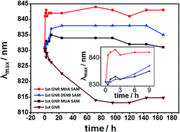
Gold nanorods (GNRs) modified with functional molecules are useful in chemical and biological sensing. In this work, the extinction spectrum of purified GNRs prepared by seed-mediated growth is systematically characterized by UV-visible absorption spectroscopy to test its stability before and after alkane-thiol modification. The results show that purification of GNRs makes the GNRs less stable, while a GNRs colloid solution functionalized by 11-mercaptoundecanoic acid (MUA), 3,3′-dithiobis[6-nitrobenzoic acid]bis(succinimide)ester (DSNB), or 16-mercaptohexadecanoic acid (MHA), respectively is stable over time. Bovine serum albumin (BSA) as a sample protein can be successfully attached to DSNB modified GNRs and form stable BSA–DSNB-GNRs probes. The red-shift observed in the extinction spectrum due to the BSA attachment is consistent over repeated experiments. Finally, anti-PSA (prostate-specific antigen) as a capture antibody is also attached to DSNB-modified GNRs. The attached anti-PSA is capable of interacting with prostate-specific antigens and induces a further red-shift, suggesting potential applications of thiol modified GNRs in bio-sensing.


Introduction

Owing to their unique optical properties, gold nanorods (GNRs) have been widely applied in biomedical sensing and imaging,1–3 cancer therapy,4,5 drug delivery,6 nano-photonics, and optoelectronics.7 One distinguishing property of GNRs is their localized surface plasmon resonances (LSPR), which are generated by coupling of light to the free electron plasma of the nanoparticles (NPs) with comparable size to the wavelength of incident light.8–10 The LSPR scattering spectrum of GNRs presents two bands depending on the polarization of the incoming light relative to the long axis of the GNR: the transverse band usually in the visible region and the lower energy longitudinal band ranging from the visible to the near-infrared region. The wavelengths of these bands are tunable with the aspect ratio of the GNR, and also sensitive to the thickness and index of refraction of any layer adsorbed on the GNRs' surface. This sensitivity to bound target molecules makes GNRs LSPR a powerful tool for chemical and biological sensing.3,11–15

Unmodified GNRs may aggregate in water, thus yielding poor performance in sensing tasks. To protect the GNRs from aggregation in water, cetyl trimethylammonium bromide (CTAB) can be used to form a bilayer on the surface of GNRs.16–18 However, further modification of GNRs with functional molecules such as thiols can facilitate the sensing of different molecules,1,13,19 and the exploring of optical devices from modified GNRs with photothermal effects.20–23 Therefore, stability of the modified GNRs by functional molecules is critical to the detection sensitivity. In this work, the formation of self-assembly monolayer (SAMs) of alkane-thiols on GNRs was studied systematically in order to investigate GNRs stability. The formed stable GNR-SAM retains its stability after further protein/antibody attachment and yield consistent result after interacting with target antigen, which is of great importance, especially for biomolecule detection.

Experimental section

Materials

Gold(III) chloride hydrate (HAuCl4·3H2O) was purchased from J & K. Sodium borohydride (NaBH4), L-ascorbic acid (AA), and AgNO3 were obtained from Alfa Aesar. 3,3-Dithiodipropionic acid di(N-hydroxysuccinimide ester) (MDAS), 11-mercaptoundecanoic acid (MUA) and 16-mercaptohexadecanoic acid (MHA) were purchased from Sigma-Aldrich. Hexadecyltrimethylammonium bromide (CTAB) was obtained from Amresco. BSA was purchased from Beyotime Biotechnology, PSA and anti-PSA were purchased from Biocell Biotechnology. GPC3 (glypican-3) and anti-GPC3 were purchased from Abnova. All materials were used without further purification. 3,3′-Dithiobis[6-nitrobenzoic acid]bis(succinimide)ester (DSNB) was synthesized according to a literature report.24 Triply distilled water was used in all processes. Chemical structures of the thiol compounds are shown in Scheme 1.
image file: c5ra22133a-s1.tif
Scheme 1 Chemical structures of the thiol compounds applied in the GNRs modification.

Seed preparation for gold nanorod growth

The preparation and growth of gold nanorods were undertaken according to a previously reported method.25

A 10 mL solution containing 0.1 M CTAB and 2.5 × 10⁻⁴ M HAuCl4·3H2O was mixed with 0.6 mL of ice-cold 0.01 M NaBH4 under vigorous stirring. After mixing, the color of the solution changed to brownish yellow, indicating the formation of seed nanoparticles.

Fabrication of gold nanorods with different aspect ratios

70 μL 0.0788 M AA was added to a 10 mL solution containing 0.1 M CTAB and 5.0 × 10 −4 M HAuCl4·3H2O. AA reduced Au(III) in the HAuCl4 solution to Au(I), which was indicated by the change of solution color from bright yellow to colorless. The above solution was then mixed with different amounts (100, 150, 200, 250 μL) of 4 mM AgNO3 solution. GNRs gradually formed in the above solution upon adding 12 μL of seed solution under gender shaking at 34 °C, indicated by the change of solution color from colorless to dark red. The reaction was terminated after 5 h by centrifugation at 14[thin space (1/6-em)]000 rpm for 15 min and removing the supernatant. The resulted GNRs were resuspended in 5 mM CTAB solution, which will be referred to herein as the 1st GNRs solution. The 1st GNRs was further centrifuged and resuspended in 5 mM CTAB solution to obtain the 2nd GNRs solution.

Functionalization of GNRs with different alkane-thiols

0.1 mL of 20 mM MDAS, MUA, MHA or DSNB solution was added to 5 mL of the 1st GNRs solution with continuous stirring for 90 min to prepare self-assembled monolayer-adsorbed nanorods (SAM-GNRs). For DSNB functionalized GNRs, the solution was filtered to remove the DSNB aggregates after stirring. The resulting solution was characterized by UV-Vis spectrometer (400–900 nm range) at different times to estimate its stability.

Modification of bovine serum albumin (BSA) to DSNB/GNR

BSA was dissolved in 2.5 mL PBS (pH = 7.4) to a final concentration of 12 μM. 3 mL of DSNB-modified GNRs was then slowly added to the above solution under gentle shaking over 30 min. The sample solution was finally characterized by UV-Vis spectrophotometer.

Results and discussion

Variations of the UV-Vis extinction spectrum of the synthesized gold nanorods with the amount of added AgNO3 were studied and the results are shown in Fig. 1. It is known that an increase in the amount of AgNO3 increases the aspect ratio of the GNRs (from ∼3 to ∼4 in Fig. 1, for example), thus resulting in a longer peak wavelength in the extinction spectrum, which is consistent with our observations. The aspect ratio of the formed GNRs can be roughly estimated by the equation proposed by Jain:26 λ = 445.4 + 90.6R (λ is the extinction peak wavelength with nm as unit, R is the aspect ratio of the GNR), to be 3.2, 3.7, 4.3 and 4.4, respectively (Fig. 1).
image file: c5ra22133a-f1.tif
Fig. 1 UV-Vis extinction spectra of GNRs formed with different amount of AgNO3. The peaks are at 734 nm, 784 nm, 831 nm and 843 nm, corresponding to different aspect ratios of 3.2, 3.7, 4.3, 4.4, respectively.

Stability of the GNRs or functionalized GNRs is of great importance for their application in bio-sensing tasks, as their extinction spectra are very sensitive to the aspect ratio and interfacial refractive index changes. However, the extinction spectra can change significantly if the GNRs are unstable. Unmodified GNRs aggregate quickly in water yielding broad extinction spectra. CTAB is widely used in the synthesis of GNRs to improve its stability. Nevertheless, a highly concentrated CTAB solution can be toxic to the attached biomolecules/systems, especially for cells bound to GNRs. Therefore, GNRs are usually purified and re-suspended in solution with low CTAB concentration before further modification. Purification can however decrease GNR stability due to the reduced presence of CTAB. We investigated carefully the stability of GNRs after both purification and modification by monitoring their extinction spectrum over time.

Further experiments were performed to determine an appropriate amount of the thiol compound for the GNRs modification, and the commercially available MDAS was chosen as a model sample since the functional NHS-group on this molecule could easily couple proteins or other target molecules with amino group. Different volumes (from 0 to 0.5 mL) of MDAS (dissolved in ethanol to a final concentration of 20 mM) were added to 5 mL of GNRs solution. The solution was stirred for 24 h before UV-Vis characterization. The λmax (peak wavelength of the extinction spectrum within the 400–900 nm range) of GNRs as a function of the added MDAS amount is shown in Fig. 2. It is clearly seen that adding 0.1 mL or more MDAS (20 mM) to 5 mL GNRs solution yields a consistent UV-Vis extinction peak, indicating a stable colloidal solution. Increasing MDAS concentration above this threshold did not facilitate further modification of GNRs. The observed red shift of the peak extinction wavelength indicated a change in the surface refractive index caused by the attachment of MDAS layer.


image file: c5ra22133a-f2.tif
Fig. 2 The extinction peak wavelength as a function of the amount of added MDAS (20 mM). Note that when the MDAS is less than 0.1 mL, the extinction spectrum is unstable and will blue shift gradually. Inset: the chemical structure of MDAS.

The variation of the peak wavelength over time for unmodified/modified 1st and 2nd GNRs, as defined in the methods part, is shown in Fig. 3. For 1st GNRs solution where more CTAB was left, the extinction spectrum was found blue shifted by ∼15 nm after 72 h, and remained stable for at least 1 month. In comparison, the 2nd GNRs solution extinction spectrum was observed to blue shift by ∼25 nm during 6–15 h and ∼40 nm after 72 h. The difference in the value of blue shift indicates that the 2nd GNRs solution is less stable than 1st GNRs due to aggregation when the amount of CTAB in solution is reduced. As a proof, SEM images (Fig. 3C) revealed the aggregation tendency of 2nd GNRs within 72 h. Introducing unstable GNRs in sensing tasks may cause experimental error when measuring the red/blue shift induced by GNRs modification, as the typical time required to modify GNRs is on the same scale in which a significant blue shift caused by aggregation may occur.


image file: c5ra22133a-f3.tif
Fig. 3 (A) λmax of 1st GNRs and GNRs SAM dependence on time (B) λmax of 2nd GNRs and GNRs SAM dependence on time (C) SEM images of 2nd GNRs revealing the aggregation over time.

Once the GNRs were successfully modified with thiols, the extinction spectrum would reach a stable state within 10 hours and remain stable for at least 6 days as shown in Fig. 3A and B. Here we chose MHA, MUA and DSNB as the target molecules considering their wide uses in modification of gold nano-materials. Among those, DSNB possesses large cross-section of Raman scattering as well as functional NHS-group, which grants it great potential in SERS-based biosensing.27,28 Red shifts were observed for GNRs functionalized by MHA, DSNB and MUA of 11 nm, 7 nm and 3 nm, respectively. The degree of red shift reflects the different structures of the thiol compounds: MHA displays the largest red shift as it has the longest chain compared to DSNB and MUA; DSNB, despite its short chain length, has an aromatic structure which is highly polarizable; meanwhile MUA is both short and lacks aromaticity so it modifies the interfacial optical response the least. Compared to the unstable 2nd GNR, it indicates that thiol modification is capable of significantly stabilizing GNRs.

Bovine serum albumin (BSA) was taken as a sample protein to attach to DSNB-bound GNRs. Fig. 4 shows the UV-Vis spectrum of GNRs before and after modification. 8.6 nm and 5.6 nm red shifts were observed after modifying GNRs with DSNB, and further with BSA, respectively. Two other batches of GNRs with different aspect ratios were modified in the same condition, and the results are shown in Table 1. It is clearly seen that the red shifts after DSNB/BSA modification for GNRs with different aspect ratio were consistent, and the extinction spectrum of the formed BSA–DSNB-GNRs was stable for at least one month. Worthy of note is that all the measurements were done after the initial GNR solution spectrum reached a steady state. For the successful refractive index shift-sensing of BSA immobilization, careful modification to form a stable GNRs solution is required.


image file: c5ra22133a-f4.tif
Fig. 4 Sample spectra of 1st GNRs, DSNB-GNRs and BSA–DSNB-GNRs.
Table 1 Peak wavelength of 1st/modifed GNRs and the red shift induced by modifications
Peak (nm) Sample 1 Sample 2 Sample 3
1st GNRs 775 796 828
DSNB-GNRs 784 803 834
BSA–DSNB 789 808 840
DSNB induced red shift 9 7 6
BSA induced red shift 5 5 6


To prove that carefully modified GNRs are capable of serving as reliable bio-sensing probes, prostate-specific antibody (PSA) and glypican-3 (GPC3) were attached to DSNB-bound GNRs and then interacted with the corresponding antigens. As shown in Fig. 5, DSNB and PSA antibody induced a 6 and 5 nm red shift, respectively, which is consistent with that in the BSA case and indicates the successful attachment of PSA to DSNB-GNRs. The spectrum of the antibody–DSNB-GNRs remains stable over time, and a red-shift was also observed when the corresponding antigen was applied to the antibody–DSNB-GNRs solution (from blue to green in Fig. 5). Note that the above red shift was not observed when the applied antigen does not match with the antibody (data not shown), indicating the red shift was the consequence of the specific antibody–antigen interaction. The detailed experimental procedure and results are provided in ESI section.


image file: c5ra22133a-f5.tif
Fig. 5 Sample spectra of 1st GNRs, DSNB/PEG–GNRs, anti-PSA–DSNB/PEG–GNRs and PSA–anti-PSA–DSNB/PEG–GNRs.

Conclusion

In this work, we investigated the stability of GNRs functionalized with thiols by means of UV-visible spectroscopy. CTAB modified GNRs can be unstable after purifying process, which may lead to experimental artifacts and poor performance in bio-sensing tasks. MUA, DSNB and MHA, at specific concentrations, are all shown to form stable SAM GNRs. The stabilized SAM GNRs can be further functionalized with bio-molecules such as BSA and other proteins to form stable sensing probes. This work helps to optimize the preparation of GNRs, improve GNRs' performance in bio-sensing applications and give better understanding to the red/blue shifts of the extinction spectrum induced by GNRs–biomolecule interactions.

Acknowledgements

This work was supported by the National Basic Research Program of China (no. 2011CB933101), National Natural Science Foundation of China (grant no. 21303208). The project was also sponsored by the Scientific Research Foundation for the Returned Overseas Chinese Scholars, State Education Ministry (no. Y31Z55) and the Start-Up Funding from the Institute of High Energy Physics of the Chinese Academy of Sciences (No. 2011IHEPYJRC504).

References

  1. C. Yu and J. Irudayaraj, Anal. Chem., 2006, 79, 572–579 CrossRef.
  2. J. S. Huang, V. Callegari, P. Geisler, C. Bruning, J. Kern, J. C. Prangsma, X. F. Wu, T. Feichtner, J. Ziegler, P. Weinmann, M. Kamp, A. Forchel, P. Biagioni, U. Sennhauser and B. Hecht, J. Am. Chem. Soc., 2006, 128, 2115 CrossRef PubMed.
  3. J. Cao, T. Sun and K. T. V. Grattan, Sens. Actuators, B, 2014, 195, 332–351 CrossRef CAS.
  4. Z. J. Zhang, L. M. Wang, J. Wang, X. M. Jiang, X. H. Li, Z. J. Hu, Y. H. Ji, X. C. Wu and C. Y. Chen, Adv. Mater., 2012, 24, 1418–1423 CrossRef CAS PubMed.
  5. M. K. Popp, I. Oubou, C. Shepherd, Z. Nager, C. Anderson and L. Pagliaro, J. Nanomater., 2014, 45067 Search PubMed.
  6. A. M. Alkilany, L. B. Thompson, S. P. Boulos, P. N. Sisco and C. J. Murphy, Adv. Drug Delivery Rev., 2012, 64, 190 CrossRef CAS.
  7. W. S. Kuo, C. N. Chang, Y. T. Chang, M. H. Yang, Y. H. Chien, S. J. Chen and C. S. Yeh, Angew. Chem., Int. Ed., 2010, 49, 2711 CrossRef CAS.
  8. S. M. Nie and S. R. Emery, Science, 1997, 275, 1102 CrossRef CAS.
  9. L. J. Sherry, S. H. Chang, G. C. Schatz, R. P. van Duyne, B. J. Wiley and Y. N. Xia, Nano Lett., 2005, 5, 2034 CrossRef CAS PubMed.
  10. A. V. Alekseeva, V. A. B. N. Bogatyrev, A. G. Mel'nikov, L. A. Dykman and N. G. Khlebtsov, Colloid J., 2006, 68, 661 CrossRef CAS.
  11. K. A. Willets and R. P. van Duyne, Annu. Rev. Phys. Chem., 2007, 58, 267 CrossRef CAS PubMed.
  12. K. M. Mayer and J. H. Hafner, Chem. Rev., 2011, 111, 3828 CrossRef CAS.
  13. W. Ma, H. Kuang, L. G. Xu, L. Ding, C. L. Xu, L. B. Wang and N. A. Kotov, Nat. Commun., 2013, 4, 2689 Search PubMed.
  14. L. Vigderman, B. P. Khanal and E. R. Zubarev, Adv. Mater., 2012, 24, 4811 CrossRef CAS.
  15. X. F. Wang, Y. Y. Mei and L. Tang, Talanta, 2015, 136, 1–8 CrossRef CAS PubMed.
  16. X. S. Ye, H. Shi, X. X. He, K. M. Wang, D. Li and P. C. Qiu, J. Mater. Chem. B, 2014, 2, 3667 RSC.
  17. J. H. Joo and J. S. Lee, Anal. Chem., 2013, 85, 6580 CrossRef CAS PubMed.
  18. D. K. Smith and B. A. Korgel, Langmuir, 2008, 24, 644–649 CrossRef CAS.
  19. Y. Y. Wang and L. Tang, Biosens. Bioelectron., 2015, 67, 18 CrossRef CAS.
  20. L. Wang, K. G. Gutierrez-Cuevas, H. K. Bisoyi, J. Xiang, G. Singh, R. S. Zola, S. Kumar, O. D. Lavrentovich, A. Urbasd and Q. Li, Chem. Commun., 2015, 51, 15039 RSC.
  21. K. G. Gutierrez-Cuevas, L. Wang, C. Xue, G. Singh, S. Kumar, A. Urbas and Q. Li, Chem. Commun., 2015, 51, 9845 RSC.
  22. C. Xue, J. Xiang, H. Nemati, H. K. Bisoyi, K. Gutierrez-Cuevas, L. Wang, M. Gao, S. Zhou, D. Yang, O. D. Lavrentovich, A. Urbas and Q. Li, ChemPhysChem, 2015, 16, 1852 CrossRef CAS.
  23. J. M. El Khoury, X. Zhou, L. Qu, L. Dai, A. Urbasc and Q. Li, Chem. Commun., 2009, 2109 RSC.
  24. D. S. Grubisha, R. J. Lipert, H. Y. Park, J. Driskell and M. D. Porter, Anal. Chem., 2003, 75, 5936 CrossRef CAS.
  25. B. Nikoobakht and M. A. El-Sayed, Chem. Mater., 2003, 15, 1957 CrossRef CAS.
  26. P. K. Jain, K. S. Lee, I. H. El-Sayed and M. A. El-Sayed, J. Phys. Chem. B, 2006, 110, 7238 CrossRef CAS PubMed.
  27. H. Zhang, M. H. Harpster, H. J. Park and P. A. Johnson, Anal. Chem., 2011, 83, 254 CrossRef CAS PubMed.
  28. G. F. Wang, H. Y. Park and R. J. Lipert, Anal. Chem., 2009, 81, 9643 CrossRef CAS.

Footnotes

Electronic supplementary information (ESI) available. See DOI: 10.1039/c5ra22133a
These authors contributed to this work equally.

This journal is © The Royal Society of Chemistry 2016
Click here to see how this site uses Cookies. View our privacy policy here.