
Hierarchical core/shell structures of ZnO nanorod@CoMoO4 nanoplates used as a high-performance electrode for supercapacitors†
Yunjiu Caoab,
Lei Ana,
Lijun Liaoa,
Xijian Liuab,
Tao Jiab,
Rujia Zoua,
Jianmao Yang*a,
Zongyi Qin*a and
Junqing Hu*a
aState Key Laboratory for Modification of Chemical Fibers and Polymer Materials, College of Materials Science and Engineering, Donghua University, Shanghai 201620, China. E-mail: yangjm@dhu.edu.cn; phqin@dhu.edu.cn; hu.junqing@dhu.edu.cn; Fax: +86-21-6779-2947; Tel: +86-21-6779-2947
bSchool of Fundamental Studies, Shanghai University of Engineering Science, Shanghai 201620, China
First published on 23rd December 2015
Abstract
Hierarchical core/shell structures of ZnO nanorod@CoMoO4 nanoplates grown directly on Ni foam were synthesized by a two-step hydrothermal process, in which ZnO nanorod arrays were first grown on Ni foam substrate, and then CoMoO4 nanoplates were grown in multiple directions on each ZnO nanorod. The as-grown ZnO@CoMoO4 core/shell structures (on Ni foam) directly used as integrated electrodes for electrochemical capacitors demonstrated prominent electrochemical performances, i.e., a high specific capacitance of 1.52 F cm−2 at a current density of 2 mA cm−2, which was higher than that (772 mF cm−2) of the pure CoMoO4 electrode, and a good long-term cycling stability, in which the electrodes retained 109% of the initial capacitance after 5000 cycles at a scan rate of 50 mV s−1. The superior electrochemical performances suggest that the ZnO@CoMoO4 core/shell structures could be considered as a prospective electrode material for supercapacitors.
1. Introduction
Supercapacitors (SCs) may be one of the most efficient storage energy devices in addressing the problems of climate change and the limited availability of fossils fuels in the future. Compared to secondary batteries and conventional dielectric capacitors, supercapacitors have been paid considerable attention over the past decade due to their high power density, fast charging/discharging characteristics and long cycle life.1–5Electrode materials as a key part of supercapacitors can be classified as three kinds: carbonaceous materials,6,7 transition metal oxides/hydroxides,8,9 and conducting polymers.10,11 Carbon-based materials absorb charge electrostatically at the electrode/electrolyte interface, leading to high power density at the cost of low energy density. By comparison, metal oxides/hydroxides and conducting polymers store charge by a faradaic or redox-type reaction similar to batteries, which enable high energy density. However, mechanical degradation of the conducting polymers due to the collapse of the polymer backbone during a limited number of charge–discharge process may lead to worse electrochemical performance and the metal oxide/hydroxide materials are not feasible application due to their poor electronic/ionic conductivity. Recently, composite pseudocapacitive transition metal oxides, such as MnMoO4, NiMoO4, CoMoO4,12–17 have been used as electrode materials, because they can supply different oxidation states for efficient redox reaction, which can obtain much higher specific capacitance and energy density.
Particularly, the CoMoO4 has been investigated as one of the most promising electrode materials owing to the low cost, non-toxic, and enhanced electrochemical properties.18,19 Xia et al. reported the CoMoO4 nanoparticles with a specific capacitance of 72 F g−1.20 Such a low capacitance may result from the poor electronic conductivity of metal oxides, and thus largely limits their practical utilization. Recently, the rational design and synthesis of unique core/shell heterostructures have been identified to be an effective means to achieve enhanced electrochemical properties.21–23 Mai et al. reported an improved the specific capacitance to 187 F g−1 from hierarchical structures of MnMoO4/CoMoO4 core/shell nanowires.13 Recent studies by other groups also demonstrated that NiO@CoMoO4 nanocomposites have a higher specific capacitance than pure CoMoO4.24 ZnO nanorod arrays have been extensively studied as a conductive support material due to their unique structure and properties, such as relatively good electrical conductivity, which provides a natural pathway for electron transport.25–27 In this regard, various core/shell nanostructures such as ZnO@Co3O4,28 ZnO@MoO3,29 ZnO@NiO/MoO2,30 have been fabricated and demonstrated their improved electrochemical performances. Therefore, CoMoO4 electrode materials supported with ZnO nanorod substrate can increase the electrical conductivity and enhance the electrochemical performance.
In this work, we have prepared hierarchical core/shell structures of ZnO nanorod@CoMoO4 nanoplates (ZnO@CoMoO4) on Ni foam for their supercapacitor applications via a two-step hydrothermal process followed by a calcination treatment. ZnO@CoMoO4 core/shell electrode materials not only retain the advantages of CoMoO4, such as the multiple valence states, environmentally benign nature, affordable cost, and natural abundance, but also improve the electron conductivity of the overall electrode. Meanwhile, the ZnO nanorod arrays serve as a scaffold, providing large surface area for the growth of CoMoO4 nanoplates. Moreover, the CoMoO4 nanoplates with large specific surface area can be beneficial for the improvement of areal specific capacitance. The ZnO nanorod arrays could also prevent the conventional aggregation of active materials that degrade the cycle stability. Here, this hierarchical core/shell structures of ZnO@CoMoO4 exhibits a high areal specific capacitance of 1.52 F cm−2 (1169 F g−1) at the current density of 2 mA cm−2, and an excellent cycle stability with 109% retention after 5000 cycles at a scan rate of 50 mV s−1. Thus, these hierarchical ZnO@CoMoO4 core/shell structures can be expected to be a promising candidate for supercapacitor applications.
2. Experimental section
2.1 Preparation of ZnO nanorod arrays on Ni foam
In this experiment, all the chemicals were analytical grade and were used without further purification. Ahead of the experiment, the Ni foam substrate (1 × 1 cm2) was ultrasonically washed with ethanol and water for 20 min each and then soaked in 0.5 M KMnO4 for 30 min to form a seed layer. The reaction solution was prepared by mixing 0.015 M zinc nitrate hexahydrate and 0.015 M hexamethylenetetramine (HMTA) together with 4 mL of ammonia in 100 mL of distilled water under constant magnetic stirring for 30 min. The Ni foam substrate was then dipped into the reaction solution and kept at 90 °C for 24 h. After the reaction, the Ni foam coated with white products was washed with deionized water, and thus the ZnO nanorods were obtained.2.2 Preparation of ZnO nanorod@CoMoO4 nanoplates
The ZnO nanorods prepared as described above were used as the scaffold for the growth of CoMoO4 via a facile hydrothermal method. First, 1 mmol of CoCl2·6H2O and 1 mmol of Na2MoO4·7H2O were dissolved in 50 mL of distilled water under constant magnetic stirring for 30 min. Second, the above solution was transferred into autoclave liner, and the as-prepared ZnO nanorods (substrate) was immersed into the reaction solution. Finally, the solution was sealed in a stainless steel autoclave and maintained at 150 °C for 5 h. After the reaction, in order to remove the residuals, the Ni foam substrate with light pink color was washed with deionized water for a few minutes and then dried at 60 °C for 12 h. Afterward, the sample was annealed at 300 °C for 2 h in air, and thus the ZnO nanorod@CoMoO4 nanoplates were obtained. For comparison, we also prepared pure CoMoO4 (without a ZnO nanorod) using the same method under the same conditions.2.3 Materials characterization
The morphology and microstructure of electrode materials were characterized using a scanning electron microscopy (SEM; S-4800) and a transmission electron microscopy (TEM; JEM-2100F) equipped with an energy dispersive spectrometer (EDS). The synthesized samples were characterized with a D/max-2550 PC X-ray diffractometer (XRD; Rigaku, Cu-Kα radiation). The mass of electrode materials was weighed on a XS analytical balance (Mettler Toledo; δ = 0.01 mg).2.4 Electrochemical characterizations
Electrochemical properties of ZnO@CoMoO4 core/shell structure materials were studied with cyclic voltammetry (CV), charge–discharge measurements, and electrochemical impedance spectroscopy (EIS) tests in an electrochemical workstation (Autolab PGSTAT302N potentiostat) using a three-electrode electrochemical configuration in 1 M KOH aqueous solution. The ZnO@CoMoO4 core/shell structure materials (1 × 1 cm2, the mass of ZnO nanorods and CoMoO4 nanoplates of ∼2 mg and 1.3 mg, respectively) were directly used as the working electrode. A platinum electrode was used as counter electrode, and a saturated calomel electrode was used as reference electrode. The pure CoMoO4 (∼1.5 mg cm−2) was also tested with electrochemical properties for comparison.3. Results and discussion
Fig. 1 illustrates the two-step synthesis of the ZnO/CoMoO4 core/shell heterostructure via hydrothermal process. First ZnO nanorod array is grown on Ni foam substrate. And then the as prepared ZnO nanorod arrays on Ni foam is subsequently coated by multidirectional and interconnected CoMoO4 nanoplates. | ||
| Fig. 1 Schematic illustration of the formation process of ZnO@CoMoO4 hierarchical core/shell heterostructure. | ||
The morphology of the ZnO@CoMoO4 core/shell heterostructures on Ni foam was first analyzed by SEM. Fig. 2a demonstrates the growth of ZnO nanorods on Ni foam. It can be clearly seen that ZnO nanorods was uniformly grown on Ni foam. The diameter and length of ZnO nanorods are about 250 nm and 6 μm, respectively (see ESI, Fig. S1†). The Ni foam is uniformly covered by ZnO@CoMoO4 core/shell heterostructures at a large scale, as shown Fig. 2b and c. It can be also seen from the high magnification images (Fig. 2d) that ZnO nanorods are uniformly coated by CoMoO4 nanoplates. The CoMoO4 nanoplates are firmly interconnected with each other to form some gaps between them on surface of ZnO nanorods. Further insight into the ZnO@CoMoO4 core/shell structures were provided by TEM studies. As shown in Fig. 3a and b the core/shell structure is easily observed from an individual ZnO@CoMoO4 core/shell structure, where the ZnO core is covered by CoMoO4 nanoplates. The HRTEM image of the CoMoO4 nanoplate shown in the insert of Fig. 3b displays an interplanar spacing of 0.244 nm, corresponding to the (400) plane of the CoMoO4.
 | ||
| Fig. 2 (a) The low magnification SEM images of ZnO nanorods. (b–d) Low and high magnification SEM images of ZnO@CoMoO4 core/shell heterostructures grown directly on Ni foam. | ||
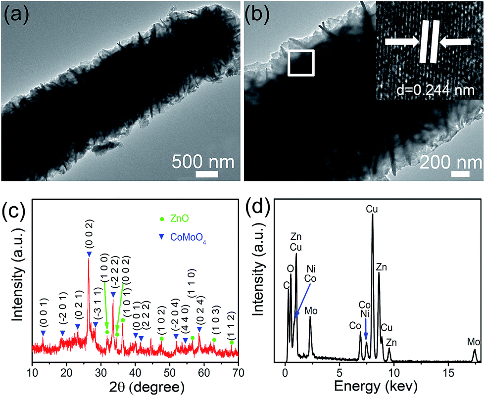 | ||
| Fig. 3 TEM (a) and high-resolution TEM (b) images of an individual ZnO@CoMoO4 core/shell structure. (c) Typical XRD pattern of ZnO@CoMoO4 core/shell structures scratched from the Ni foam. (d) EDS pattern of ZnO@CoMoO4 core/shell structure. | ||
XRD analysis was employed to further investigate the crystal structure and phase composition, as shown in Fig. 3c. The ZnO@CoMoO4 core/shell structures were scratched from Ni foam in order to avoid the strong peaks from the Ni foam substrate. It is obvious that the diffraction signal of the powders can be indexed to the standard diffraction patterns of ZnO (JCPDS no. 79-2205) and CoMoO4 (JCPDS no. 21-0868). The energy dispersive spectrometry (EDS) was carried out in TEM to further verify the elemental composition of the individual ZnO@CoMoO4 core/shell structures. The EDS result reveals the presence of elements of Zn, Co, Mo and O, as shown in Fig. 3d.
The obtained ZnO@CoMoO4 core/shell heterostructures on Ni foam were directly used as binder-free electrodes for the electrochemical performance evaluation. The electrochemical measurements were carried out in a three-electrode system with a 1 M KOH aqueous solution as electrolyte. The cyclic voltammogram (CV) curves of the ZnO@CoMoO4 electrodes were recorded at various scan rates within the potential range of 0 to 0.7 V as shown in Fig. 4a. A pair of redox peaks observed in all the CV curves is apparently different from the CV curves of electric double layer capacitors (EDLCs) that are similar to an ideal rectangular shape, indicating that the electrochemical performance of electrodes results from the pseudocapacitive capacitance.31,32 As the scan rates increases, the oxidation and reduction peaks shift to higher and lower potential, respectively, which could be attributed to the internal resistance of the electrode. Fig. S2 (see ESI†) shows the CV comparison of the ZnO@CoMoO4 and CoMoO4 electrodes recorded at the scan rate of 50 mV s−1. The CV curve integral area of the ZnO@CoMoO4 electrode is much larger than that of CoMoO4 electrode. It suggests that the ZnO nanorods used as conductive support for CoMoO4 material can greatly enhance electrochemical performance of CoMoO4 material.
 | ||
| Fig. 4 (a) CV curves of the ZnO@CoMoO4 core/shell hierarchical electrode at various scan rates. (b) Galvanostatic discharge curves of the ZnO@CoMoO4 on Ni foam electrode at various current densities. (c) A comparison of galvanostatic discharge curves of the ZnO@CoMoO4 and pure CoMoO4 electrodes at discharge current densities of 2 mA cm−2. (d) A comparison of specific capacitances for the ZnO@CoMoO4 and pure CoMoO4 as a function of current density. | ||
Galvanostatic charge–discharge measurements were performed at various currents from 2 mA cm−2 to 20 mA cm−2. The typical discharge curves of the ZnO@CoMoO4 electrodes at different current densities within the potential window of 0–0.5 V are presented in Fig. 4b. The areal capacitance for the ZnO@CoMoO4 electrodes can be calculated based on the discharge curves. There is a distinct plateau region in each discharge process, indicating that the capacitance derives from redox reaction, which is consistent with the CV curves. By comparison, Fig. 4c shows the discharge profiles of the ZnO@CoMoO4 electrodes and the pure CoMoO4 electrode at 2 mA cm−2. As can be seen, the discharge time of ZnO@CoMoO4 electrodes is much longer than that the pure CoMoO4 electrode, which indicates that ZnO@CoMoO4 electrodes exhibits a higher areal specific capacitance. The calculated results are displayed in Fig. 4d. The ZnO@CoMoO4 electrode displays high areal capacitance values of 1.52, 1.27, 1.05, 0.92 and 0.78 F cm−2 at different current densities of 2, 4, 8, 12 and 20 mA cm−2, respectively. The capacitance can retain 51.3% of its initial value even if the current density increases by 10 times, indicating a good rate capability. However the pure CoMoO4 electrode exhibits lower areal capacitance values of 772, 678, 581, 528 and 424 mF cm−2 at the same current densities. It is obvious that ZnO@CoMoO4 electrode delivers higher areal specific capacitance than the pure CoMoO4 electrode. The result may be attributed to the special role of the ZnO nanorod arrays within these core/shell structure providing a large surface area for the growth of CoMoO4 nanoplates which thus improved the surface area of active materials and avoided the conventional aggregation of the active materials. Furthermore, the capacitance of the ZnO@CoMoO4 electrode is also superior to previously reported CoMoO4 electrodes, such as CoMoO4·0.9H2O nanorods (326 F g−1 at 5 mA cm−2),33 CoMoO4 nanorods (286 F g−1 at 5 mA cm−2),34 MnMoO4/CoMoO4 heterostructured nanowires (187.1 F g−1 at 1 A g−1),13 porous ZnO/NiO composite micropolyhedrons (649 F g−1 at 5.8 A g−1),35 and Co3O4/NiO core/shell nanowire arrays (452 F g−1 at 2 A g−1).22
The long-term cycling stability of electrodes is also an important requirement for practical supercapacitor applications. The electrochemical stability of the ZnO@CoMoO4 core/shell electrode is evaluated by repeated CV processes at a scan rate of 50 mV s−1 for 5000 cycles as shown in Fig. 5a. The CV curves before and after cycling are displayed in Fig. S3,† where integral area of the 5000th cycle was larger than that of the first cycle. It is observed that the cycle stability is greatly enhanced in the ZnO@CoMoO4 electrode and maintains 109% of its initial value after 5000 cycles. The possible explanation is that only a part of electrode material is active at first. More and more sites become activated and contribute to the capacitance increasing as the electrolyte gradually penetrates into the inner region of the electrode.36 Moreover, the ZnO nanorods possess the chemical stability and are tightly integrated with the CoMoO4, making the structure stable and strengthening the structural integrity of the core during the charge–discharge process, resulting in enhanced cycle stability. This result shows that the combination of complex metal oxide (CoMoO4) nanoplates with ZnO nanorods provides high electrochemical stability for long cycle life applications.37,38
 | ||
| Fig. 5 (a) Cycling performance of the ZnO@CoMoO4 core/shell structure on Ni foam at a scan rate of 50 mV s−1. (b) Electrochemical impedance spectra before and after cycling. | ||
The electrochemical impedance spectroscopy measurements were further employed to discuss the electrochemical behavior of the ZnO@CoMoO4 electrode before and after cycling. The corresponding Nyquist plots were shown at Fig. 5b. The internal resistance of the electrochemical system (Rs) can be estimated from the intercept of the curve at Z′-axis at the high frequency, which includes the intrinsic resistance of the substrate, ionic resistance of electrolyte and contact resistance between current collector and active material. Warburg resistance (W) is estimated by the slope of the linear portion of the curve on the right of the semicircle, which is a result of the frequency dependent ion diffusion/transport in the electrolyte toward the electrode surface. It can be seen that both Rs and W of the ZnO@CoMoO4 electrode exhibit subtle changes after a long time cycling, indicating that the 3D structure of the ZnO@CoMoO4 was favorable for the penetration of the electrolyte within the electrode, the enhanced utilization of the electrode materials, and higher areal specific capacitance value, which can well explain the increase of the capacitance as described above.
The high areal specific capacitance, good rate capability, and excellent cycle stability of the ZnO@CoMoO4 could be attributed to the following factors: first, the 3D structure of ZnO nanorod arrays acted as a scaffold, providing a large surface area for the growth of the ZnO@CoMoO4 and accelerating electron transport for the faradaic reaction. Second, the gaps between the neighboring ZnO@CoMoO4 made the electrolyte fully penetrate into the inner region of the electrode, facilitating the transport of the electrolyte and ensuring efficient contact between the electrode material and the electrolyte, thus increasing the utilization of the active materials. Third, the ZnO@CoMoO4 core/shell structures were directly grown on the current collector (Ni foam), avoiding the use of polymer binders and conducting additives, which improves the utilization of the electrode material. Most importantly, the ZnO nanorods were grown directly on the Ni foam (substrate) with a robust adhesion and bridged the CoMoO4 nanoplates with the current collector, which can facilitate electron transport during the processes of charging and discharging because of its high electrical conductivity.
4. Conclusions
In sum, we have adopted a two step hydrothermal method to synthesize hierarchical core/shell ZnO@CoMoO4 nanostructures on the 3D continuous porous Ni foam and used these nanostructures as high performance electrochemical capacitor electrode. The electrochemical examinations show that as-fabricated electrodes made of such a nanomaterial possessed a high area specific capacitance of 1.57 F cm−2 (1169 F g−1) at the current density of 2 mA cm−2 and a good cycling stability of 109% of the initial specific capacitance after the 5000 repetitive cycle processes. These results suggest that such architecture is a promising electrode material for high-performance electrochemical capacitor.Acknowledgements
This work was financially supported by the National Natural Science Foundation of China (Grant no. 21171035, 51472049 and 51302035), the Key Grant Project of Chinese Ministry of Education (Grant no. 313015), the PhD Programs Foundation of the Ministry of Education of China (Grant no. 20110075110008 and 20130075120001), the National 863 Program of China (Grant no. 2013AA031903), the Science and Technology Commission of Shanghai Municipality (Grant no. 13ZR1451200), the Fundamental Research Funds for the Central Universities, the Program Innovative Research Team in University (IRT1221), the Shanghai Leading Academic Discipline Project (Grant no. B603), the Program of Introducing Talents of Discipline to Universities (no. 111-2-04) and the State Key Laboratory for Modification of Chemical Fibers and Polymer Materials (Grant no. LK1218), Donghua University.Notes and references
- J. R. Miller and P. Simon, Science, 2008, 321, 651 CrossRef CAS PubMed.
- P. Simon and Y. Gogotsi, Nat. Mater., 2008, 7, 845 CrossRef CAS PubMed.
- Y. Sun, Q. Wu and G. Shi, Energy Environ. Sci., 2011, 4, 1113 CAS.
- X. Yu, B. Lu and Z. Xu, Adv. Mater., 2014, 26, 1044 CrossRef CAS PubMed.
- Y. Huang, J. Liang and Y. Chen, Small, 2012, 8, 1805 CrossRef CAS PubMed.
- Y. Zhu, S. Murali, M. D. Stoller, K. J. Ganesh, W. Cai, P. J. Ferreira, A. Pirkle, R. M. Wallace, K. A. Cychosz, M. Thommes, D. Su, E. A. Stach and R. S. Ruoff, Science, 2011, 332, 1537 CrossRef CAS PubMed.
- A. B. Fuertes and M. Sevilla, ACS Appl. Mater. Interfaces, 2015, 7, 4344 CAS.
- C. H. Tang, Z. Tang and H. Gong, J. Electrochem. Soc., 2012, 159, A651 CrossRef CAS.
- Y. J. Zhang, C. T. Sun, P. Lu, K. Y. Li, S. Y. Song and D. F. Xue, CrystEngComm, 2012, 14, 5892 RSC.
- K. Zhang, L. L. Zhang, X. S. Zhao and J. Wu, Chem. Mater., 2010, 22, 1392 CrossRef CAS.
- A. Bahloul, B. Nessark, E. Briot, H. Groult, A. Mauger, K. Zaghib and C. M. Julien, J. Power Sources, 2013, 240, 267 CrossRef CAS.
- K. K. Purushothaman, M. Cuba and G. Muralidharan, Mater. Res. Bull., 2012, 47, 3348 CrossRef CAS.
- L. Q. Mai, F. Yang, Y. L. Zhao, X. Xu, L. Xu and Y. Z. Luo, Nat. Commun., 2011, 2, 381 CrossRef PubMed.
- M. C. Liu, L. Kang, L. B. Kong, C. Lu, X. J. Ma, X. M. Lia and Y. C. Luo, RSC Adv., 2013, 3, 6472 RSC.
- B. Senthilkumar, K. V. Sankar, R. K. Selvan, M. Danielleb and M. Manickamb, RSC Adv., 2013, 3, 352 RSC.
- G. K. Veerasubramani, K. Krishnamoorthy, S. Radhakrishnan, N. J. Kim and S. J. Kim, Int. J. Hydrogen Energy, 2014, 39, 5186 CrossRef CAS.
- Z. Xu, Z. Li, X. Tan, C. M. B. Holt, L. Zhang, B. S. Amirkhiz and D. Mitlin, RSC Adv., 2012, 2, 2753 RSC.
- Y. Ding, Y. Wan, L. Min, W. Zhang and S. H. Yu, Inorg. Chem., 2008, 47, 7813 CrossRef CAS PubMed.
- J. Zhao, Q. S. Wu and M. Wen, J. Mater. Sci., 2009, 44, 6356 CrossRef CAS.
- X. Xia, W. Lei, L. Hao, W. Wang and X. Wang, Electrochim. Acta, 2013, 99, 253 CrossRef CAS.
- X. Xia, J. Tu, Y. Zhang, X. Wang, C. Gu, X. Zhao and H. J. Fan, ACS Nano, 2012, 6, 5531 CrossRef CAS PubMed.
- C. Zhou, Y. Zhang, Y. Li and J. Liu, Nano Lett., 2013, 13, 2078 CrossRef CAS PubMed.
- X. Y. Xue, Z. H. Chen, L. L. Xing, S. Yuan and Y. J. Chen, Chem. Commun., 2011, 47, 5205 RSC.
- X. J. Ma, L. B. Kong, W. B. Zhang, M. C. Liu, Y. C. Luo and L. Kang, RSC Adv., 2014, 4, 17884 RSC.
- J. Liu, X. Huang, Y. Li, X. Ji, Z. Li, X. He and F. Sun, J. Phys. Chem. C, 2007, 111, 4990 CAS.
- H. J. Fan, P. Werner and M. Zacharias, Small, 2006, 2, 700 CrossRef CAS PubMed.
- J. Jiang, Y. Li, J. Liu and X. Huang, Nanoscale, 2011, 3, 45 RSC.
- X. Xia, J. Tu, Y. Zhang, X. Wang, C. Gu, X. B. Zhao and H. J. Fan, ACS Nano, 2012, 6, 5531 CrossRef CAS PubMed.
- G. R. Li, Z. L. Wang, F. L. Zheng, Y. N. Ou and Y. X. Tong, J. Mater. Chem., 2011, 21, 4217 RSC.
- S. C. Hou, G. H. Zhang, W. Zeng, J. Zhu, F. L. Gong, F. Li and H. G. Duan, ACS Appl. Mater. Interfaces, 2014, 6, 13564 CAS.
- C. Yuan, L. Yang, L. Hou, L. Shen, X. Zhang and X. W. Lou, Energy Environ. Sci., 2012, 5, 7883 CAS.
- G. Q. Zhang, H. B. Wu, H. E. Hoster, M. B. Chan Park and X. W. Lou, Energy Environ. Sci., 2012, 5, 9453 CAS.
- M. C. Liu, L. B. Kong, X. J. Ma, C. Lu, X. M. Li, Y. C. Luo and L. Kang, New J. Chem., 2012, 36, 1713 RSC.
- C. Z. Yuan, L. Yang, L. R. Hou, J. Y. Li, Y. X. Sun, X. G. Zhang, L. F. Shen, X. J. Lu, S. L. Xiong and X. W. Lou, Adv. Funct. Mater., 2012, 22, 2560 CrossRef CAS.
- H. Pang, Y. H. Ma, G. C. Li, J. Chen, J. S. Zhang, H. H. Zheng and W. M. Du, Dalton Trans., 2012, 41, 13284 RSC.
- X. Y. Liu, Y. Q. Zhang, X. H. Xia, S. J. Shi, Y. Lu, X. L. Wang, C. D. Gu and J. P. Tu, J. Power Sources, 2013, 239, 157 CrossRef CAS.
- Z. Xing, Q. Chu, X. Ren, C. Ge, A. H. Qusti, A. M. Asiri, A. O. Al-Youbi and X. Sun, J. Power Sources, 2014, 245, 463 CrossRef CAS.
- Z. Pu, Q. Liu, A. H. Qusti, A. M. Asiri, A. O. Al-Youbi and X. Sun, Electrochim. Acta, 2013, 109, 252 CrossRef CAS.
Footnote |
| † Electronic supplementary information (ESI) available: Experimental process, supplementary figure and specific capacitance calculation. See DOI: 10.1039/c5ra21953a |
| This journal is © The Royal Society of Chemistry 2016 |