DOI:
10.1039/C5RA21607A
(Paper)
RSC Adv., 2015,
5, 96855-96861
Crystal structures and luminescent properties of new lanthanide(III) complexes derived from 2-phenyl-4-pyrimidinecarboxylate†
Received
16th October 2015
, Accepted 3rd November 2015
First published on
6th November 2015
Abstract
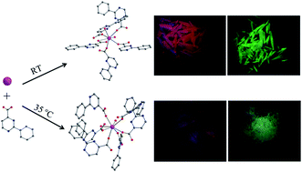
In this work, five novel lanthanide(III) coordination polymers derived from 2-phenylpyrimidine-4-carboxylic acid (Hppmc), namely, [Ln(ppmc)3(H2O)2]·2H2O [Ln = Eu (1), Tb (2)] and [Ln(ppmc)3(H2O)2] [Ln = Eu (3), Gd (4), Tb (5)] were successfully synthesized by a facile solution method and characterized by single-crystal X-ray diffraction, power X-ray diffraction (PXRD), infrared (IR) spectroscopy, elemental analysis, and thermogravimetric analysis (TGA). It was found that subtly different reaction conditions result in disparate structural characteristics. For example, by combining Hppmc with lanthanide(III) ions at room temperature, compounds 1 and 2 featuring a carboxylate-bridging chain structure, in which the carboxylates adopt both chelating and bridging modes, are isolated. However, the reaction at 35 °C generates three isostructural compounds 3–5 with a distinct chain structure, in which the lanthanide ions are connected by carboxylates via syn–syn and syn–anti modes. Photoluminescent studies of the Eu3+ and Tb3+ complexes reveal that the Hppmc ligand is a better sensitizer for Tb3+ ion than for Eu3+ ion. The investigation of the relationship between the crystal structures and the photoluminescence properties indicate that the coordination environments of lanthanide ions and the arrangement of the ligands are the dominating factors that affect the luminescence behaviors of the solid samples.
Introduction
Current interest in the design and construction of lanthanide-based coordination polymers has led to increasing attention in this area not only because of their aesthetically appealing architectures but also because of their intriguing photoluminescence properties.1 Their pure emission colors, sharp emission lines, and long excited-state lifetimes2 have received much attention and inspired numerous applications in various technological and research areas such as electroluminescent devices,3 analytical tools,4 and medical imaging devices.5 The luminescence properties displayed by almost all lanthanide ions are due to the f–f transitions. However, the transition of the 4f electron into the 4f* excited state is forbidden by the selection rules, leading to weak or non-existent emission bands. Thus, the use of organic chromophores as “antenna” ligands is often necessary and desirable. The ligands absorb energy in the ultraviolet (UV) region of spectrum, transferring it to stimulate Ln3+ emission in a process known as the antenna effect.6 The energy match for the intramolecular energy migration efficiency from the lowest triplet energy level of the ligand to the resonant energy level of the lanthanide ion plays the most important role.7 Furthermore, the photoluminescent properties can be affected by other factors, including the coordination environment of the central lanthanide,8 the arrangement of chromophoric units,9 the oscillation of –OH, –NH, C–H bonds10 and so on. So far, a lot of efforts have been made to improve the fluorescence of lanthanide coordination polymers, such as selecting organic ligands with conjugated motifs,11 synthesizing mixed-lanthanide12 or mixed lanthanide-transition-metal compounds,13 and regulating the structures and the photoluminescent properties through postsynthetic methods.14 Though the final structure of lanthanide compounds can be influenced by reaction conditions, such as solvents,15 reaction time,16 and temperature,17 the lanthanide compounds have a degree of structural predictability and tunability which provide a new platform for studying the relationship between the crystal structures and the photoluminescent properties. In our initial reports, 2-phenylpyrimidine-4-carboxylic acid (Hppmc) proved suitable for the construction of lanthanide coordination polymers with interesting magnetic and photoluminescent properties.18,19 In order to further study the relationship between the crystal structures and the photoluminescent properties, we explore in the present study the potential for improving the photoluminescence properties of the lanthanide coordination polymers with different structures by changing the synthetic conditions. Herein, we report a series of new lanthanide coordination polymers obtained under different synthetic conditions, which exemplify two unique structural types: [Ln(ppmc)3(H2O)2]·2H2O [Ln = Eu (1), Tb (2)] for type I and [Ln(ppmc)3(H2O)2] [Ln = Eu (3), Gd (4), Tb (5)] for type II. These compounds were characterized by infrared (IR) spectroscopy, elemental analysis (EA), thermogravimetric analysis (TGA), and X-ray single-crystal and power structural analyses. Furthermore, the fluorescence properties of the compounds were studied in detail. The distinct characteristics of the two structural types of these new compounds provided an excellent opportunity for probing the influence factors of the luminescence properties of the solid samples, including the type and the coordination environment of the metal centers and the arrangement of lumophores.
Experimental section
Synthesis
All the starting materials were commercially available reagents of analytical grade and used without further purification. The Hppmc ligand was prepared according to literature methods.20
[Eu(ppmc)3(H2O)2]·2H2O (1).
Eu(Ac)3·6H2O (32.9 mg, 0.1 mmol) was added to a 3 mL aqueous solution containing Hppmc (60.0 mg, 0.3 mmol) and NaOH (3 mL, 0.1 mol L−1). The resulting solution was stirred for another 10 min to give white precipitate. After adding water/ethanol (1
:
5, 15 mL), the resulting mixture was heated for another 5 min to give a clear solution. Upon filtration, the resulting colorless solution was left to evaporate at room temperature for several days before giving rise to colorless columnar crystals of complex 1, which were filtered off and air-dried. Yield, 32.0 mg (38.4% based on Eu3+). Elemental analysis for C33H39EuN6O10 (%): C, 48.24; H, 3.56; N, 10.23. Found (%): C, 47.93; H, 3.70; N, 9.76. Selected IR (KBr, cm−1): 3412(w), 1602(s), 1552(s), 1462(s), 1407(s), 743(s).
[Tb(ppmc)3(H2O)2]·2H2O (2).
Compound 2 was synthesized following a procedure similar to the synthesis of 1, with the only exception that Tb(Ac)3·6H2O was used instead of Eu(Ac)3·6H2O. Yield, 33.0 mg (38.5% based on Tb3+). Elemental analysis for C33H39TbN6O10 (%): C, 47.84; H, 3.53; N, 10.14. Found (%): C, 47.86; H, 3.47; N, 9.95. Selected IR (KBr, cm−1): 3412(w), 1600(s), 1549(s), 1460(m), 1408(s), 741(s).
[Ln(ppmc)3(H2O)2] [Ln = Eu (3), Gd (4), Tb (5)].
Ln(NO3)3·6H2O (0.1 mmol) was added to a 3 mL aqueous solution containing Hppmc (60.0 mg, 0.3 mmol) and NaOH (3 mL, 0.1 mol L−1). The resulting solution was stirred for another 10 min, water/ethanol (1
:
1, 18 mL) was added, and the mixture was heated until the precipitate disappeared. Upon filtration, the resulting solution was kept at 35 °C in the oven for several days before giving rise to colorless needle-shaped crystals of complexes 3–5, which were filtered off and air-dried. Yield, 50.7%, 50.7% and 47.8% based on lanthanide ions, for 3, 4 and 5, respectively. Elemental analysis for 3 (%): C, 50.46; H, 3.21; N, 10.70. Found (%): C, 50.42; H, 3.13; N, 10.51. Selected IR for 3 (KBr, cm−1): 3398(w), 1702(m), 1632(s), 1548(s), 1459(m), 1407(s), 743(s). Elemental analysis for 4 (%): C, 50.12; H, 3.19; N, 10.63. Found (%): C, 50.06; H, 3.16; N, 10.43. Selected IR for 4 (KBr, cm−1): 3399(w), 1703(m), 1632(s), 1548(s), 1459(m), 1408(s), 743(s). Elemental analysis for 5 (%): C, 50.01; H, 3.18; N, 10.60. Found (%): C, 49.97; H, 3.11; N, 10.36. Selected IR for 5 (KBr, cm−1): 3399(w), 1706(m), 1633(s), 1548(s), 1459(m), 1407(s), 743(s).
X-ray crystallography and physical measurement
Single-crystal X-ray diffraction data of compounds 1–5 were collected at 293 K using a Bruker Smart Apex II CCD diffractometer with graphite-monochromated Mo Kα radiation (λ = 0.71073 Å). All of the structures were solved by direct methods and refined with the full-matrix least-squares technique based on F2 using the SHELXL program.21 All non-hydrogen atoms in the complexes were refined with anisotropic displacement parameters. Hydrogen atoms of the water molecules were located from the difference Fourier maps and refined with restraint of the O–H and H⋯H distances (0.96 and 1.52 Å, respectively). Other hydrogen atoms were placed at the calculation positions. A summary of the crystallographic data and details of the structure refinements are given in Table 1. Selected bond lengths and bond angles are listed in Tables S1 and S2.† Power X-ray diffraction data for all samples were collected at room temperature on bulk samples with Cu Kα radiation (λ = 1.54059 Å).
Table 1 Crystal data and structure refinement parameters for complexes 1–5
Compound |
1
|
2
|
3
|
4
|
5
|
Formula |
C33H29EuN6O10 |
C33H29TbN6O10 |
C33H25EuN6O8 |
C33H25GdN6O8 |
C33H25TbN6O8 |
F
w
|
821.58 |
828.54 |
785.55 |
790.84 |
792.51 |
Crystal system |
Monoclinic |
Monoclinic |
Monoclinic |
Monoclinic |
Monoclinic |
Space group |
P21/n |
P21/n |
P21/c |
P21/c |
P21/c |
a (Å) |
13.329(1) |
13.376(1) |
15.873(1) |
15.896(1) |
15.860(2) |
b (Å) |
10.348(1) |
10.305(1) |
22.954(2) |
22.952(2) |
22.849(3) |
c (Å) |
23.849(2) |
23.900(2) |
8.926(1) |
8.930(1) |
8.889(1) |
α (°) |
90 |
90 |
90 |
90 |
90 |
β (°) |
92.199(2) |
92.146(1) |
104.803(1) |
104.757(1) |
104.730(2) |
γ (°) |
90 |
90 |
90 |
90 |
90 |
V (Å3) |
3287.0(6) |
3291.9(4) |
3144.2(4) |
3150.9(5) |
3115.4(6) |
Z
|
4 |
4 |
4 |
4 |
4 |
µ (mm−1) |
1.976 |
2.216 |
2.057 |
2.167 |
2.333 |
F(000) |
1648 |
1656 |
1568 |
1572 |
1576 |
GOF |
0.965 |
1.007 |
1.024 |
1.064 |
1.039 |
Data collected |
19 708 |
19 630 |
19 003 |
19 100 |
18 881 |
Unique |
7537 |
7489 |
7230 |
7211 |
7200 |
R
int
|
0.0656 |
0.0514 |
0.0317 |
0.0350 |
0.0360 |
R
1 [I > 2σ(I)] |
0.0482 |
0.0472 |
0.0280 |
0.0279 |
0.0294 |
wR2 [I > 2σ(I)] |
0.0994 |
0.1064 |
0.0280 |
0.0657 |
0.0625 |
R
1 [all data] |
0.0982 |
0.0828 |
0.0371 |
0.0429 |
0.0424 |
wR2 [all data] |
0.1182 |
0.1228 |
0.0693 |
0.0811 |
0.0669 |
Elemental analyses of carbon, hydrogen and nitrogen were performed on an ElementarVario El analyzer. The FTIR spectra were recorded with a Nicolet Magna 750 FT/IR spectrometer with pressed KBr pellets in the 4000–400 cm−1 regions. Thermogravimetric analyses (TGA) were carried out using a Mettler-Toledo TGA/DSC1. Fluorescence spectra were recorded on an RF-5301PC spectrophotometer, phosphorescence spectra were measured with F-7000 and the emission quantum yields (ΦQY) were measured with an Edinburgh Instrument fls920 spectrometer.
Result and discussion
Crystal structures
Structure of [Ln(ppmc)3(H2O)2]·2H2O [Ln = Eu (1), Tb (2)].
X-ray structure analyses reveal that 1 and 2 are isostructural and crystallize in the monoclinic space group P21/n. As a representative example, the crystal structure of 1 is described in detail. As shown in Fig. 1a, the asymmetric unit of 1 contains one crystallographically independent Eu3+ ion, three ppmc− ligands, two coordinating water molecules, and two solvating water molecules. Each Eu3+ ion is eight-coordinated by two water molecules and six carboxylate oxygen atoms from five different ppmc− ligands. The coordination geometry around Eu3+ ion is a slightly distorted square-antiprism (Fig. S1a†). The Eu–O bond lengths range from 2.325 to 2.505 Å and the bond angles of O–Eu–O are in the range of 65.65–160.0°, comparable to those in other Eu3+ carboxylate-based complexes observed in previous reports.22 Owing to the effect of lanthanide contraction,23 the Tb–O bonds in 2 are slightly shorter than the corresponding Eu–O bonds in 1. The ppmc− ligands are divided into two types, one acting as a chelating terminal ligand while the other serving as a bridging one. Two bridging ppmc− ligands connect adjacent Eu3+ ions in a syn–anti mode, forming a one-dimensional (1D) chain running along the b direction with a Ln⋯Ln distance of 5.530 and 5.503 Å for 1 and 2, respectively (Fig. 1b). The adjacent chains are connected to form a two-dimensional (2D) layer through π–π interactions between the bridging ppmc− ligands with a centroid⋯centroid distance of 3.656 and 3.660 Å for 1 and 2. Moreover, neighboring layers are further linked, also by π–π stacking between the terminal ppmc− ligands, and they feature a centroid⋯centroid separation of 4.172 and 4.161 Å for 1 and 2, thus producing a 3D supramolecular structure (Fig. S1b†).
 |
| Fig. 1 Coordination environment of Eu3+ (a) and the 1D chain connected by ppmc− (b) in 1 (symmetry code: a: 3/2 − x, y − 1/2, 3/2 − z; b: 3/2 − x, y + 1/2, 3/2 − z; the hydrogen atoms are omitted for clarity). | |
Structure of [Ln(ppmc)3(H2O)2] [Ln = Eu (3), Gd (4), Tb (5)].
Compounds 3–5 are isostructural and crystallize in the monoclinic P21/c space group. Here, the crystal structure of the compound 3 is described as an example. The asymmetric unit of 3 is composed of one lanthanide ion, two coordination water molecules and three ppmc− ligands (Fig. 2a). Similar to 1 and 2, the lanthanide ions in 3 are also eight-coordinated by eight oxygen atoms (six from six ppmc− ligands and two from water molecules), resulting in a distorted square antiprism coordination geometry (Fig. S2a†). The Eu–O bond lengths range from 2.330 to 2.546 Å, which are typical for Eu3+ complexes.24 The mean Ln–O bond lengths in 3–5 follow the order of Eu–O (2.410 Å) > Gd–O (2.400 Å) > Tb–O (2.379 Å), consistent with the lanthanide contraction effect (Table S2†).23 In contrast to those found in compounds 1 and 2, all the ppmc− ligands in 3–5 adopt bidentate bridging mode. Three ppmc− ligands (one in syn–anti mode and the other two in syn–syn mode) link lanthanide ions to form a 1D chain extending along the c-axis direction (Fig. 2b). The distances between the neighboring Ln3+ ions are 4.592 and 4.953 Å for 3 and 4, respectively, which are significantly shorter than the Ln⋯Ln separation in complexes 1 and 2. This discrepancy is probably due to the different amounts of bridging ligands between two mental ions.25 The more bridging ligands, the shorter the metal–metal distance, since the ligands are expected to reduce cation–cation repulsion.25 The adjacent chains are linked to each other via hydrogen bond between the nitrogen atom of pyrimidine ring and coordination water molecules (N4⋯H7C = 1.951(0) Å, O7⋯N4 = 2.868(0) Å, ∠O7–H7C⋯N4 = 166.4(3)°, 1 − x, y + 1/2, 3/2 − z) to yield a slab extending along the ab plane (Fig. S2b†).
 |
| Fig. 2 Coordination environment of Eu3+ (a) and the 1D chain connected by ppmc− (b) in 3 (symmetry code: a: x, 1/2 − y, z + 1/2; b: x, 1/2 − y, z − 1/2; the hydrogen atoms, nitrogen atoms and partial carbon atoms are omitted for clarity). | |
Powder XRD pattern, TGA data and IR
The simulated and experimental PXRD patterns of compounds 1–5 obtained at room temperature are presented in Fig. 3. Their peak positions correspond well with the simulated PXRD patterns calculated based on single crystal diffraction data, indicating that all of the samples were in a pure phase. The differences in intensity may be due to the preferred orientation of the power samples.26
 |
| Fig. 3 Powder X-ray diffraction profiles of 1–5 together with the corresponding patterns derived from the single crystal data. | |
The thermal stability of 1–3 and 5 was examined by TGA in a N2 atmosphere using single-phase polycrystalline samples at a heating rate of 5 °C min−1. As shown in Fig. S3,† all samples showed two obvious steps of weight loss from 25 °C to 800 °C. Compounds 1 and 2 are isomorphous and exhibit similar thermal behavior. They gradually lost their lattice water molecules at about 60 °C. After the loss of the lattice water molecules, the coordinating water molecules begin to be liberated. A total weight loss of 8.8% was observed for 1 and 2 (calculated 8.8% for 1 and 8.7% for 2). In contrast, for compounds 3 and 5, the weight loss associated with the coordinating water molecules occurs between 75 °C and 155 °C (weight loss 4.7%, calculated 4.6%). The weight loss above 270 °C is attributed to the thermal decomposition of the organic components. The remaining weight of 21.1–22.6% indicates that the final products are Eu2O3 for 1 and 3 and Tb4O7 for 2 and 5 (calculated 21.4%, 22.6%, 22.4%, 23.6% for 1, 2, 3 and 5, respectively).
The IR spectra of 1–5 are quite similar. They all show strong and broad absorption bands around 3400 cm−1, which may be assigned to the characteristic peaks of OH stretching vibration. The strong vibrations located at around 1630, 1550 and 1407 cm−1 correspond to the asymmetric and symmetric stretching vibrations of the carboxylate group of ppmc− ligands, respectively.
Photoluminescence properties
It is well-known that the luminescence of Ln3+ ions is greatly influenced by the intramolecular energy transfer from the triplet state of ligands to the resonance level of Ln3+ ions.27 Herein, the triplet-state energy of Hppmc ligands of the Eu3+ and Tb3+ complexes was estimated from the Gd3+ complex by measuring the phosphorescence spectra at the low temperature (77 K).28 The phosphorescence spectrum of 4 was measured and shown in Fig. S4.† Judging from the onset of the emission bands (433 nm),25 the energy of lowest triplet state can be determined to be 23
094 cm−1 for Hppmc. This energy is higher than the resonant energy level of Eu3+ (18
674 cm−1) and Tb3+ (20
500 cm−1), confirming the suitability of the ligands as sensitizers for the excitation of Eu3+ and Tb3+. The four compounds examined show characteristic red or green emission attributed to Eu3+ and Tb3+ under the ultraviolet light.
The solid-state luminescent property of 1–3 and 5 were investigated at room temperature. Fig. 4 and S5† show the normalized excitation and emission spectra registered at room temperature. The broad excitation band in the 250–450 nm range can be attributed to the π–π* transition of the conjugated organic ligand and possible ligand-to-metal charge transfer (LMCT) state.18,29 When 1 and 3 were excited at 354 nm and 332 nm, respectively, their solid-state emission spectra exhibited typical emission patterns characteristic of Eu3+ ions with narrow, sharp and well-separated bands which are assigned to the intra-4f6 electron transitions of Eu3+. The emissions that appeared in the range of 450–800 nm can be ascribed to the 5D0 → 7Fn (n = 1, 2, 3, 4) transitions of the Eu3+ ion. The 5D0 → 7F1 (580 nm) transition is a magnetic dipole transition, the intensity of which is insensitive to the coordination environment. The 5D0 → 7F2 (616 nm) is an electric dipole transition, which is extremely sensitive to the coordination environment.30 The integrated intensity ratio of the 5D0 → 7F2 to 5D0 → 7F1 transitions is 4.0 and 2.3 for 1 and 3 respectively, indicating that the Eu3+ ions occupy a low-symmetry site without an inversion center,31 which is in good agreement with the crystallographic analysis.
 |
| Fig. 4 Solid-state excitation/emission spectra and emission decay patterns (insets) of 1 (a) and 2 (b). | |
Under excitation of 334 nm, complexes 2 and 5 exhibit spectra characteristic of Tb3+ ion from the emitting level (5D4) to the ground multiplet (7F6–2). The emission positions of 2 and 5 are similar except for slight differences in the photoluminescence intensities. The emission spectrum shows five emission bands at 490, 543, 585, 619 and 646 nm, respectively. The most intense emission is dominated by the 5D4 → 7F5 transition at 543 nm, which results in green fluorescence.
To better understand the luminescent properties of the four compounds, the lifetime and quantum yield were determined from the luminescence decay profiles. For all compounds, the decay curves were well fitted to a single-exponential function (see the insets of Fig. 4 and S5†), indicating the preference of a single chemical environment around the Ln3+ ions. The result fits well with the X-ray structure analyses. Moreover, as listed in Table 2, the lifetimes for Tb3+-containing complexes 2 and 5 are longer than those found for Eu3+-containing compounds 1 and 3. The result of lifetime measurements appeared to follow the same trend as the quantum yield, which can be attributed to the energy difference between the triplet energy level of ppmc− ligands and the resonance level of Ln3+ ions. The energy difference for Eu3+ is significantly larger (4420 cm−1) than that of Tb3+ ion (2594 cm−1),32 indicating that ppmc− ligand is more suitable for tuning the photoluminescence of Tb3+ than Eu3+. It is worth noting that this result is in sharp contrast to the prediction we previously made,19 which omitted the role that the secondary ligand plays in the intramolecular energy transfer process. Since the triplet state of ppmc− is higher than that of 1,10-phenanthroline (phen) (22
132 cm−1), the secondary ligand used in our earlier study, it is reasonable to presume that the phen molecule may act as an energy channel to transfer the energy from the triplet state of ppmc− to the resonance level of Ln3+ ions.33 The lowest triplet energy level of phen ligand matches better the resonance levels of Eu3+ than those of Tb3+.
Table 2 Lifetimes and quantum yields of complexes 1–3 and 5
|
Φ
QY
|
λ
ex (nm) |
τ (ms) |
1
|
7.5% |
354 |
0.34(3) |
2
|
36.1% |
334 |
0.51(1) |
3
|
6.6% |
332 |
0.40(2) |
5
|
19.5% |
334 |
1.03(2) |
Interestingly, compounds 5 has a much longer lifetime than 2, which can be attributed to the distinct coordination environments of the metal centers in these two compounds. The Tb3+ ions in 5 coordinate to six ppmc− ligands and two water molecules, while the asymmetric unit of 2 comprises five ppmc− ligands, two coordination water molecules, and two guest water molecules. Thus the Tb3+ in 5 has more “antenna” ligands that can absorb more light energy. In addition, there is no guest water molecular in the structure of 5 that virtually decreases the nonradiative decay rate and increases the life time of the sample.34 Therefore, we reason that a higher content of “antenna” ligands and less deactivation, allowing 5 to exhibit a longer lifetime than compound 2.25,35 However, it is surprising that complex 2 has a lower content of “antenna” ligands and more water molecules, and yet shows a relatively higher quantum yield than 5. The result is also supported by the photographs of the compounds taken under the same UV lamp (Fig. S6†). This seemingly contradicting result might be due to the π–π interactions between interdigitating ppmc− ligands in 2, which bring the luminophores closer and enable energy transition more effectively.36 In addition, the phenyl substituent of the ligands might be locked into fixed positions, which reduces the elevation of the nonradiative transition.19 These are also likely reasons for compound 1 having a shorter lifetime and higher quantum yield than 3.
Conclusions
In summary, by virtue of fine-tuning the reaction condition, we have employed 2-phenylpyrimidine-4-carboxylic acid as an antenna ligand to successfully synthesize five Ln3+ coordination polymers featuring distinct structural motifs. The carboxylate groups of the Hppmc ligand in these compounds adopt various coordination or bridging mode. The photophysical properties, including the excitation and emission spectra, low temperature phosphorescence spectra, lifetime, and quantum yield of these compounds have been thoroughly studied. It is worth highlighting that the different photoluminescent properties are attributed to the different structural characteristics of the compounds, such as the number of the “antenna” and water molecules and their supramolecular architectures. These factors affect the luminescence behavior of lanthanide coordination polymers in a manner that is amenable to synthetic manipulations. This work thus provides important insight into understanding the relationship between structure and luminescence behavior of these systems. We are currently applying these principles to design new generations of compounds with enhanced photoluminescent properties that may lead to a range of practical applications.
Acknowledgements
This work was supported by the National Natural Science Foundation of China (No. 21171023), National Key Basic Research Program of China (2013CB933402), and Beijing Higher Education Young Elite Teacher Project. Z. W. acknowledges the financial support of the University of South Dakota and South Dakota EPSCoR.
Notes and references
-
(a) A. Thirumurugan, S. K. Pati, M. A. Greenc and S. Natarajan, J. Mater. Chem., 2003, 13, 2937–2941 RSC;
(b) R. C. Evans, P. Douglas and C. J. Winscom, Coord. Chem. Rev., 2006, 250, 2093–2126 CrossRef CAS;
(c) K. Binnemans, Chem. Rev., 2009, 109, 4283–4374 CrossRef CAS PubMed;
(d) P. F. Shi, G. Xiong, B. Zhao, Z. Y. Zhang and P. Cheng, Chem. Commun., 2013, 49, 2338–2340 RSC;
(e) J. Y. Zou, N. Xu, W. Shi, H. L. Gao, J. Z. Cui and P. Cheng, RSC Adv., 2013, 3, 21511–21516 RSC;
(f) Q. P. Li and S. W. Du, RSC Adv., 2014, 4, 30963–30967 RSC;
(g) Y. Hasegawa and T. Nakanishi, RSC Adv., 2015, 5, 338–353 RSC.
-
(a) A. P. D. Silva, D. B. Fox, A. J. M. Huxley and T. S. Moody, Coord. Chem. Rev., 2000, 205, 41–57 CrossRef;
(b) D. Parker, Coord. Chem. Rev., 2000, 205, 109–130 CrossRef CAS;
(c) A. D. B. Dias, Dalton Trans., 2007, 36, 2229–2241 RSC;
(d) C. Maxim, D. G. Branzea, C. Tiseanu, M. Rouzières, R. Clérac, M. Andruh and N. Avarvari, Inorg. Chem., 2014, 53, 2708–2717 CrossRef CAS PubMed.
-
(a) N. Marques, A. Sella and J. Takats, Chem. Rev., 2002, 102, 2137–2160 CrossRef CAS PubMed;
(b) J. Kido and Y. Okamoto, Chem. Rev., 2002, 102, 2357–2368 CrossRef CAS PubMed;
(c) M. D. Allendorf, C. A. Bauer, R. K. Bhakta and R. J. Houka, Chem. Soc. Rev., 2009, 38, 1330–1352 RSC;
(d) Y. J. Cui, Y. F. Yue, G. D. Qian and B. L. Chen, Chem. Rev., 2012, 112, 1126–1162 CrossRef CAS PubMed.
-
(a) J. M. Bryson, J. W. Reineke and T. M. Reineke, Macromolecules, 2012, 45, 8939–8952 CrossRef CAS PubMed;
(b) J. A. Peters and K. Djanashvili, Eur. J. Inorg. Chem., 2012, 1961–1974 CrossRef CAS;
(c) P. Harvey, I. Kuprov and D. Parker, Eur. J. Inorg. Chem., 2012, 2015–2022 CrossRef CAS;
(d) S. V. Eliseeva and J. C. G. Bünzli, Chem. Soc. Rev., 2010, 39, 189–227 RSC.
-
(a) M. D. Allendorf, C. A. Bauer, R. K. Bhakta and R. J. T. Houk, Chem. Soc. Rev., 2009, 38, 1330–1352 RSC;
(b) J. C. G. Bunzli, Chem. Rev., 2010, 110, 2729–2755 CrossRef PubMed;
(c) X. J. Zhou, X. Q. Zhao, Y. J. Wang, B. Wu, J. Shen, L. Li and Q. X. Li, Inorg. Chem., 2014, 53, 12275–12282 CrossRef CAS PubMed.
-
(a) J. M. Lehn, Angew. Chem., Int. Ed., 1990, 29, 1304–1319 CrossRef;
(b) Z. A. Taha, A. M. Ajlouni, A. K. Hijazi, N. A. A. Rawashdeh, K. A. A. Hassan, Y. A. A. Haj, M. A. Ebjaai and A. Y. Altalafha, J. Lumin., 2015, 161, 229–238 CrossRef CAS.
-
(a) H. P. Wang, H. G. Li, G. N. Lu, N. Tang, W. S. Liu and Y. Tang, Inorg. Chem. Commun., 2010, 13, 882–886 CrossRef CAS;
(b) X. H. Yan, C. L. Yi, X. G. Huang, W. S. Liu, Y. Tang and M. Y. Tan, Inorg. Chem. Commun., 2011, 14, 654–658 CrossRef CAS;
(c) L. Zhang, Y. F. Ji, X. B. Xu, Z. L. Liu and J. K. Tang, J. Lumin., 2012, 132, 1906–1909 CrossRef CAS.
-
(a) K. A. White, D. A. Chengelis, M. Zeller, S. J. Geib, J. Szakos, S. Petoud and N. L. Rosi, Chem. Commun., 2009, 30, 4506–4508 RSC;
(b) X. Yan, Z. Cai, C. Yi, W. Liu, M. Tan and Y. Tang, Inorg. Chem., 2011, 50, 2346–2353 CrossRef CAS PubMed.
- L. X. You, S. J. Wang, G. Xiong, F. Ding, K. W. Meert, D. Poelman, P. F. Smet, B. Y. Ren, Y. W. Tian and Y. G. Sun, Dalton Trans., 2014, 43, 17385–17394 RSC.
-
(a) G. Mancino, A. J. Ferguson, A. Beeby, N. J. Long and T. S. Jones, J. Am. Chem. Soc., 2005, 127, 524–525 CrossRef CAS PubMed;
(b) W. G. Lu, D. C. Zhong, L. Jiang and T. B. Lu, Cryst. Growth Des., 2012, 12, 3675–3683 CrossRef CAS;
(c) J. Cuan and B. Yan, RSC Adv., 2013, 3, 20077–20084 RSC;
(d) Y. E. Cha, X. Li, D. Ma and R. Huo, Eur. J. Inorg. Chem., 2014, 2969–2975 CrossRef CAS.
-
(a) T. M. Reineke, M. Eddaoudi, M. Fehr, D. Kelley and O. M. Yaghi, J. Am. Chem. Soc., 1999, 121, 1651–1657 CrossRef CAS;
(b) F. S. Liang, Q. G. Zhou, Y. X. Cheng and L. X. Wang, Chem. Mater., 2003, 15, 1935–1937 CrossRef CAS;
(c) X. P. Yang, R. A. Jones, J. H. Rivers, R. Lai and I. Pen, Dalton Trans., 2007, 3936–3942 RSC;
(d) C. J. Li, M. X. Peng, J. D. Leng, M. M. Yang, Z. J. Lin and M. L. Tong, CrystEngComm, 2008, 10, 1645–1652 RSC.
-
(a) D. T. de Lill, A. de Bettencourt-Dias and C. L. Cahill, Inorg. Chem., 2007, 46, 3960–3965 CrossRef CAS PubMed;
(b) B. L. Chen, L. B. Wang, Y. Q. Xiao, F. R. Fronczek, M. Xue, Y. J. Cui and G. D. Qian, Angew. Chem., Int. Ed., 2008, 47, 1–5 CrossRef;
(c) X. J. Zhao, J. H. Yang, Y. Liu, P. F. Gao and Y. F. Li, RSC Adv., 2014, 4, 2573–2576 RSC.
-
(a) J. M. Herrera, S. J. A. Pope, H. Adams, S. Faulkner and M. D. Ward, Inorg. Chem., 2006, 45, 3895–3904 CrossRef CAS PubMed;
(b) J. W. Cheng, S. T. Zheng and G. Y. Yang, Inorg. Chem., 2008, 47, 4930–4935 CrossRef CAS PubMed;
(c) Q. Liu, F. Wan, L. X. Qiu, Y. Q. Sun and Y. P. Chen, RSC Adv., 2014, 4, 27013–27021 RSC.
-
(a) H. L. Sun, D. D. Yin, Q. Chen and Z. Q. Wang, Inorg. Chem., 2013, 52, 3582–3584 CrossRef CAS PubMed;
(b) R. M. Abdelhameed, L. D. Carlos, P. Rabu, S. M. Santos, A. M. S. Silva and J. Rocha, Eur. J. Inorg. Chem., 2014, 5285–5295 CrossRef CAS;
(c) S. N. Zhao, X. Z. Song, M. Zhu, X. Meng, L. L. Wu, J. Feng, S. Y. Song and H. J. Zhang, Chem.–Eur. J., 2015, 21, 1–6 CrossRef.
-
(a) J. L. C. Rowsell and O. M. Yaghi, Microporous Mesoporous Mater., 2004, 73, 3–14 CrossRef CAS;
(b) J. H. He, J. H. Yu, Y. T. Zhang, Q. H. Pan and R. R. Xu, Inorg. Chem., 2005, 44, 9279–9282 CrossRef CAS PubMed;
(c) W. X. Chen, Y. P. Ren, L. S. Long, R. B. Huang and L. S. Zheng, CrystEngComm, 2009, 11, 1522–1525 RSC;
(d) S. W. Zhang, E. Duan, Z. S. Han, L. L. Li and P. Cheng, Inorg. Chem., 2015, 54, 6498–6503 CrossRef CAS PubMed.
-
(a) M. Y. Zhang, W. J. Shan and Z. B. Han, CrystEngComm, 2012, 14, 1568–1574 RSC;
(b) J. Xiao, B. Y. Liu, G. Wei and X. C. Huang, Inorg. Chem., 2011, 50, 11032–11038 CrossRef CAS PubMed;
(c) J. Xu, W. P. Su and M. C. Hong, Cryst. Growth Des., 2011, 11, 337–346 CrossRef CAS;
(d) X. C. Chai, Y. Q. Sun, R. Lei, Y. P. Chen, S. Zhang, Y. N. Cao and H. H. Zhang, Cryst. Growth Des., 2010, 10, 658–668 CrossRef CAS.
-
(a) J. Zhang, L. Wojtas, R. W. Larsen, M. Eddaoudi and M. J. Zaworotko, J. Am. Chem. Soc., 2009, 131, 17040–17041 CrossRef CAS PubMed;
(b) L. X. You, S. J. Wang, G. Xiong, F. Ding, K. W. Meert, D. Poelman, P. F. Smet, B. Y. Ren, Y. W. Tian and Y. G. Sun, Dalton Trans., 2014, 43, 17385–17394 RSC;
(c) L. L. Liu, Z. G. Ren, L. W. Zhu, H. F. Wang, W. Y. Yan and J. P. Lang, Cryst. Growth Des., 2011, 11, 3479–3488 CrossRef CAS.
-
(a) L. Jia, Q. Chen, Y. S. Meng, H. L. Sun and S. Gao, Chem. Commun., 2014, 50, 6052–6055 RSC;
(b) D. D. Yin, Q. Chen, Y. S. Meng, H. L. Sun, Y. Q. Zhang and S. Gao, Chem. Sci., 2015, 6, 3095–3101 RSC.
- L. Jia, Y. C. Hui, Z. S. Li, H. L. Sun and Z. Q. Wang, CrystEngComm, 2014, 16, 6483–6490 RSC.
-
(a) N. Nickita, G. Gasser, P. Pearson, M. J. Belousoff, L. Y. Goh, A. M. Bond, G. B. Deacon and L. Spiccia, Inorg. Chem., 2009, 48, 68–81 CrossRef CAS PubMed;
(b) R. R. Hunt, J. F. W. McOmie and E. R. Sayer, J. Chem. Soc., 1959, 525–530 RSC.
-
(a)
G. M. Sheldrick, SHELXTL Version 5.1, Bruker Analytical X-ray Instruments Inc., Madison, Wisconsin, USA, 1998 Search PubMed;
(b)
G. M. Sheldrick, SHELX-97, PCVersion, University of Göttingen, Germany, 1997 Search PubMed.
-
(a) L. Sun, G. Z. Li, M. H. Xu, X. J. Li, J. R. Li and H. Deng, Eur. J. Inorg. Chem., 2012, 1764–1772 CrossRef CAS;
(b) J. J. Zhou, X. Yu, Y. C. Zhao, X. Xiao, Y. Q. Zhang, S. F. Xue, Z. Tao, J. X. Liu and Q. J. Zhu, CrystEngComm, 2014, 16, 10674–10680 RSC;
(c) L. F. Marques, C. C. Correa, S. J. L. Ribeiro, M. V. dos Santos, J. D. L. Dutra, R. O. Freire and F. C. Machado, J. Solid State Chem., 2015, 227, 68–78 CrossRef CAS.
-
(a) L. Pan, X. Huang, J. Li, Y. Wu and N. Zheng, Angew. Chem., Int. Ed., 2000, 39, 527–530 CrossRef CAS;
(b) Z. He, E. Gao, Z. Wang, C. Yan and M. Kurmoo, Inorg. Chem., 2005, 44, 862–874 CrossRef CAS PubMed;
(c) J. W. Cheng, S. T. Zheng and G. Y. Yang, Dalton Trans., 2007, 4059–4066 RSC;
(d) Y. T. Liu, Y. Q. Du, X. Wu, Z. P. Zheng, X. M. Lin, L. C. Zhu and Y. P. Cai, CrystEngComm, 2014, 16, 6797–6802 RSC.
-
(a) X. D. Zhu, J. Lu, X. J. Li, S. Y. Gao, G. L. Li, F. X. Xiao and R. Cao, Cryst. Growth Des., 2008, 8, 1897–1901 CrossRef CAS;
(b) S. J. Liu, Y. B. Huang, Z. J. Lin, X. F. Li and R. Cao, RSC Adv., 2013, 3, 9279–9287 RSC;
(c) Q. P. Li and S. W. Du, RSC Adv., 2014, 4, 30963–30967 RSC;
(d) C. Maxim, D. G. Branzea, C. Tiseanu, M. Rouzières, R. Clérac, M. Andruh and N. Avarvari, Inorg. Chem., 2014, 53, 2708–2717 CrossRef CAS PubMed;
(e) N. Wei, M. Y. Zhang, X. N. Zhang, G. M. Li, X. D. Zhang and Z. B. Han, Cryst. Growth Des., 2014, 14, 3002–3009 CrossRef CAS;
(f) C. Feng, J. W. Sun, P. F. Yan, Y. X. Li, T. Q. Liu, Q. Y. Sun and G. M. Li, Dalton Trans., 2015, 44, 4640–4647 RSC;
(g) B. M. Ji, D. S. Deng, J. Y. Ma, C. W. Sun and B. Zhao, RSC Adv., 2015, 5, 2239–2248 RSC.
- W. T. Chen and S. Fukuzumi, Inorg. Chem., 2009, 48, 3800–3807 CrossRef CAS PubMed.
-
(a) X. L. Zhao, X. Y. Wang, S. N. Wang, J. M. Dou, P. P. Cui, Z. Chen, D. Sun, X. P. Wang and D. F. Sun, Cryst. Growth Des., 2012, 12, 2736–2739 CrossRef CAS;
(b) X. Y. Li, Z. J. Lin, Y. Y. Yang and R. Cao, CrystEngComm, 2014, 16, 6425–6432 RSC.
-
(a) C. J. Li, M. X. Peng, J. D. Leng, M. M. Yang, Z. J. Lin and M. L. Tong, CrystEngComm, 2008, 10, 1645–1652 RSC;
(b) K. S. Kisel, G. Linti, G. l. Stariva, V. V. Sizov, A. S. Melnikov, A. P. Pushkarev, M. N. Bochkarev, E. V. Grachova and S. P. Tunik, Eur. J. Inorg. Chem., 2015, 1734–1743 CrossRef CAS.
- R. D. Archer and H. Y. Chen, Inorg. Chem., 1998, 37, 2089–2095 CrossRef CAS.
-
(a) D. F. Weng, X. J. Zheng, X. B. Chen, L. C. Liand and L. P. Jin, Eur. J. Inorg. Chem., 2007, 3410–3415 CrossRef CAS;
(b) P. C. R. S. Santos, L. C. Silva, F. A. A. Paz, R. A. S. Ferreira, J. Rocha, L. D. Carlos and H. I. S. Nogueira, Inorg. Chem., 2010, 49, 3428–3440 CrossRef PubMed;
(c) H. M. Wang, H. P. Liu, T. S. Chu, Y. Y. Yang, Y. S. Hu, W. T. Liu and S. W. Ng, RSC Adv., 2014, 4, 14035–14041 RSC.
-
(a) Y. B. Lu, S. Jin, S. D. Zhu, S. Y. Zhang, G. T. Lou and Y. R. Xie, Inorg. Chem. Commun., 2014, 49, 120–123 CrossRef CAS;
(b) C. H. Zeng, F. L. Zhao, Y. Y. Yang, M. Y. Xie, X. M. Ding, D. J. Hou and S. W. Ng, Dalton Trans., 2013, 42, 2052–2061 RSC.
-
J. C. G. BNnzli and G. R. Choppin, in Lanthanide Probes in left, Chemical and Earth Sciences, Theory and Practice, Elsevier Scientific Publishers, Amsterdam, 1989, ch. 7 Search PubMed.
-
(a) J. Cornil, D. A. D. Santos, X. Crispin, R. Silbey and J. L. Brédas, J. Am. Chem. Soc., 1998, 120, 1289–1299 CrossRef CAS;
(b) S. Sato and M. Wada, Bull. Chem. Soc. Jpn., 1970, 43, 1955–1962 CrossRef CAS.
-
H. G. Yan, Fundamental and Application of Rare Earth Luminescent Materials, Scientific Publishers, Beijing, 2011, ch. 11 Search PubMed.
-
(a) F. S. Richardson, Chem. Rev., 1982, 82, 541–545 CrossRef CAS;
(b) R. Feng, F. L. Jiang, M. Y. Wu, L. Chen, C. F. Yan and M. C. Hong, Cryst. Growth Des., 2010, 10, 2306–2313 CrossRef CAS.
- J. L. Song, C. Lei and J. G. Mao, Inorg. Chem., 2004, 43, 5630–5634 CrossRef CAS PubMed.
- T. F. Liu, W. J. Zhang, W. H. Sun and R. Cao, Inorg. Chem., 2011, 50, 5242–5248 CrossRef CAS PubMed.
Footnote |
† Electronic supplementary information (ESI) available: The detail of the selected bond lengths and detail of hydrogen bonds, coordination environment, 3D supramolecular structure and the photoluminescence data, and X-ray crystallographic data in CIF format of compounds 1–5. CCDC 1420293–1420297. For ESI and crystallographic data in CIF or other electronic format see DOI: 10.1039/c5ra21607a |
|
This journal is © The Royal Society of Chemistry 2015 |
Click here to see how this site uses Cookies. View our privacy policy here.