Jing Zhangab and
Yongjun Liu*a
aKey Laboratory of Colloid and Interface Chemistry, Ministry of Education, School of Chemistry and Chemical Engineering, Shandong University, Jinan, Shandong 250100, China. E-mail: yongjunliu_1@sdu.edu.cn; Fax: +86 531 885 644 64; Tel: +86 531 883 655 76
bKey Laboratory of Inorganic Chemistry in Universities of Shandong (Jining University), Qufu, Shandong 273155, China
First published on 20th November 2015
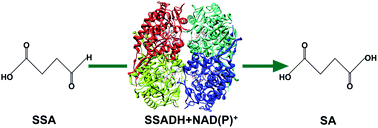
Succinic semialdehyde dehydrogenase (SSADH) belongs to the aldehyde dehydrogenase (ALDH) superfamily, which oxidizes succinic semialdehyde (SSA) to succinate (SA) in the final step of the degradation of the inhibitory neurotransmitter γ-aminobutyric acid (GABA). In this article, the catalytic mechanism of SSADH has been studied using a combined quantum mechanics and molecular mechanics (QM/MM) approach on the basis of the crystal structures of SSADH from Synechococcus sp. PCC 7002 (SySSADH) and Salmonella typhimurium (StSSADH). Our calculations reveal that, for SySSADH, the acylation process of substrate SSA is relatively difficult owing to the fact that the catalytic cysteine residue has already formed an adduct with the cofactor (NADP+), which corresponds to an overall energy barrier of 18.2 kcal mol−1. However for StSSADH, the cysteine residue exists as the thiolate ion and the acylation process is easily occurs, corresponding to an overall energy barrier of 9.6 kcal mol−1. In the subsequent deacylation process, using SySSADH to construct the computational model, the activation of the hydrolytic water molecule is concerted with the formation of a thioester intermediate, which is the rate-limiting step for the deacylation process, corresponding to an energy barrier of 18.2 kcal mol−1. Thus, for SySSADH, both the acylation and deacylation are possible rate-limiting steps. The pocket residues such as S261, C262 and S419/S425 play an important role in stabilizing the substrate and involved intermediates. Our calculation results may provide useful information for further understanding the catalytic mechanism of SSADH.
To date, several crystal structures of SSADHs from different species, such as human (HsSSADH), Escherichia coli (EcSSADH), Salmonella typhimurium (StSSADH) and Synechococcus sp. PCC 7002 (SySSADH), have been solved.17–22 The crystal structures of HsSSADH and EcSSADH, both in oxidized and reduced conditions, reveal that the reactivity of SSADHs is regulated by redox-switch modulation owing to the existence of two cysteine residues in the catalytic loop, i.e., these SSADHs are active in the reduced condition and inactive in the oxidized condition.17–19 Moreover, when the environment switches from oxidized condition to reduced one, their activity will be recovered. Under oxidized condition, a disulfide bond is formed between the two cysteine residues, leading to a closed conformation of the catalytic loop, which blocks the access of the substrate and cofactor into the binding site.17–19 Reduction of the disulfide bond causes a large conformational change of the binding site, by which the catalytic loop switches to an open conformation, permitting the access of the substrate and cofactor into the binding sites.17–19 But for SSADHs with only one cysteine residue in the catalytic loop, such as StSSADH and SySSADH, the redox-dependent regulatory mechanism is unavailable due to the lack of a second cysteine.20–22 Jinseo Park et al. suggested that SySSADH adopts the cofactor-dependent oxidation protection mechanism because the catalytic cysteine forms a cysteine–NADP adduct with the cofactor NADP+, which protects the catalytic cysteine from H2O2-dependent oxidative stress.20 However, the functional role of the covalent adduct was generally considered to recruit the cofactor in the vicinity of the catalytic nucleophile for catalysis, particularly for hydride transfer from the substrate to NAD(P)+. In addition, the crystal structure of StSSADH does not show the formation of a cysteine–NAD(P) adduct. Instead, the catalytic cysteine exists in a free form.21 So far, the role of the cysteine–cofactor adduct and the regulatory mechanism of 1-cysteine SSADHs still remain unclear.
As has been suggested for the ALDH superfamily, the catalytic mechanism of SSADH involves two processes, acylation and deacylation, as shown in Scheme 2.23–28 In the acylation process, the deprotonated cysteine residue attacks on the carbonyl carbon of SSA to form a thiohemiacetal intermediate. Then the hydride ion of the tetrahedric intermediate transfers to the C4 atom of the nicotinamide ring of NAD(P)+, generating the thioester intermediate. In the following deacylation process, the hydrolytic water molecule is activated by a nearby glutamate residue and the generated hydroxyl attacks the carbonyl carbon of SSA to form the second tetrahedral intermediate. Then the S–C bond of the intermediate cleaves to produce the final product SA and regenerate the cysteine side chain. Experimental data and crystal structures have provided meaningful information for understanding the catalytic mechanism.17–30 For example, it has been found that SSADHs from various species have different cofactor specificity (NADP+ or NAD+) and some special residues in the cofactor-binding site play important roles in the selective recognition of cofactor.21,29,31 In addition, the catalytic cysteine residue must be firstly deprotonated by a glutamate residue before its nucleophilic attack on the substrate, which is a required step for acylation process.23 The generated thiolate ion is supposed to be stabilized by the positively charged nicotinamide ring of the cofactor NAD(P)+ and/or adjacent main chain amide groups.21 Alternatively, the thiolate ion forms a covalent linkage with the C4 of the nicotinamide ring of NAD(P)+ to form a cysteine–NAD(P) adduct.20,32 Crystal structures and NMR experiments have shown multiple conformations of the nicotinamide portion of the NAD(P)+ and the catalytic glutamate residue.20–23,31–33 As shown in Fig. S1 of ESI,† after NAD(P)+ enters the active site, its nicotinamide occupies the hydride transfer channel, causing the side chain of the catalytic glutamate residue to depart away from the active site and block its contact with the catalytic cysteine residue. When NAD(P)+ is reduced to NAD(P)H, the nicotinamide moiety is away from the active site, allowing the catalytic glutamate residue to rotate back and restore its contact with the cysteine residue. In this conformation, the catalytic glutamate residue locates in a favorable position for activating the water molecule to hydrolyze the thioester intermediate. This conjugate movement of the catalytic glutamate residue and nicotinamide moiety of NAD(P)+ is common among the ALDH superfamily.31–33
Despite this wealth of knowledge, several important aspects still remain undetermined. For instance, the deprotonated catalytic cysteine residue may exist as thiolate ion form, such as in StSSADH, or form a cysteine–NAD(P) adduct with the cofactor, such as in SySSADH.20–23 In addition, it is no idea how the nucleophilic attack will take place when the deprotonated cysteine residue forms an adduct with NAD(P)+. Jinseo Park et al. have suggested that the cysteine–NAD(P) adduct may readily dissociate in the presence of the substrate SSA, but no detailed description was provided.20 Furthermore, it is still unknown the existing form of the carbonyl oxygen in the thiohemiacetal intermediate, although the oxyanion form has been proposed in the reaction scheme of other ALDH enzymes.31 However, another suggestion that the carbonyl oxygen exists in the hydroxyl form was also proposed, such as in rat ALDH3 and human mitochondrial ALDH2.31,32,34 So far, there is still no detailed description at the atomic level on the catalytic reaction of SSADH and other ALDH enzymes. To gain insight into the catalytic mechanism of SSADH, a hybrid quantum mechanics and molecular mechanics (QM/MM) calculations has been performed on the basis of crystal structures of SySSADH and StSSADH. The key point of QM/MM method is to divide the entire system into two portions, the QM region and the MM region. The QM region which involves the formation and cleavage of chemical bonds is described quantum mechanically while the MM region which represents the surrounding protein is treated by a MM force field. In this scheme, both the chemical reactions occurred in active site and the effect of surrounding enzymatic residues and solvent environment can be considered.35–38 This method has been extensively applied in studying enzymatic reaction system and has been testified to be successful in recent years.39–43 Based on our QM/MM calculations, the detailed reaction pathway, the roles of pocket residues involved in stabilizing the reaction intermediates and transition states, as well as the energetics of the whole catalytic cycle have been illuminated.
![]() | ||
Fig. 1 The crystal structure (a) and active site (b) of SySSADH in complex with NADP+ (PDB code: 4ITA). |
![]() | ||
Fig. 2 The crystal structure (a) and active site (b) of StSSADH in complex with NAD+ (PDB code: 3EFV). |
Since the present study aims to elucidate the reaction pathway of SSADH, the calculations should start from the enzyme–cofactor–substrate ternary complex. To acquire the enzyme in complex with the cofactor and substrate, the substrate (SSA) was preliminarily optimized with the Gaussian 03 package at the level of B3LYP/6-31G(d,p),44 and then was docked into the active site using the Autodock 4.0 program.45 The representative structure from docking was chosen for the following molecular dynamics (MD) simulations, which were performed with CHARMM22/CMAP all-atom force field implemented in the CHARMM program.46 The protonation states of all titrable residues were determined on the basis of the experimental condition and the pKa values were predicted by the empirical PROPKA 3.1 program,47 and then verified by the VMD program.48 The catalytic cysteine residue is assumed to be the initial nucleophile, it was therefore set to its deprotonated state. The missing hydrogen atoms were added using the HBUILD facility in the CHARMM package.49 The whole system was solvated into a water sphere centered on the S atom of the catalytic cysteine residue, while the crystallographic water molecules were kept at their original positions. The solvent was relaxed by a short MD simulation while all the other atoms were fixed. All water molecules were treated as TIP3P residues.50 In addition, some Na+ ions were added randomly to neutralize the system. Preliminary geometrical optimization was performed to remove the bad contacts between atoms and to relax the system. Subsequently, 10 ns MD simulations were carried out to equilibrate the system with stochastic boundary conditions at 298 K and 1 atm. During the MD simulation, the system was divided into two regions: an inner reaction region where the simulation was performed by Newton's equations of motion, and an outer buffer region where the atoms were described by Langevin dynamics with friction and random force. The system can be kept at thermal equilibrium by using this hybrid method which couples the water molecules in the buffer region to a heat bath. The root-mean-squared deviations (RMSDs) of the two proteins were derived from the MD simulation, which are shown in Fig. S2 and S3 of ESI.† The dynamics trajectories were basically stable after 6 ns with the RMSD values of 1.6 Å and 2.5 Å, indicating that the backbones of the two proteins only changed slightly during the MD simulations.
When NAD(P)+ is reduced to NAD(P)H, the nicotinamide ring moves away from the active site, allowing the catalytic glutamate residue to rotate back and restore its contact with the catalytic cysteine residue. This conformation (SSADH in complex with NAD(P)H) is different from that of SSADH in complex with NAD(P)+, therefore, the crystal structure of SySSADH in complex with NADPH and SSA (PDB ID: 4ITB) was used to construct the computational model for studying the deacylation process.20 The crystal structure and active site are shown in Fig. 3. One can see from Fig. 3b that the catalytic cysteine residue (C262) forms a covalent linkage with the substrate SSA, generating a thiohemiacetal intermediate. This intermediate was manually changed to a thioester intermediate, which acts as the starting structure for studying the deacylation process. Then, 10 ns MD simulation was performed to equilibrate the system, and the RMSD of the protein during the MD simulation is shown in Fig. S4 of ESI.† The dynamics trajectory was stable after 6 ns with a RMSD value of 1.8 Å. The final snapshot was used for the following QM/MM calculations.
![]() | ||
Fig. 3 The crystal structure (a) and active pocket (b) of SySSADH in complex with NADPH and SSA (PDB code: 4ITB). |
![]() | ||
Fig. 4 (a) The QM/MM optimized active site structure of SySADH in complex with NADP+ (PDB code: 4ITA); (b) the superposition of the optimized structure with that of the crystal structure. The key bond distances are shown in angstrom. |
On the basis of our calculations, the acylation process contains two elementary steps: the nucleophilic attack, which is coupled with the cleavage of the covalent linkage between the thiolate ion and C4 atom of the nicotinamide ring of NADP+, and the hydride transfer from SSA to C4. The optimized structures and key parameters of the reactant (RA), transition states (TS1A and TS2A) and intermediates (IM1A and IM2A) are shown in Fig. 5. Firstly, the catalytic cysteine residue (C262) conducts a nucleophilic attack on the carbonyl carbon of SSA to form a thiohemiacetal intermediate (IM1A). In this elementary step, the distance between the thiolate ion and the carbonyl carbon of SSA decreases from 3.69 Å in RA to 2.50 Å in TS1A, and finally to 2.17 Å in IM1A. In the meantime, the covalent linkage between the thiolate ion and C4 atom of the nicotinamide ring of NADP+ cleaves, during which the length of C4–S changes from 1.95 Å in RA to 3.85 Å in TS1A. The distance of 3.85 Å means the C4–S covalent linkage has already broken in TS1A. Therefore, we can conclude that, although the nucleophilic attack is concerted with the cleavage of the covalent linkage, they are highly unsynchronized. The cleavage of C4–S bond is much earlier than the nucleophilic attack. To further explore the possibility of stepwise pathway, we also scanned the reaction coordinate defined by RC = R(SC262–C4NADP+) for the cleavage of the covalent linkage. Along the reaction coordinate, the calculated energies were continuously increased, and no saddle points were found (as shown in Fig. S5†), which implies that the single cleavage of the covalent linkage corresponds to very high energy barrier.
![]() | ||
Fig. 5 Optimized geometries of various species for the acylation process when cysteine–NADP adduct acts as initial nucleophile. The key bond distances are shown in angstrom. |
Fig. 5 also shows that the carbonyl oxygen atom of the substrate SSA in IM1A exists in the oxyanion form, which forms two hydrogen bonds with the main chain amide of C262 and the side chain of S261, with lengths of 1.71 and 1.84 Å, respectively. This configuration is often called the “oxyanion hole” which has the function to stabilize the negative charge of the oxyanion. However, some studies suggested the proton of the main chain amide of the catalytic cysteine may transfer to the oxygen anion of the intermediate to form a hydroxyl.31,32,34 Therefore, to explore the proton transfer process, we also scanned the reaction coordinates defined by RC = R(Othiohemiacetal–HC262) from the amide of C262 and RC = R(Othiohemiacetal–HS261) from the side chain hydroxyl of S261 to the oxygen anion of the intermediate. As shown in Fig. S6 and S7,† along the reaction coordinates, the calculated energies were continuously increased and no saddle points were recognized, which indicate that the proton transfer is energetically unfavorable and the thiohemiacetal intermediate exists in its oxyanion form. Furthermore, the S–C bond length in the thiohemiacetal intermediate is 2.17 Å, which is much longer than the typical S–C single bond and can be classified as a coordinate covalent bond. This case was also found in the previous quantum mechanical calculations.32,34
In the following step, the carbonyl hydride of SSA transfers to the C4 atom of the nicotinamide ring of NADP+, causing the collapse of the thiohemiacetal to a thioester intermediate and the formation of NADPH. The length of the C–H bond changes from 2.44 Å in IM1A to 1.10 Å in IM2A via 1.46 Å in TS2A. The sp3 hybridized carbon atom of SSA returns back to sp2 hybridization, and the hybridization of C4 atom changes from sp2 to sp3. Meanwhile, the S–C length changes to the typical S–C single bond of 1.81 Å.
The energy profile of the acylation process is shown in Fig. 6. One can see that the nucleophilic attack of C262 is calculated to be endothermic by 11.4 kcal mol−1 with an energy barrier of 13.6 kcal mol−1. The following hydride transfer process corresponds to a low energy barrier of 6.8 kcal mol−1. However, the overall energy barrier of the acylation process is 18.2 kcal mol−1, to which the nucleophilic attack of C262 contributes a lot. In general, the acylation process is calculated to be exothermic by 3.7 kcal mol−1. Therefore, as had been suggested by Jinseo Park et al.20 the cysteine–NAD(P) adduct can dissociate in the reaction pathway, but it is concerted with the nucleophilic attack of the cysteine residue on the carbonyl carbon of SSA.
![]() | ||
Fig. 7 (a) The QM/MM optimized structure taken from the active site when the crystal structure of StSSADH in complex with NAD+ (PDB code: 3EFV) was chosen as initial model to study the acylation process; (b) the superposition of the active site taken from the QM/MM optimized structure with that of the crystal structure. The key bond distances are shown in angstrom. |
Fig. 7a shows that the catalytic thiolate anion is stabilized by the positively charged nicotinamide ring of the NAD+. In addition, two hydrogen bonds are formed between C268 and the adjacent main chain amide groups of A269 and C268, with lengths of 2.35 and 2.40 Å, respectively. The carbonyl oxygen of SSA forms one hydrogen bond with the water molecule with length of 1.96 Å. The carboxyl group of SSA forms three hydrogen bonds with the side chains of S425, K92 and a water molecule, with lengths of 1.67, 1.90 and 1.70 Å, respectively. The catalytic glutamate residue (E234) forms two hydrogen bonds with NAD+ and residue R145, with lengths of 1.75 and 1.62 Å, respectively. These hydrogen bonds form a large hydrogen bonding network in the active site, which stabilize the substrate and cofactor. Mutagenesis experiments have also provided some evidences about the roles of these active pocket residues.21 The E234Q and C268A mutant proteins were basally inactive, which suggests that these residues are likely to play catalytic roles as in other SSADH enzymes.21 Alanine replacement of other conserved residues in the active site, such as S425A, K92A and Q142A, produced mutant proteins with reduced activity.21
The optimized structures of the reactant (RB), transition states (TS1B and TS2B) and intermediates (IM1B and IM2B) are shown in Fig. 8. One can see that the catalytic cysteine residue (C268) in RB exists in the thiolate ion form, which is different from that of RA in Fig. 5. To explore whether the catalytic C268 can form a cysteine–NAD adduct with the cofactor NAD+, we scanned the reaction coordinate defined by RC = R(SC268–C4NAD+) for this covalent linkage, and then optimized the structure of the cysteine–NAD adduct and performed high-level single point energy calculations. The relative energies of the system were almost unchanged along the reaction coordinate, no matter the catalytic cysteine residue exists in the thiolate ion form or forms cysteine–NAD adduct with the cofactor NAD+, as shown in Fig. S8.† Therefore, the two states of catalytic C268 can easily transform each other. In the subsequent QM/MM calculations, the catalytic C268 thiolate ion was chosen as the initial reactant (RB). As shown in Fig. 8, the carbonyl carbon of SSA is 3.45 Å away from the thiolate ion, which changes to 2.27 Å in TS1B. In IM1B, this distance further shortens to 2.10 Å, indicating the complete formation of the thiohemiacetal intermediate. In addition, the oxygen atom in the thiohemiacetal intermediate also exists in an oxyanion form and forms two hydrogen bonds with the main chain amide of C268 and a water molecule, with lengths of 1.71 and 1.69 Å (not shown in Fig. 8), respectively. In the subsequent step, the carbonyl hydride is transferred from SSA to the C4 atom of the nicotinamide ring of NAD+, generating a thioester intermediate and NADH. From IM1B to IM2B, the distance between the carbonyl hydride of SSA and C4 atom of the nicotinamide ring of NAD+ decreases from 2.79 to 1.10 Å via 1.36 Å, indicating the formation of a thioester intermediate and NADH.
![]() | ||
Fig. 8 Optimized geometries of various species for the acylation process when cysteine thiolate ion acts as initial nucleophile. The key bond distances are shown in angstrom. |
The energy profile of the acylation process is shown in Fig. 9. It can be seen that the nucleophilic attack of C268 can proceed easily with an energy barrier of 2.0 kcal mol−1, which is much lower than that of the nucleophilic attack of the cysteine–NADP adduct (2.0 vs. 13.6 kcal mol−1). This difference is reasonable considering the fact that the nucleophilic attack of the cysteine–NADP adduct is concerted with the cleavage of the covalent bond between the cysteine residue and NADP+. The following hydride transfer process corresponds to an energy barrier of 11.5 kcal mol−1. In general, the acylation process when cysteine thiolate ion acts as initial nucleophile is calculated to be exothermic by 5.9 kcal mol−1.
In the crystal structure of SySSADH in complex with NADPH and SSA, the catalytic cysteine residue forms a thiohemiacetal with SSA.20 Therefore, to construct the computational model, the thiohemiacetal was changed to a thioester, and a hydrolytic water molecule was manually added from the bulk solvent to the catalytic site. Then, a serial of MD simulations were performed to equilibrate the system. In the subsequent QM/MM calculations, the QM subsystem contains 153 atoms, including the side chains of F132, S419, W135, F425, R139, Q136, E228, N131, S261 and C262, the main chain of C262, the cofactor NADPH, and the substrate SSA which is covalently bonded to C262. All other atoms of the system were set as MM subsystem. The QM/MM optimized active site structure is shown in Fig. 10a, and its superposition with that of the crystal structure is shown in Fig. 10b. Some slight deviations can be found in the two structures, which can be explained by the fact that the thioester in the QM/MM optimized structure was changed from the thiohemiacetal in crystal structure and the water molecule was added. From Fig. 10a we can see that the nicotinamide moiety of NADPH moves away from the active site, and the catalytic glutamate residue rotates back. The catalytic cysteine residue (C262) forms a thioester with the substrate SSA, which acts as the initial structure of the deacylation process. The carbonyl oxygen of the substrate SSA forms one hydrogen bond with the main chain amide of C262 with length of 1.81 Å. The carboxyl group of the substrate SSA forms two hydrogen bonds with residues S419 and W135, with lengths of 1.57 and 2.30 Å, respectively. In addition, the hydrolytic water molecule is fixed by two hydrogen bonds (Fig. 10a).
![]() | ||
Fig. 10 (a) The QM/MM optimized structure taken from the active site when the crystal structure of SySSADH in complex with NADPH and SSA (PDB code: 4ITB) was chosen as initial model to study the deacylation process; (b) the superposition of the active site taken from the QM/MM optimized structure with that taken from the crystal structure. The key bond distances are shown in angstrom. |
On the basis of our calculations, the deacylation process contains two elementary steps: the nucleophilic attack of the activated water molecule on the thioester intermediate, and the cleavage of the S–C bond to produce the product SA. The optimized structures and key parameters of the intermediates (IM3A and IM4A), transition states (TS3A and TS4A) and product (PA) are shown in Fig. 11. Firstly, the water molecule is activated by the catalytic glutamate residue, and simultaneously the generated hydroxyl conducts a nucleophilic attack on the thioester intermediate. This concerted process corresponds to an energy barrier of 18.2 kcal mol−1 (Fig. 6). The formed intermediate (IM4A) is 14.2 kcal mol−1 higher than IM3A in energy, and the following S–C bond cleavage corresponds to a very low energy barrier (1.1 kcal mol−1). From IM4A to PA, the S–C bond length increases from 2.09 Å to 3.07 Å via 2.47 Å in TS4A. The calculated energy of PA is 8.3 kcal mol−1 higher than that of IM3A, indicating that the overall deacylation process is endothermic.
![]() | ||
Fig. 11 Optimized geometries of various species for the deacylation process. The key bond distances are shown in angstrom. |
Footnote |
† Electronic supplementary information (ESI) available. See DOI: 10.1039/c5ra21535h |
This journal is © The Royal Society of Chemistry 2015 |