J. S. Boateng*,
H. V. Pawar and
J. Tetteh
Department of Pharmaceutical, Chemical and Environmental Sciences, Faculty of Engineering and Science, University of Greenwich at Medway, Chatham Maritime, Kent, UK ME4 4TB. E-mail: j.s.boateng@gre.ac.UK; joshboat40@gmail.com; Tel: +44 (0) 208 331 8980
First published on 7th December 2015
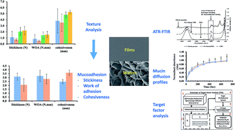
The adhesive properties of two dressing types, solvent cast films and freeze-dried wafers have been determined and compared using two analytical techniques, combined with chemometrics data analysis. Films and wafers were prepared from gels containing polyox (POL) combined with carrageenan (CAR) or sodium alginate (SA), glycerol (GLY) as plasticiser (films) with streptomycin and diclofenac as model drugs. The gels were dried in an oven at 40 °C or freeze-dried to obtain films and wafers respectively. The adhesive performance of the films and wafers was assessed with 6.67% w/v gel using a texture analyser to measure the stickiness, work of adhesion and cohesiveness. The effect of viscosity of simulated wound fluid [containing (2% w/w or 5% w/w bovine serum albumin)] and mucin solution (2% w/w) present on the gelatin surface on texture analyser profiles was investigated. Furthermore, the adhesive properties were estimated and evaluated using attenuated total reflectance Fourier transform infrared spectroscopy by monitoring the diffusion of mucin solution [2% w/w in phosphate buffered saline (PBS) pH 7.4] through the formulations. The diffusion data was analysed using target factor analysis (chemometrics approach) to establish proof of concept for predicting adhesion by measuring mucin interaction and its diffusion through films and wafers. There was a significant effect of simulated wound fluid, viscosity, plasticizer (for films) and drug loading on the adhesive performance of both films and wafers. POL-SA films showed higher mucoadhesive performance in the presence of viscous simulated wound fluid containing 5% bovine serum albumin. Wafers and plasticised films demonstrated high detachment force indicating strong interactions between the chains of the polymers (POL, SA and CAR) and the model wound surface (gelatin). ATR-FTIR spectroscopy showed that mucin diffused independently through the solvent and across the films and wafers. POL-CAR films generally showed slower diffusion of mucin when compared with POL-SA films whilst the opposite effect was observed for diffusion through POL-CAR wafers and POL-SA wafers. Generally, diffusion through wafers was faster than the corresponding films.
In the case of wounds, prolonged residence time of the dressing is an essential functionality since frequent dressing changes, which also causes pain, is a major source of patient non-compliance, and can result in complications and delays in wound healing.8,9
It has been suggested that the interaction between a mucosal (moist) surface and mucoadhesive polymers is a result of physical entanglement and secondary interactive forces, mainly due to hydrogen bonding and van der Waals attraction forces which depend upon the chemical structure of the polymer.10 Peppas and Buri proposed certain characteristics which are necessary for effective mucoadhesion. These include polymers containing strong H-bonding groups, strong anionic charges, high molecular weight, sufficient chain flexibility, and surface energy properties which favour spreading of the polymer onto the mucosal surface.11 For the purpose of wound dressings, adhesive properties of the polymers could be affected by degree of hydration, amount of exudate released from the wound, exudate viscosity, salts and proteins present in exudate, presence of microorganisms as well as the depth and area of the wound.12
There are different approaches used to evaluate the adhesive performance of polymers and polymeric dosage forms. These include texture analyser,9,10,13–15 rheometric measurements16 and attenuated total reflection-Fourier transform infrared (ATR-FTIR) spectroscopy.2,17 Recently, texture analyser has been used for studying properties of mucoadhesive polymers and dosage forms using tensile mode of testing. These in vitro experiments involve attaching the dosage form to a probe and force applied to bring the sample in contact with a representative mucosal substrate for a specific time (contact time) and a mechanical force applied to detach the probe from the mucosal substrate.3,18 The adhesive strength (stickiness) is evaluated by the force (Fmax) required to detach the sample from the model mucosal substrate after mucoadhesive bonding has been established.19 Total work of adhesion represents the total amount of energy involved in the withdrawal of the probe from the mucosal surface and determined by the area under the force versus distance curve. Cohesiveness determines the ability of the sample to resist the separation from the mucosal (wound) substrate due to the intermolecular forces (such as those from hydrogen bonding and van der Waals forces). It is determined through the distance travelled by sample before being detached.10,20–22
Mucoadhesion by means of a texture analyser usually involves the measurement of mechanical force required to fracture the interface between a substrate or mucin and polymer and therefore depends largely upon the fracture theory of mucoadhesion.17 Saiano and co-workers2 reported that variations in the experimental parameters such as contact time, contact force, test speed and rate of removal from the adhesive test surface consequently results in variations in experimental muco-(bio)-adhesive results. Such variations make it very difficult to compare data from different investigators to assign exact values representing bioadhesive/mucoadhesive performance.2 Further, Jabbari and co-workers reported that though mucoadhesion studies using a texture analyser is advantageous for classification of mucoadhesive polymers, it is not an accurate technique for determining mechanisms of adhesion at the biointerface.17 As a result, alternative spectroscopic analysis techniques have been implemented and adopted to investigate the interaction between substrates and the polymer matrix to help evaluate mucoadhesion mechanisms. In particular, ATR-FTIR has been applied effectively to study the interpenetration and entanglement of polymer chains and mucous which is fundamentally based on the diffusion theory of mucoadhesion.2,17 ATR-FTIR can also be used to study mucoadhesion properties and diffusion profiles of solvent through different membrane surfaces such as biological tissues, films and silicon membranes2,23 since it can provide real time information of diffusion of materials such as mucin through the membranes.
However, measurement of kinetic diffusion of mucin solution across the polymer based on FTIR spectroscopy generally produces large quantities of often complex multivariate data sets that require appropriate chemometric analysis. The employment of chemometric techniques generally involves calibration, validation and extraction of maximum chemical information from the analytical data and its usefulness has previously been reviewed.24 Among the chemometric techniques used to resolve complex spectral data, factor analysis or principal component analysis25,26 based on singular value decomposition are the most common. Factor analysis is a multivariate technique for reduction of data matrices into its lowest dimensionality by the use of orthogonal factor space and transformation that yields predictions and/or recognisable factors which influence the data matrix.
In this paper, we report on the in vitro wound adhesion properties of two different dressing formulations (films and wafers) both comprising two different polymers (composite) and two drugs, using texture analysis and ATR-FTIR spectroscopy. Initially, the mucoadhesive performance of the films and wafers was assessed using a texture analyser to measure the stickiness, work of adhesion and cohesiveness as well as the effect of viscosity of simulated wound fluid [(2% w/w or 5% w/w bovine serum albumin), 2% w/w mucin solution, drugs (streptomycin and diclofenac) and glycerol (for films) on the adhesive properties of the composite films and wafers]. The adhesion properties of the formulations have been further evaluated using ATR-FTIR spectroscopy to measure and compare the diffusion of mucin through the two formulations to establish proof of concept for measuring mucin interaction with the films and wafers as indication of their adhesive performance. Here the bovine serum albumin and mucin were used as model proteins to simulate wound exudate and moist wound surfaces respectively.
The various formulations (films and wafers) prepared are summarised in Table 1.
Number | Formulation |
---|---|
1 | POL-CAR-BLK-film |
2 | POL-CAR-DL-film |
3 | POL-CAR-BLK-20%GLY-film |
4 | POL-CAR-DL-20%GLY-film |
5 | POL-SA-BLK-film |
6 | POL-SA-DL-film |
7 | POL-SA-BLK-9%GLY-film |
8 | POL-SA-DL-9%GLY-film |
9 | POL-SA-BLK-wafer |
10 | POL-CAR-BLK-wafer |
11 | POL-SA-DL-wafer |
12 | POL-CAR-DL-wafer |
Simulated wound fluid containing 2% w/w and 5% w/w bovine serum albumin represented thin and viscous exudate respectively. In addition, a separate gelatin gel was also equilibrated with 2% w/w solution of mucin (model protein) in PBS of pH 7.4 for direct comparison with the ATR-FTIR mucin diffusion experiment (described below). The probe, lined with film or wafer was set to approach the model wound surface with the following pre-set conditions: pre-test speed 0.5 mm s−1; test speed 0.5 mm s−1; post-test speed 1.0 mm s−1; applied force 1 N; contact time 60.0 s; trigger type auto; trigger force 0.05 N and return distance of 10.0 mm. The adhesive strength (stickiness), total work of adhesion and cohesiveness were calculated using the Texture Exponent 32® software.
![]() | ||
Fig. 1 ATR assembly for in situ measurement of inter-diffusion of mucin solution through the films and wafers. This experimental set up is similar to that published previously.2 |
Target factor analysis was used to deconvolute the spectral profiles of the 2% w/w mucin solution together with those of the film and wafer formulations in the wavenumber range 1400–1700 cm−1. The 1400–1700 cm−1 range was used because mucin has the amino group which can interact with polymers through hydrogen bond formation and used to follow the diffusion of mucin.30 The relative rate of diffusion was then deduced from the data generated after subjecting to target factor analysis using InSight® software (InSight 2009, DiKnow Ltd, Rochester, UK) (see Fig. S2 and S3 in the ESI† data section).
Fig. 2 shows the stickiness, work of adhesion and cohesiveness of POL-CAR and POL-SA films upon being detached from the model wound surface (gelatine) equilibrated with thin exudate. POL-SA-BLK films showed higher stickiness (1.9 ± 0.4 N) and work of adhesion (1.1 ± 0.7 N mm) values compared to those for POL-CAR-BLK films at (0.6 ± 0.1 N) and (0.4 ± 0.1 N mm) respectively. The observed cohesiveness for both films was similar (POL-CAR-BLK 1.2 ± 0.1 mm, POL-SA-BLK 1.3 ± 0.2 mm). In the presence of viscous exudate, POL-CAR-BLK films showed higher values of stickiness (2.5 ± 0.7 N) and work of adhesion (1.5 ± 0.5 N mm) which were decreased for POL-SA-BLK films (1.7 ± 0.7 N and 1.1 ± 0.5 N mm respectively) (Fig. 3). This may be associated with the fact that the increased viscosity due to increased concentration of bovine serum albumin results in the formation of a gel like structure and helps more intimate contact with the substrate and therefore requires a stronger force to detach the POL-CAR-BLK films. In the presence of viscous exudate CAR gets hydrated to form a gel and is responsible for increased mucoadhesive performance. The increased intra-molecular attraction of the viscous exudate and the formation of internal cross-linkages on the gelatin surface might limit solvent diffusion into the polymeric matrix resulting in decreased mucoadhesion in the case of POL-SA-BLK film.
Various factors can influence mucoadhesive performance of formulations including structural constituents of polymers which affect degree of solvent diffusion and polymer chain entanglement. Roy and co-authors reported that hydrophilic polymers such as CAR and SA with anionic charges, exhibited high adhesion performance.35 This is because such charged groups have an impact on the degree of hydration of the polymer when in contact with the mucosal surface.36 In addition, polymer hydration and swelling polymer enhance the inter-diffusion process, allowing physical entanglement and increased surface availability for hydrogen bonding and electrostatic interaction between the polymer and the mucous network.36 This might be the reason for the variation in the mucoadhesive performance of films comprising two different polymers blended together in a single formulation.
The plasticised POL-CAR films (both blank and drug loaded) showed less detachment forces (stickiness). The presence of glycerol could possibly have interacted with CAR and POL and an increase in number and strength of hydrogen bonds between POL, CAR and glycerol could result in the formation of a strong network structure that resists the rapid penetration of water which might result in decreased mucoadhesive performance in the presence of thin exudate. In the presence of viscous simulated wound fluid (5% bovine serum albumin) the stickiness of blank [POL-CAR-20%GLY (2.1 ± 0.5 N)] was increased and [POL-SA-9%GLY (1.7 ± 0.8 N)] was decreased whilst work of adhesion of BLK [POL-CAR-20%GLY (2.1 ± 0.9 N) and POL-SA-9%GLY (1.1 ± 0.5 N)] and cohesiveness [POL-CAR (3.4 ± 0.4 N) and POL-SA (2.8 ± 1.0 N)] was increased. This may be due to the fact that in the presence of glycerol, simulated wound fluid in higher concentration behaves as a slippery mucilage which results in reduced net mucoadhesive performance.
The decrease in the work of adhesion in the presence of added drug can be attributed to two main reasons. Firstly, ionic interactions occurred between the anionic polymers (CAR and SA) and cationic streptomycin which had an effect on the hydrogen bonding mechanism between the polymers and simulated wound fluid which contains salts and proteins.
Secondly, Tobyn and co-workers reported that increased ionic strength of the media and the presence of sodium and potassium ions resulted in decreased adhesion.37 Further, the sodium sulphate formed during gel preparation and ultimately present in the films further increased the ionic strength of the simulated wound fluid resulting in decreased adhesion. In the case of the films, the presence of sodium sulphate may have interfered with the physical properties of the delivery matrix and reduced the extent of adhesion to the gelatin substrate equilibrated with simulated wound fluid.
Usually thin watery serous type exudate in a wound signifies possible bacterial infection, such as Staphylococcus aureus which produce staphylokinase, a known fibrinolytic, and degrades fibrin clots resulting in thin watery exudate.38,39 The drug loaded POL-CAR and POL-SA wafers can help to manage such exudate due to their porous nature. Haemorrhagic and haemopurulent (viscous, sticky and thick) exudate signifies both infection and trauma and POL-CAR wafers can provide prolonged retention of wafers at the site of injury.
Overall, mucoadhesion results from wafers (both blank and drug loaded) confirmed that the porosity plays a critical role due to the ability to absorb simulated wound fluid and hydration of the polymeric network (POL, SA and CAR). The decreased stickiness in the drug loaded wafers was associated with the decreased porosity of these wafers due to the added drugs and subsequent salt (sodium sulphate) formation which inhibit rapid hydration of the wafers. From the results obtained it can be concluded that the wafers generally possessed good adhesive strength with the wound substrate containing two different types of exudate compared to the films. Therefore these wafers can adhere to the wound site and protect the wound from the external environment with the absorption of large amounts of exudate, whilst maintaining their structure, which is a primary requirement for a formulation to function as an ideal wound dressing.
Overall, it appears that the POL-SA films and wafers can be used in the presence of normal exudate where concentration of protein is less to achieve prolonged retention time and bioavailability. POL-CAR films and wafers (POL-CAR and POL-SA) on the other hand can be used for wounds which produce viscous exudate to achieve better adhesive performance.
![]() | ||
Fig. 4 Mucoadhesion performance of films and wafers (POL-CAR and POL-SA) using texture analyser in the presence of 2% mucin solution showing stickiness, WOA and cohesiveness profiles. |
Addition of glycerol had a marked effect on the mucoadhesion performance of the films. Stickiness for the plasticised POL-CAR based films ranged from 2.05 to 2.21 N and from 1.91 to 2.37 N for plasticised POL-SA based films. Overall, the adhesiveness for POL-SA based films was found in the descending order of POL-SA-BLK-9%GLY > POL-SA-DL > POL-SA-DL-9%GLY > POL-SA-BLK. The adhesiveness for POL-CAR films was found in the decreasing order of POL-CAR-BLK-20%GLY > POL-CAR-DL-20%GLY > POL-CAR-BLK > POL-CAR-DL.
In ATR-FTIR spectroscopy, wavelength of the incident radiation affects the depth of penetration of IR radiation into the sample. Time dependent spectral data from ATR-FTIR enabled detailed characterisation of the films and wafers. There were however, some challenging factors related to ATR-FTIR set-up that required careful consideration when analysing diffusion of mucin through the films and wafers. These included quality of contact of the formulations with the ATR crystal across the focal plane of the detector. This is important because absence of proper contact with the ATR crystal will produce inaccurate spectral profiles and can affect the diffusion results. Poor contact can be a function of the smoothness, porous (wafers) and thin (films) nature of the formulations.23
McAuley reported that the wavelength influences the depth of penetration of IR radiation into the membrane.28 This could therefore affect the interpretation of relative diffusion rates of the mucin dissolved in PBS when different windows of IR spectrum are analysed for the films and wafers. For that purpose, the spectral window of 1400–1700 cm−1 was selected to evaluate the diffusion of mucin through the formulations, where the peak of mucin is most prominent. There could be other factors that can interfere with the diffusion of mucin (due to spectral overlap) which could be avoided by using chemometric data analysis by selecting a particular reference spectral window of mucin. Chemometrics involves the extraction of information from chemical systems by data-driven means using methods frequently employed in core data-analytic disciplines such as multivariate statistics, applied mathematics, and computer science, in order to address problems in chemistry, biochemistry, medicine, biology and chemical engineering. Multivariate curve resolution techniques are particularly very efficient in the analysis and modelling of such data sets. The basic principle of multivariate curve resolution is based on the fact that each component in the multivariate spectral data contributes additively and linearly to the absorbance at each spectral wavenumber.
In addition, the observed absorbance is directly proportional to the concentration based on the Beer Lambert law. In the current study, the multivariate curve resolution technique used was based on target factor analysis and the theoretical details are discussed elsewhere.44,45 The advantage of this approach is that models of real factors (here mucin peak at 1650 cm−1 is considered as a real factor which can form hydrogen bonds with polymeric films and wafers) can be systematically pieced together. This will ultimately give information about the mucoadhesion occurring between polymeric films and wafers. The profiles were normalised by setting the time frame of 780 s and a representative profile of three replicates are shown in Fig. 6. Such reproducibility measurements for the various samples was important given that the diffusion was measured over small distances as well as the combined effects of the many formulation variables (plasticiser, drugs, polymers, formed sodium sulphate and viscosity of simulated wound fluid) impacting on the mucoadhesive profiles being measured. The average of three profiles is shown on the same scale for comparison. As can be seen, though differences exist between the three replicates, the profiles were largely reproducible and shows the ability of the chemometrics approach combined with ATR-FTIR to measure the diffusion profiles of mucin through the films and wafers. In the figures plotted for the diffusion of mucin across the representative formulation, error bars were added for only positive values for clarity and comparison purposes as was the case in previous literature.2,17
Table 2 represents a summary of all the data from diffusion (relative intensity) of mucin across the various formulations used in the study. This data was used to study the effect of polymer, drug, and glycerol (only films) on the diffusion of mucin simultaneously monitoring the changes in the IR spectrum in the region of 1400–1700 cm−1. The observed diffusion coefficient of mucin through the formulations are presented in Table 3 below.
Formulations | Time (s) | |||||||
---|---|---|---|---|---|---|---|---|
50 | 100 | 200 | 300 | 400 | 500 | 600 | 700 | |
POL-CAR-BLK-20%GLY | 0.57 ± 0.05 | 0.68 ± 0.09 | 0.80 ± 0.20 | 0.84 ± 0.28 | 0.86 ± 0.39 | 0.89 ± 0.50 | 0.89 ± 0.59 | 0.93 ± 0.67 |
POL-CAR-BLK | 0.24 ± 0.05 | 0.28 ± 0.06 | 0.47 ± 0.07 | 0.75 ± 0.08 | 0.84 ± 0.12 | 0.94 ± 0.20 | 0.92 ± 0.28 | 0.94 ± 0.35 |
POL-CAR-DL | 0.05 ± 0.05 | 0.20 ± 0.07 | 0.39 ± 0.14 | 0.62 ± 0.24 | 0.72 ± 0.37 | 0.82 ± 0.49 | 0.91 ± 0.90 | 0.98 ± 1.03 |
POL-CAR-DL-20%GLY | 0.66 ± 0.14 | 0.28 ± 0.35 | 0.09 ± 0.55 | 0.18 ± 0.52 | 0.32 ± 0.65 | 0.42 ± 0.76 | 0.57 ± 0.84 | 0.75 ± 0.99 |
POL-CAR-DL-wafer | 0.34 ± 0.09 | 0.21 ± 0.17 | 0.16 ± 0.28 | 0.42 ± 0.31 | 0.68 ± 0.33 | 0.81 ± 0.35 | 0.72 ± 0.43 | 0.87 ± 0.51 |
POL-CAR-BLK-wafer | 0.40 ± 0.16 | 0.58 ± 0.25 | 0.62 ± 0.32 | 0.77 ± 0.33 | 0.76 ± 0.37 | 0.79 ± 0.42 | 0.86 ± 0.51 | 0.80 ± 0.64 |
POL-SA-BLK-9%GLY | 0.42 ± 0.02 | 0.62 ± 0.04 | 0.76 ± 0.05 | 0.87 ± 0.06 | 0.91 ± 0.08 | 0.93 ± 0.11 | 0.92 ± 0.14 | 0.94 ± 0.24 |
POL-SA-BLK | 0.37 ± 0.05 | 0.54 ± 0.07 | 0.77 ± 0.11 | 0.81 ± 0.14 | 0.91 ± 0.16 | 0.90 ± 0.19 | 0.93 ± 0.22 | 0.97 ± 0.30 |
POL-SA-DL-wafer | 0.05 ± 0.04 | 0.25 ± 0.07 | 0.65 ± 0.12 | 0.72 ± 0.15 | 0.86 ± 0.19 | 0.75 ± 0.23 | 0.80 ± 0.31 | 0.86 ± 0.43 |
POL-SA-DL-9%GLY | 0.36 ± 0.03 | 0.49 ± 0.04 | 0.71 ± 0.06 | 0.80 ± 0.08 | 0.87 ± 0.10 | 0.91 ± 0.11 | 0.92 ± 0.13 | 0.93 ± 0.16 |
POL-SA-DL | 0.35 ± 0.03 | 0.50 ± 0.04 | 0.70 ± 0.07 | 0.79 ± 0.09 | 0.88 ± 0.11 | 0.91 ± 0.13 | 0.95 ± 0.15 | 0.97 ± 0.19 |
POL-SA-BLK-wafer | 0.30 ± 0.10 | 0.50 ± 0.12 | 0.68 ± 0.17 | 0.64 ± 0.25 | 0.81 ± 0.32 | 0.70 ± 0.40 | 0.86 ± 0.46 | 0.82 ± 0.66 |
POL-CAR films/wafers | Diffusion coefficient “D” (cm2 s−1) | POL-SA films/wafers | Diffusion coefficient “D” (cm2 s−1) |
---|---|---|---|
POL-CAR-BLK | 4.0 × 10⁻³ | POL-SA-BLK | 6.2 × 10⁻³ |
POL-CAR-DL | 2.8 × 10⁻³ | POL-SA-DL | 6.5 × 10⁻³ |
POL-CAR-BLK-20%GLY | 9.7 × 10⁻³ | POL-SA-BLK-9%GLY | 7.4 × 10⁻³ |
POL-CAR-DL-20%GLY | 0.7 × 10⁻³ | POL-SA-DL-9%GLY | 6.9 × 10⁻³ |
POL-CAR-BLK-wafer | 7.8 × 10⁻³ | POL-SA-BLK-wafer | 6.1 × 10⁻³ |
POL-CAR-DL-wafer | 0.8 × 10⁻³ | POL-SA-DL-wafer | 0.8 × 10⁻³ |
Unplasticised POL-CAR-BLK films showed slower rate of diffusion of mucin through the films when compared to the plasticised films (Fig. 9). The initial absorbance for unplasticised POL-CAR-BLK film was 0.24 ± 0.05 which increased to 0.57 ± 0.05 in the first 50 s due to quicker diffusion of mucin solution through plasticised POL-CAR-BLK-20%GLY films. This may be due to the plasticizing effect of glycerol which has a higher affinity for water and increasing the mobility and elasticity of the films. Such a high water transfer inside the plasticised film matrix, has already been reported for glycerol plasticised CAR films.46 POL-CAR-BLK films showed a steady increase in relative concentration and this is supported by the swelling studies previously reported.8 Further, unplasticised POL-CAR-BLK films showed slower and steady diffusion of mucin which is due to the slow hydration of the POL and CAR to form a gel. After 400 s, both plasticised and unplasticised POL-CAR films showed constant diffusion of mucin which may be associated with the saturation of mucin solution within the films and absorbance values ranged from 0.84–0.94.
All blank POL-SA films showed relatively faster swelling which ultimately increased diffusion of mucin through the BLK films when compared with the corresponding DL films. The individual profiles of mucin diffusion through the POL-SA films are shown in Fig. 8.
The overall trend for the diffusion of mucin through the films and wafers as described above is summarised in Table 3. This shows that in general, diffusion was faster through the POL-SA films than the corresponding POL-CAR films with the exception of blank plasticised films where the POL-CAR-BLK-20%GLY showed a higher diffusion than the POL-SA-BLK-9%GLY. This is due to the higher amounts of glycerol present in the POL-CAR films (20%GLY) compared to the POL-SA films (9%GLY).
If the formulation has higher adhesional wetting, it results in higher mucoadhesion.47 This trend was observed in the blank films and wafers which showed higher mucoadhesion compared to drug loaded films and wafers where wettability or hydration of the formulation was reduced due to the added drugs (sodium sulphate formed).
The ATR-FTIR detection of diffusion of mucin on the other hand is based on the diffusion theory which represents inter-diffusion of polymer chains across an adhesive interface. It is driven by concentration gradients and affected by the available molecular chain lengths and their mobilities. The diffusion of mucin is dependent upon the diffusion coefficient and the time of contact. Smart reported that if the depth of penetration is sufficient, it creates a semi-permanent adhesive bond.13 Another advantage of the FTIR approach is that unlike the texture analysis, the samples and target molecule can be directly analysed without the need for the gelatin mucosal substrate equilibrated with simulated fluid as well as need for a probe to ensure contact with the substrate followed by probe withdrawal. This introduces further steps to the measurement of mucoadhesive performance which can consequently introduce further variability.
Overall, the mucoadhesion data generated from these two techniques were therefore not directly comparable due to the different theories associated with these techniques. However, it gives an overview and proof of principle of precise parameters (such as effect of polymer, plasticizer and added drug) affecting the potential wound adhesion of films and wafer dressing.
Tamburic and Craig, in their study, compared three different methods (oscillatory rheometry, texture analysis in penetration mode and texture analysis in tensile detachment mode) to measure mucoadhesive performance of polymeric discs in the presence of mucin solutions.16 The effect of different variables including neutralization states of the polymers, physical states of the formulation (hydrogel and compact discs) as well as mucin solution on mucoadhesive performance using the three in vitro approaches were investigated. In the present study, we have compared the use of texture analyser and ATR-FTIR spectroscopy to determine mucoadhesive performance of solvent cast films and freeze-dried wafers in the presence of simulated wound fluid (texture analyser) and mucin (ATR-FTIR). Though the techniques employed were different in both studies (except texture analyser), it is evident that such physico-mechanical analytical techniques are useful tools for predicting mucoadhesive performance. Each technique provides unique information relevant for explaining the many variables that impact on this important characteristic (mucoadhesion) essential for functional performance at mucosal and moist surfaces such as buccal mucosa and exuding wound surfaces.
Brako et al. in a study similar to the current research, reported the relationship between viscosities (gel strengths) of polymer–mucin systems with mucoadhesion properties of polymers.49 However, the authors used lower molecular weight of POL (200 kDa) to prepare nanofibers using pressured gyration, whereas in our study, films and wafers were prepared using high molecular weight POL combined with CAR or SA. They showed that POL on its own exhibited weaker interaction with mucin while blends of POL with carboxymethylcellulose demonstrated higher mucoadhesive properties. This is interesting as we have previously shown that blending of POL with either CAR or SA improved its functional performance (including swelling and adhesion) compared to POL on its own.8 Further, whilst we employed texture analysis and ATR-FTIR combined with chemometrics to investigate adhesive performance as potential wound dressings, they employed a combination of texture analysis and atomic force microscopy to characterise their composite nanofibers for their mucoadhesive potential in vaginal applications. It is evident from both studies that employing more than one technique as well as composite formulations is an appropriate means of determining appropriate adhesive performance.
A number of characteristics, including polymer chain entanglement related to molecular weight42 and net charge distribution, (cationic, neutral or anionic) have often been used to explain the mucoadhesive behaviour of polymers. In addition, several theories have been used to explain how mucoadhesion occurs. Two of these theories,13 the wetting theory including hydration also largely considered a prerequisite for facilitating hydrogen bonding for molecular interaction and the diffusion theory where interpenetration of polymer chains across an adhesive interface, must have occurred prior to gel formation between the fibres and mucin (or exudate in the case of wound dressings).
ATR-FTIR spectroscopy combined with chemometrics (target factor analysis) showed that mucin diffused independently through the solvent and across the films and wafers. POL-CAR films showed slower diffusion when compared with POL-SA films whilst POL-CAR wafers showed higher diffusion than the POL-SA wafers which decreased in the presence of drug. Plasticized films (POL-CAR and POL-SA) showed higher diffusion of mucin than the blank and drug loaded films. Finally, the results show a proof of principle of specific formulation parameters such as polymer, plasticizer and added drug affecting the potential wound adhesion of films and wafer dressings.
Footnote |
† Electronic supplementary information (ESI) available. See DOI: 10.1039/c5ra20787h |
This journal is © The Royal Society of Chemistry 2015 |