DOI:
10.1039/C5RA20678B
(Paper)
RSC Adv., 2015,
5, 106912-106917
Label-assisted laser desorption/ionization mass spectrometry (LA-LDI-MS): use of pyrene aldehyde for detection of biogenic amines, amino acids and peptides†
Received
6th October 2015
, Accepted 30th November 2015
First published on 1st December 2015
Abstract
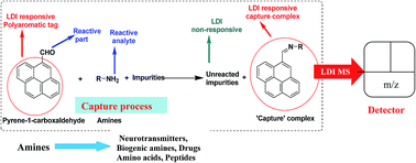
Label-assisted laser desorption/ionization (LA-LDI) technique has recently been applied to the detection of small molecules through a time of flight (TOF) mass spectrometric measurement. By excluding the external matrix, the mass spectrum becomes much cleaner being free of matrix related peaks and noises. Peaks corresponding to compounds/complexes formed between the analytes and the label are mostly seen in the spectrum. In this paper we report a LA-LDI mass spectrometry based method for detection of amines, including the ones ubiquitous to biological samples like biogenic amines, amino acids, di- and tripeptides using 1-pyrene carboxaldehyde as the LDI label.
Introduction
Laser desorption/ionization (LDI) is a soft ionization mass spectrometric (MS) method that has become an important technique in the analysis of a wide range of macromolecules including proteins, DNA, sugars, synthetic polymers etc.1 One of the most important LDI-MS methods is matrix-assisted laser desorption/ionization mass spectrometry (MALDI-MS).2 The matrix absorbs the laser light and causes the analyte to ionize and fly along with the matrix materials thus allowing recording of the molecular ion via the time of flight (TOF) measurement. Very often, the biopolymers are fragile and thus tend to fragment when ionized by more conventional ionization methods.3 The success of MALDI depends on many factors like the ability of the analyte to co-crystallise with the matrix. This requirement has made selection of matrix for MALDI somewhat analyte specific and choice of proper matrix often becomes a matter of trial and error. Also, the non-uniform nature of co-crystallization process causes “hot spots” to occur within the matrix film. In addition, MALDI is also rarely used for analytes of low molecular weight, because of appearance of matrix peak clusters which complicate the spectrum and hence the analysis. Quite a few variants of MALDI have been developed like SELDI, SIMS, DESI, DIOS etc. which differ mainly in technical aspects.4–7 SIMS was one of the first matrix-free desorption/ionization approaches used to analyze metabolites from biological samples. However, the technique suffers from its limited sensitivity at >500 Da and from analyte fragmentation generated by the high-energy primary ion beam. Desorption electrospray ionization (DESI) is a matrix-free technique for analyzing biological samples that uses a charged solvent spray to desorb ions from a surface. The main limitation of DESI is spatial resolution because of the difficulty in focusing the charged solvent spray.8 Desorption/ionization on silicon (DIOS), is another matrix-free laser induced desorption/ionization approach applicable for detection of small molecules and has the advantages of low chemical noise and high sensitivity in the low-mass range. While the technique has reached an impressive level of detection of small molecules,9a it has certain disadvantages like the fragility of porous silicon structure and lack of selectivity as all small molecules in a mixture are going to show up in the spectrum.9b On the other hand, Label Assisted Laser Desorption/Ionization Mass Spectrometry (LA-LDI MS), reported in recent years, has come into prominence which is used for the detection of small molecules and metal ions. A suitable tag is introduced to the analyte via the formation of a covalent bond or chelation. The tag absorbs the laser power to help desorption cum ionization of the tag-analyte combine, thereby eliminating the need of external matrix. Polyaromatic moieties are most versatile in this regard because they have high molar absorptivity, thus allowing efficient absorption of laser radiation and their facile ionisation to form molecular ion. Kozmin et al.10 have recently introduced this matrix free technique for rapid mass spectrometric detection of products originating from such labeled reactants in complex reaction mixtures without any chromatographic separation. Encouraged by Kozmin's work, we have reported the use of LA-LDI MS technique for selective detection of metal ions (like Zn2+)11 and also catechol amines12 like dopamine and related 1,2-diols. The labeled molecules used were an anthracene based oxine derivative and a pyrene boronic acid respectively. The working principle for LA-LDI MS is shown in Fig. 1. In the present work, we have selected 1-pyrene carboxaldehyde (PA) as a selective probe for identification of molecules with primary amine functionality. Our earlier work on the use of pyrene boronic acid for detection of 1,2-diols is limited by the number of 1,2-diols present in biological systems or as environmental pollutants. In order to broaden the scope of this detection technique, we targeted small molecules bearing primary amine functionality by capturing with PA in the form of imines which should be LDI responsive (Fig. 2). Primary amines are ubiquitous in biology. The breakdown of amino acids releases primary amines such as tyramine, ethanolamine, histamine and neurotransmitters like dopamine, serotonin, norepinephrine. Besides, amino groups are the most common moieties in amino acids, peptides, proteins. Some important drugs also contain primary amine functionality (e.g. ampicillin). Imine formation is quite common in biology as exemplified in the chemistry of PLP. It may be mentioned here that traditional techniques like UV-Vis spectroscopy,13 fluorescence spectroscopy,14 Liquid Chromatography-Mass Spectrometry (LC-MS)15 and High Performance Liquid Chromatography (HPLC),16 surface plasmon resonance based optical sensors17 and microfluidics based sensors18 have been used for detection of small molecules. However, these methods have their own limitations like interference from other analytes. For example electrospray mass spectrometry (ESI MS) will be difficult for mixture of analytes unless these are pre-separated via liquid chromatography. Otherwise, all the analytes will show up in the spectrum and will make the analysis difficult. The present method offers a possible alternative where there is no need for separation.
 |
| Fig. 1 Principle of LA-LDI MS. | |
 |
| Fig. 2 Present work. | |
Results and discussion
The commercially available 1-pyrenecarboxaldehyde (PA) was first subjected to LDI TOF spectrometry.19 Like the previously reported cases, because of the high molar absorptivity of (54
000 M−1 cm−1 at 335 nm), the pyrene moiety acted as LDI tag to form radical cations and showed a strong molecular ion peak at m/z 230 without any external matrix, besides other peaks like m/z 203 (MH+–CO) (Fig. 3). The origin of peak at m/z 400 is not clear at this moment.
 |
| Fig. 3 LDI-TOF mass spectrum of 1-pyrenecarboxaldehyde (m/z = 230) in positive ion mode. | |
The initial screening was carried out with simple amines like n-butyl amine, benzyl amine, ethylene diamine and 1,3-diamino propane. In all cases, the LDI spectra showed strong molecular ion peaks for the corresponding iminium ions (bis imine for ethylene diamine and 1,3-diaminopropane). We then turned our attention to the detection of biogenic amines like dopamine, histamine, tyramine, and ethanolamine each of which plays important role in various ways in the biological system. Thus aqueous solution (0.1 mM) of the respective amine hydrochloride salts was incubated with PA (0.1 mM in methanol) in presence of excess amount of triethyl amine for 30 min at room temperature. An aliquot of 1 μL from each reaction mixture was spotted in an LDI plate and analyzed by a TOF mass spectrometer in positive ion mode. In each case, a strong peak for the protonated iminium ion MH+ was observed in the spectrum.20 We have also extended this methodology for the detection of primary amine containing drugs like ampicillin. The results are summarized in the Table 1. A representative spectrum for dopamine is shown in Fig. 4 (for other spectra, see ESI†). For comparison, the spectra obtained for only the matrix (CHCA) as well as the MALDI MS of dopamine are shown (Fig. 4a and b).
Table 1 LA-LDI MS results for various amines
Amines |
Expected imine peak (m/z) |
Obtained (imine + 1) peak (m/z) |
 |
285 |
286 |
 |
319 |
320 |
 |
484 |
485 (bisimine + 1) |
 |
498 |
499 (bisimine + 1) |
 |
365 |
366 |
 |
349 |
350 |
 |
323 |
324 |
 |
273 |
274 |
 |
561 |
562 (imine + 1) peak, 318 (benzylic cleavage of imine) |
 |
| Fig. 4 Comparison of LDI mass spectrum of dopamine with and without matrix in positive ion mode (a) CHCA matrix; (b) CHCA matrix + dopamine (m/z = 153) [matrix : analyte = 100 : 1 molar ratio]; (c) dopamine + 1-pyrenecarboxaldehyde (m/z = 366). | |
Sensitivity of detection of the present protocol was tested using dopamine. This was evaluated by incubating dopamine solution of different concentration with a fixed concentration of PA. A series of solutions of PA (1 mM in methanol) and varying concentration of dopamine (50 μM, 25 μM, 15 μM, 10 μM, 5 μM, 2 μM, and 1 μM) were incubated at room temperature for 2 h and analyzed (TOF-MS in positive ion mode). The peak of the iminium ion corresponding to dopamine was clearly detectable up to 2 μM i.e. up to 0.28 nanogram μL−1 (Fig. 5a and b). The method could also detect respective amines in a mixture of dopamine, tyramine and histamine in presence of 10 fold excess of common biomolecule glucose. These three amines were clearly detectable by LDI MS up to a concentration of 10 μM without any interference from glucose thus demonstrating the selectivity of the present technique (Fig. 6).
 |
| Fig. 5 Comparison of LA-LDI-TOF mass spectrum of capture complex of dopamine at different concentrations in positive ion mode (a) 50–1 μM concentration (b) expanding intensity scale for lower concentrations (5–1 μM). | |
 |
| Fig. 6 Comparison of LA-LDI-TOF mass spectrum of capture complex of dopamine, tyramine, and histamine from their mixture at different concentrations in presence of excess glucose in positive ion mode. | |
We next turned our attention to the detection of amino acids. For this, several categories of amino acids, namely α, β, γ and ε were selected. The α-amino acids screened included glycine, alanine, valine, methionine, phenyl alanine, tryptophan, tyrosine, lysine, glutamic acid and aspartic acid. In the β-amino acid category, β-alanine was chosen while gabapentin, a γ-amino acid that is used as an anticonvulsant and analgesic and 6-amino caproic acid, an ε-amino acid were included in this assay. Keeping in view of the fact that amino acids are capable of existing in zwitterionic forms and hence pH needs to be adjusted to generate the free amine, we performed our experiment for screening these four types of amino acids in three different conditions. In the 1st condition, an aqueous solution of amino acid was incubated with methanolic solution PA in absence of any external base. The 2nd condition involved incubating the aqueous solution of amino acids and methanolic solution of PA in presence of excess of Et3N. In the 3rd protocol, the aqueous solution of amino acid was first neutralized by NaOH and then incubated with methanolic solution of PA. An aliquot of 1 μL were taken from each reaction mixture and LDI TOF-MS was recorded in positive ion mode. The results are summarized in the Table 2.
Table 2 LA-LDI MS results for amino acids
Amino acid type |
Obtained peaks (m/z) |
Neutralisation by NaOH (final pH ∼ 11) |
Neutralisation by Et3N (final pH ∼ 8) |
Without any neutralisation |
α-Amino acids with neutral side chain |
(Imine + 45) peak |
No significant peak |
α-Amino acids with basic side chain |
(Imine + 45) peak |
No significant peak |
α-Amino acids with acidic side chain |
(Imine + 1) peak |
No significant peak |
β-Amino acid |
(Imine + 45) peak |
No significant peak |
γ-Amino acid |
(Imine + 45) peak |
(Imine + 1) peak |
No significant peak |
ε-Amino acid |
(Imine + 45) peak |
(Imine + 1) peak |
(Imine + 1) peak |
Amongst all amino acids, only 6-amino caproic acid could be detected (appearance of M+ + 1 peak) in absence of any external base like Et3N or NaOH. In presence of excess Et3N, gabapentin was captured but α- amino acids and β-amino acid failed to show up in the spectrum. All types of amino acids including α- and β-amino acids were captured in presence of NaOH. An interesting result was observed in the LDI-MS recorded for amino acids treated with NaOH. In these cases, (iminium ion + 44) peak was obtained except for acidic amino acids like aspartic acid and glutamic acid. We believe the peak originated from the capture of CO2 by the iminium ions derived from amino acids and PA during incubation under basic condition. A representative spectrum is shown for alanine (Fig. 7). The method works equally well for detection of amino acids in a mixture in presence of other interfering analytes like glucose, ascorbic acid, tartaric acid, citric acid and α-ketoglutaric acid (10 fold excess) (Fig. 8). Literature survey21 revealed several reports of CO2 capture by amino acids. To the best of our knowledge, ours is the first report of an imine acting as a CO2 absorbing agent. The striking feature of these amino acid imines is the absence of peak corresponding to free imines (M+) or protonated iminium ions (MH+) which were observed for compounds with only amine functionality. This indicated high efficiency of the pyrene–amino acid imines to absorb CO2. This observation is noteworthy because of the importance of CO2 absorption and merits further exploration.
 |
| Fig. 7 LDI-TOF mass spectrum of capture complex of alanine (m/z = 346) in positive ion mode (in presence of NaOH). | |
 |
| Fig. 8 LDI-TOF mass spectrum of capture complex of glycine, alanine, valine (m/z = 332, 346, 374) from in mixture with excess ascorbic acid, tartaric acid, citric acid, glucose, α-keto glutaric acid in positive ion mode (in presence of NaOH). | |
This LA LDI-MS methodology is also able to detect dipeptides (glycyl–L-leucine, glycyl–L-isoleucine), L-carnosine (β-alanyl-L-histidine) and tripeptide (glycyl–glycyl–L-leucine) (Table 3). Here also the peak appeared at M+ + 45 indicating possible absorption of CO2. The method can also detect these peptides in a mixture (Fig. 9) and hence may be useful for small peptide profiling of a mixture.
 |
| Fig. 9 LDI-TOF mass spectrum of capture complex of L-carnosine and glycyl–glycyl-L-leucine (m/z = 483 and 502) from their mixture in positive ion mode (in presence of NaOH). | |
Table 3 LA-LDI MS results for peptide
Peptides |
Expected imine peak (m/z) |
Obtained (imine + 45) peak (m/z) |
 |
400 |
445 |
 |
400 |
445 |
 |
457 |
502 |
 |
438 |
483 |
Conclusion
In conclusion, we have expanded the scope of the LA LDI-TOF MS technique to detect a variety of biogenic amines, amino acids and peptides using PA as the label.22 The observation of CO2 capture by the imines derived from amino acids and peptides from PA is also noteworthy and needs further exploration. Presently we are working on extending this methodology in quantitative analysis of analytes using room temperature ionic liquids to avoid the occurrence of hot spots.
Acknowledgements
DST is acknowledged for an SERC grant to AB and AKD (grant no. SB/S1/C-94/2013) and for the JC Bose National Fellowship to AB which supported this research. AM is grateful to IIT Kharagpur for a Junior Research Fellowship. We also thank Dr P. S. Addy for helpful discussion.
Notes and references
- T. Koichi, H. Waki, Y. Ido, S. Akita, Y. Yoshida, T. Yoshida and T. Matsuo, Rapid Commun. Mass Spectrom., 1998, 2, 151 Search PubMed.
-
(a) K. Dreisewerd, Chem. Rev., 2003, 103, 395 CrossRef CAS PubMed;
(b) D. C. Muddiman, R. Bakhtiar, S. A. Hofstadler and R. D. Smith, J. Chem. Educ., 1997, 74, 1288 CrossRef CAS.
- C. S. Ho, C. W. K. Lam, M. H. M. Chan, R. C. K. Cheung, L. K. Law, L. C. W. Lit, K. F. Ng, M. W. M. Suen and H. L. Tai, Clin. Biochem. Rev., 2003, 24, 1 Search PubMed.
- N. Tang, P. Tornatore and S. R. Weinberger, Mass Spectrom. Rev., 2004, 23, 34 CrossRef CAS PubMed.
- W. G. Lewis, J. Shen, M. G. Finn and G. Siuzdac, Int. J. Mass Spectrom., 2003, 226, 107 CrossRef CAS.
-
(a) B. M. Harless, S. Trimpin, C. A. Lutomski, B. Wang and T. J. El-Baba, Mass Spectrom., 2014,(5), 32 Search PubMed;
(b) J. Takats, J. M. Wiseman, B. Gologan and R. G. Cooks, Science, 2004, 306, 471 CrossRef PubMed.
- J. J. Thomas, Z. Shen, J. E. Crowell, M. G. Finn and G. Siuzdak, Proc. Natl. Acad. Sci. U. S. A., 2001, 98, 4923 Search PubMed.
- M. P. Greving, G. J. Patti and G. Siuzdak, Anal. Chem., 2011, 83, 2 CrossRef CAS PubMed.
-
(a) A. B. Jemere, L. W. Bezuidenhout, M. J. Brett and D. J. Harrison, Rapid Commun. Mass Spectrom., 2010, 24, 2305 CrossRef CAS PubMed;
(b) H. Sato, T. Seino, A. Yamamoto, M. Toimura and H. Tao, Abstract Book, 17th International Mass Spectrometry Conference, Prague, 2006, Abstract No. TuP-142 Search PubMed.
- J. R. Cabrera-Pardo, D. I. Chai, S. Liu, M. Mrksich and S. A. Kozmin, Nat. Chem., 2013, 5, 423 CrossRef CAS PubMed.
- P. S. Addy, S. Basu Roy, S. M. Mandal and A. Basak, RSC Adv., 2014, 4, 23314 RSC.
- P. S. Addy, A. Bhattacharya, S. M. Mandal and A. Basak, RSC Adv., 2014, 4, 46555 RSC.
-
(a) A. Manz, N. Graber and N. M. Widmer, Sens. Actuators, B, 1990, 1, 244 CrossRef CAS;
(b) D. J. Harrison, A. Manz, Z. Fan, H. Ludi and M. H. Widmer, Anal. Chem., 1992, 64, 1926 CrossRef CAS;
(c) S. Hiki, K. Mawatari, A. Hibara, M. Tokeshi and T. Kitamori, Anal. Chem., 2006, 78, 2859 CrossRef CAS PubMed.
-
(a) Z. Dai and J. W. Canary, New J. Chem., 2007, 31, 1708 RSC;
(b) J. R. Lakowicz, Principles of Fluorescence Spectroscopy, Springer, New York, 2006 Search PubMed; B. Valeur, Molecular Fluorescence Principles and Applications, Wiley-VCH, New York, 2002 Search PubMed;
(c) Y. Choi, Y. Park, T. Kang and L. P. Lee, Nat. Nanotechnol., 2009, 4, 742 CrossRef CAS PubMed.
-
(a) J. J. Pitt, Clin. Biochem. Rev., 2009, 30, 19 Search PubMed;
(b) C. Lauridsen, S. W. Leonard, D. A. Griffin, D. C. Liebler, T. D. McClure and M. G. Traber, Anal. Biochem., 2001, 289, 89 CrossRef CAS PubMed.
- G. J. van Berkel and V. Kertesz, Rapid Commun. Mass Spectrom., 2013, 27, 1329 CrossRef CAS.
-
(a) K. V. Kong, Z. Lam, W. K. O. Lau, W. K. Leong and M. Olivo, J. Am. Chem. Soc., 2013, 135, 18028 CrossRef CAS;
(b) S. K. Srivastava, R. Verma and B. D. Gupta, Sens. Actuators, B, 2011, 153, 194 CrossRef CAS.
- H. F. Li and J. M. Lin, Anal. Bioanal. Chem., 2009, 393, 555 CrossRef CAS PubMed.
- All the experiments were carried out using ultra Flextrene MALDI Time-of-Flight Mass Spectrophotometer from Bruker. UV laser: smart beam II laser, 355 nm wavelength; laser rep rate 2000 Hz, reflector mode. Dopamine was procured in injectable form as dopamine hydrochloride. The mass spectra were recorded in positive ion mode.
- In some cases, we have also observed additional peaks at m/z 242 and 215 which are fragmentations arising from the original iminium ion (the mechanism of fragmentation is included in ESI†)
. - D. M. Mũnoz, A. F. Portugal, A. E. Lozano, J. G. Campaa and J. Abajo, Energy Environ. Sci., 2009, 2, 883 Search PubMed.
- Considering the general instability of the imines, we have recorded the LDI-MS (spectra included in ESI†) after reducing the imines with NaBH4 or NaCNBH3. We could observe the peak for [M + 2] indicating successful reduction to the amine. However, the spectral quality was not any better as compared to that of imine.
Footnote |
† Electronic supplementary information (ESI) available. See DOI: 10.1039/c5ra20678b |
|
This journal is © The Royal Society of Chemistry 2015 |
Click here to see how this site uses Cookies. View our privacy policy here.