DOI:
10.1039/C5RA20103A
(Paper)
RSC Adv., 2015,
5, 104062-104070
A comprehensive comparison of bacterial and fungal aerobic granules: formation, properties, surface modification, and biosorption of metals†
Received
29th September 2015
, Accepted 2nd December 2015
First published on 3rd December 2015
Abstract
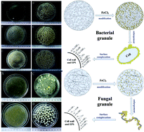
Aerobic granules, a relatively new form of microbial aggregate, can be formed with bacteria or fungi as the dominant population, depending on operational conditions. In this study, a comprehensive comparison is conducted between these two kinds of granules. Bacterial granules (BG) were formed after 3 weeks of cultivation, and exhibited a settling velocity and ash content of 5.6 cm s−1 and 24%, respectively. In contrast, fungal granule (FG) cultivation took only 5 days, and their settling velocity and ash content were 45% and 22% of BG, respectively. BG and FG selectively enriched calcium and potassium, respectively, and these elements were replaced by iron upon Fe(III) modification. Original BG had 7 times larger surface areas and 9 times higher pore volumes than FG, but Fe(III) modification reversed the trend. Original and modified granules were used as biosorbents for removal of Zn(II), Cu(II), Ni(II) and Sb(OH)6− at various pH levels. The original granules removed the cations efficiently (BG better than FG), but had no affinity for Sb(V) under all pH values. Fe(III) modification significantly enhanced Sb(V) removal by both granules, with the best performance achieved under pH 3.4. FG, though it had lower Fe loading, exhibited a comparable ultimate Sb(V) adsorption capacity to BG (111 vs. 125 mg g−1), and 3 times higher initial adsorption rate. In addition, FG was more stable at higher pH and showed better adsorption performance under pH > 4.3. Pseudo-second-order kinetics and a Langmuir isotherm model described the adsorption processes well for both kinds of modified granules, which proved to be good biosorbents.
1. Introduction
Aerobic granules are a promising wastewater treatment technology, which can efficiently remove organics, nitrogen, phosphorus, and toxic substances.1 Mature granules are usually compact in structure and high in density, and harboring various functional populations like heterotrophic, nitrifying, denitrifying and phosphorous-accumulating bacteria.2 However, researchers have discovered that some operation conditions could lead to overgrowth of filamentous microorganisms in aerobic granules.3–7 For example, carbohydrates like glucose and citric acid, and some other readily biodegradable organics are believed to favor the growth of the filamentous microorganisms; acidic pH could also shift the structure of the microbial community in the granules towards filamentous growth. Some of these filamentous cells were later confirmed to be fungi populations.6,7 The filamentous granules formed are characterized by their loose structure and low density, which can cause severe biomass washout from reactors. Therefore, fungal granules are often considered useless in practice, and detrimental to process stability.
However, some recent studies have proposed a potential application for the fungal granules, as they had exhibited considerable biosorption capacity, especially after surface modification.7 Biosorption is an eco-friendly and cost-effective way to removal heavy metals from aqueous medium,8 and aerobic granule has been investigated as a promising biosorbent due to its excellent settleability and compact structure.9 Up to now, mainly aerobic granules had been applied, while only a few have dealt with biosorption by both bacterial and fungal granules, utilizing Fe(III) imbedding as a surface modification method. Biosorption of Sb(V) onto Fe-modified bacterial granules was first experimented, and the kinetics, isotherm, thermodynamic, influencing factors and adsorption mechanism were studied in detail.10,11 Moreover, the microbial community of fungal granules and use of Fe(III)-modified fungal granules for Sb(V) biosorption were also briefly discussed.7
These studies, though rigorous and detailed, are somewhat isolated and cannot provide a full picture of these two kinds of granules. Therefore in this work, it is intended that a comprehensive comparison should be made, based on previous and new results, to give an overview of the characteristics and applicability of bacterial and fungal aerobic granules. The objectives of this work are: (I) to study in detail the differences in the cultivation conditions, morphology, physical structure, and chemical composition of bacterial and fungal granules; (II) to perform Fe(III) surface modification on these granules, and to investigate the respective changes in their properties; (III) using Cu(II), Zn(II) and Ni(II) as model cations to test the biosorption capacities of unmodified granules; (IV) using Sb(V) as the model oxyanion and kinetic modeling as tools, to study the adsorption capacity, velocity, and maximum quantity of modified granules. Finally a comprehensive appraisal on the bacterial and fungal granules is presented, which has not been attempted before, and would provide valuable information in both fundamental research and practical applications.
2. Experimental
2.1 Cultivation and preparation of aerobic granules
Two identical sequencing batch reactors (SBRs), i.e., R1 and R2 were used to cultivate bacterial and fungal granules, respectively. Activated sludge obtained from a nearby domestic wastewater treatment plant was inoculated into the reactors for granule cultivation. They were both operated at 4 h cycles, with air pumped through diffusers at the bottom (volumetric flow rate 5 L min−1). Each cycle included 3 min filling, 227 min aeration and settling, 5 min effluent decanting, and 5 min idling. The settling time was reduced from 30 min to 1 min for R1, but was fixed at 5 min for R2. R1 was fed with a synthetic wastewater with the composition of NH4Cl (0.2 g L−1), KH2PO4 (0.66 g L−1), NaHCO3 (0.013 g L−1), MgSO4·7H2O (0.025 g L−1), CaCl2 (0.03 g L−1), FeSO4·5H2O (0.02 g L−1), peptone (0.4 g L−1), and yeast extract (0.25 g L−1). The synthetic wastewater for R2 contained the same components, except for KH2PO4 (0.2 g L−1), peptone (0.04 g L−1), and no yeast extract. Carbon sources for R1 were sodium acetate and propionate at a combined chemical oxygen demand (COD) concentration of 1000–3000 mg L−1 in the influent, while glucose (COD concentration 1000 mg L−1) was provided for R2 as the sole carbon source. These differences in the wastewater composition aimed to promote the formation of different granules. The reduced KH2PO4 concentration resulted in lower buffering capacity, which, combined with the lower COD loading in R2, could promote the growth of fungal granules. After granule formation, both granules were harvested from the SBRs, and washed with MilliQ water three times before tests.
2.2 Surface modification by Fe(III)
The granules were modified using the same method as reported in previous work.7,11 Specifically, bacterial or fungal granules (25 g on wet basis) were incubated with 250 mL 0.1 M FeCl3 solution. The suspension was continuously shaken on an orbital shaker for 24 h at 35 °C, pH 2.0 and 160 rpm. Subsequently, the Fe-modified granules were collected and repeatedly washed with MilliQ water until no Fe(III) can be detected in the supernatant, then stored in tap water at 4 °C until use. The original aerobic granules were designated as OAGS-B (bacterial) and OAGS-F (fungal), and the modified aerobic granules MAGS-B and MAGS-F, respectively.
2.3 Adsorption tests
Cu(II), Zn(II) and Ni(II) were used as the target metal cations for biosorption experiments by unmodified granules. The standard stock solutions of these ions (1000 mg L−1) were prepared by dissolving CuSO4, ZnCl2 or NiSO4 in MilliQ water, which were further diluted and their pH adjusted with 0.1 M HCl. OAGS-B or OAGS-F of 0.5 g (wet basis, average water content 94.2 ± 0.2%) were mixed with 10 mg L−1 solution of each ion, and the removal was studied under pH 1.9, 2.6, 3.4, 4.8, 6.5 and 7.6.
Sb(V) was applied as the model oxyanionic metal for adsorption tests with both the original and modified granules. The Sb(V) standard stock solution of 1000 mg L−1 was prepared by dissolving KSb(OH)6 in MilliQ water, and was further diluted to the desired concentrations. 0.1 M HCl or NaOH was added to adjust pH in the range of 2 to 11.4 to study the pH effect. For adsorption kinetics, initial Sb(V) concentrations of 20, 60 and 100 mg L−1 were used. For the adsorption isotherms, the concentrations varied from 10 to 500 mg L−1, and the thermodynamics analysis was conducted under temperatures from 10 °C to 45 °C. The contact time for all the tests was 300 min.
In the adsorption tests, the supernatant of the solution was collected at fixed time intervals and centrifuged at 10
000 rpm for the subsequent measurement of metal concentrations. The adsorption quantity, qt was expressed as: qt = (C0 − Ce) × v/m, where C0 and Ce are the initial and equilibrium concentrations (mg L−1), respectively; v represents the solution volume (L), and m is the dried weight of the sorbent. The amount of Sb adsorbed by the flask was measured as the blank, and was found to be negligible. All tests were performed in triplicate, and all the results are shown as mean values with the error bars as the standard deviation.
2.4 Sampling and analytical methods
Various physical and chemical characteristics of the granules were studied, including their morphology by digital camera imaging and scanning electron microscopy (SEM) observation, metal composition, surface area, functional groups, and microbial population. For electron microscopy, granules were sequentially dehydrated by a serial of ethanol solutions of increasing concentrations and dried overnight, followed by coating with aurum and observation by a Philips XL30 electron microscope. Microanalysis by transmission electron microscopy (TEM) equipped with energy-dispersive X-ray spectrometry (EDS) was conducted on a JEOL JEM2011 instrument. The surface areas of the samples were determined with a BET (Brunauer–Emmett–Teller) system (Quadrasorb SI instrument) from the nitrogen sorption isotherm at −196 °C. Functional groups were studied by Fourier transformation spectroscopy (FTIR, Nicolet Nexus-470), which was performed on KBr pellets pressed from the mixture of 1 mg dried granules and 100 mg spectrometry grade KBr under vacuum. X-ray diffraction (XRD) analysis was conducted on a Bruker D8 Advance X-ray diffractometer using nickel-filtered Cu Kα radiation at 40 kV and 40 mA. The concentrations of Sb and other common metals were analyzed by an Inductively Coupled Plasma Atomic Emission Spectrometer (ICP-AES, HITACHI P-4010). The concentrations in the solutions were directly measured, while those in the biomass were determined after its digestion and dissolution in acid. The concentrations of Cu, Zn and Ni were measured using flame atomic absorption spectroscopy (AAS, HITACHI Z-5000).
2.5 Adsorption models and mechanism
The kinetics, isotherms and thermodynamics were studied using widely accepted models, including pseudo-first-order and pseudo-second-order kinetics, and Langmuir and Freundlich isotherms. The thermodynamic parameters of entropy (ΔS), enthalpy (ΔH) and Gibbs free energy (ΔG) were also obtained. The detailed models, parameters and calculations are shown in the ESI materials (Tables S2–S4†).
3. Results and discussion
3.1 Granule cultivation and morphology characteristics
Bacterial granules of size 3–5 mm were formed after 20 days in R1, the progress of which is showed in Fig. S1.† During cultivation, the morphology of the biomass changed from highly dispersed flocs to structure-compact aggregates, and the COD removal efficiency was usually higher than 98%. Further study indentified various bacteria as the dominating populations.12 In contrast, filamentous granules with fluffy surface were observed in R2 after only 5 days, and the changes in biomass morphology can be seen in Fig. S2.† The size of the filamentous granules ranged from 1 to 7 mm, and their COD removal capacity was comparable to those in R1. These granules were found to be dominated by one fungal species, Geotrichum fragrans.7 During the aeration phrase in a cycle, the pH increased from 7 to 8.5 in R1, but decreased from 7 to 4.5 in R2. The different carbon sources used in the cultivation very likely caused the different trends of pH change, by producing various intermediates during substrate degradation. Glucose in R2 could be degraded to pyruvate following a glycolytic pathway, and further transformed into volatile fatty acids, which would lead to an acidic condition.6 Moreover, KH2PO4 content in R2 was significantly lower, resulting in a weaker buffer capacity. These factors in combination contributed to the sustained low pH in R2, which in turn influenced the microbial community structure of the granules. It has been reported that pH was the determining parameter in the type of granules formed, and low pH caused the overgrowth of filamentous microorganisms and formation of fungal granules.6
Significant differences in the morphology were observed between OAGS-B and OGSB-F, as shown in Fig. 1(A) and (F). OAGS-B had light-yellow surface and irregular shape, while OGSB-F was white and near-spherical. Further observation of the granules surface suggested that OAGS-B had compact structure, and OGSB-F was loose, with abundant mycelia entangled with each other (Fig. 1(C) and (H)). Some physicochemical characteristics are shown in Table 1. Sludge volume index (SVI) reflects the sludge settling ability, and the SVI of OAGS-F was twice that of OGSB-B. In addition, the settling speed of OGSB-B was 124% higher than that of OAGS-F, which indicated that OAGS-F settled more slowly, and when settled, formed less compact sludge bed. This is in accordance with the fluffy structure of the fungal granules. The two kinds of granules had similar moisture contents, but the ash content in OGSB-B (24%) was significantly higher than that in OAGS-F (5.2%). It seemed that bacterial granules were more inclined to incorporate mineral elements into their structure. It was suggested that addition of Ca2+ or Mg2+ enhanced aerobic granulation,13 which is in agreement with the higher ash content and settling ability found with OAGS-B in this study. However, if, and how does the lower ash content correlate to the formation and properties of fungal granules requires further study, as the same Ca2+ and Mg2+ concentrations were provided in the cultivation medium.
 |
| Fig. 1 Morphology observations of the bacterial and fungal granules. (A) Original bacterial granules (B) Fe(III) modified bacterial granules; (C) original bacterial granules surface; (D) original bacterial granules surface (detailed); (E) bacterial granules surface after modification; (F) original fungal granules; (G) Fe(III) modified fungal granules; (H) original fungal granules surface; (I) original fungal granules surface (detailed); (J) fungal granules surface after modification. | |
Table 1 Physicochemical characteristics of the original bacterial and fungal aerobic granules
|
SVI (mL g−1) |
Settling velocity (cm s−1) |
Moisture content (%) |
Ash content (%) |
OAGS-B (R1) |
48 |
5.6 |
92 |
24 |
OAGS-F (R2) |
74 |
2.5 |
93 |
5.2 |
3.2 Fe(III) modification on the granules
Surface modification is a practical technique for metal removal by biosorption, and iron is a promising modification agent, as it is cheap and non-toxic.14 In this study, the granules were subjected to modification by Fe(III), in the hope of expanding their applicability in biosorption. Both granules exhibited color change to brownish after Fe(III) embedding, but their general shapes seemed to be unchanged, as shown in Fig. 1(B) and (G). However, different pH significantly affected the integrity of the modified granules and the iron speciation on their surface, the details of which were described in previous work.7,11 In short, strong acid led to the dissolution of iron hydro(oxide) on the surface, and disintegration of MAGS-B was observed under strong alkaline conditions. However, the integrity of MAGS-F seemed to be unaffected by high pH, though the change of the iron speciation was similar.
Finer details on the granules' surface were observed using SEM and TEM with EDS. OAGS-B consisted of cocci and bacilli of size 0.5–2 μm closely grouped together, while OAGS-F was made up of intertwined mycelia with the diameter of around 2 μm, and presented a more loose and hollow structure (Fig. 1(D) and (I)). After FeCl3 treatment, the surface of the granules became coarse and wrinkled and nano-precipitates were observed (Fig. 1(E) and (J)), which are also seen in the TEM images (Fig. 2(B) and (D)). EDS spectra confirmed the particles to be iron compound. The spectra also showed that OAGS-B and OAGS-F enriched elements Ca and K, respectively, which were replaced by iron after Fe(III) modification (Fig. 2(E–H)).
 |
| Fig. 2 TEM images and EDS spectra of bacterial and fungal granules. (A) Original bacterial granules; (B) modified bacterial granules; (C) original fungal granules; (D) modified fungal granules; (E), (F), (G) and (H) were the corresponding EDS spectra. | |
3.3 Further characterization of the granules' physiological properties
3.3.1 Metal composition. As mentioned in Section 3.2, it was possible that the granules selectively enriched certain metal elements in the biomass. To further investigate this phenomenon, elemental analysis of some common metals was conducted on both granules before and after Fe(III) modification and the results are summarized in Table S1.†7,11 Elements Ca and K were selectively enriched in OAGS-B and OAGS-F, respectively, with the corresponding contents of 8.8% and 2.5%. The iron contents in OAGS-B and OAGS-F were insignificant. However after Fe(III) modification, they increased to 5.9% and 1.6% for MAGS-B and MAGS-F (0.059 and 0.016 mg per mg granule), respectively. This indicated that Fe was indeed taken up by the granules, and the BG had almost 4 times Fe loading capacity of FG. Meanwhile, a sharp decrease in all other common metals (K, Na, Ca, and Mg) was observed, one reason of which could be that iron modification was accompanied by the hydrolysis of FeCl3. The resultant acidic condition in the solution would dissolve these metals in the granules. Ion exchange between Ca/K and Fe could also play a role.
3.3.2 Analysis of surface area. Specific surface area is an important parameter reflecting the adsorption capacity of an adsorbent. The granules' surface area was analyzed by BET, and the results are presented in Table 2. OAGS-B and OAGS-F had specific surface areas of 65.4 and 9.1 m2 g−1, respectively, which changed to 8.52 and 29.7 m2 g−1 after modification. Accordingly, the pore volume of BG decreased from 0.196 to 0.029 mL g−1, whereas that of FG increased from 0.021 to 0.054 mL g−1. This significant difference could probably be attributed to the distinct physical structures of the granules. Upon contact with FeCl3, bacterial granules shrank slightly, which might have prevented foreign ions from permeating into their interior, and formation of iron-precipitate might further block the pores. These factors in combination resulted in a decrease in surface area and pore volume. Similar results were also found in a study of activated carbon modified with iron to be used as adsorbent for arsenate removal.15 However, OAGS-F mainly contained long intertwined mycelia, which corresponded to relative small surface area. The nano-particle precipitation onto the mycelia in turn increased the specific surface area, which might enhance their adsorption capacity.
Table 2 Analysis of the granules' surface area and pore volume by BET before and after Fe(III) modification
|
Specific surface area (m2 g−1) |
Pore volume (mL g−1) |
Pore size (nm) |
OAGS-B |
65.4 |
0.196 |
12.38 |
Fe-MAGS-B |
8.52 |
0.029 |
14.81 |
OAGS-F |
9.10 |
0.021 |
10.81 |
Fe-MAGS-F |
29.7 |
0.054 |
6.97 |
3.3.3 Analysis of granule surface by FTIR and XRD. Functional groups analysis was conducted by FTIR, and the results are shown in Fig. 3 with all the major groups marked. The bands in the region of 3200 to 3600 cm−1 reflect the overlapping of stretching vibration of OH and NH, indicating the presence of hydroxyl and amine groups on both the granules' surface. The bands at 2925 and 2856 cm−1 are indicative of asymmetric and symmetric stretching vibration of CH2, respectively. The band at 1261 cm−1 results from the deformation vibration of C
O, while that at 1056 cm−1 is attributed to the stretching vibration of OH from polysaccharides.9 After Fe(III) modification, for both granules the band at 3399 cm−1 shifted to 3321 and 3367 cm−1, while that at 1056 cm−1 shifted to 1076 and 1065 cm−1, respectively. However, hardly any shifts were observed for the bands at 1261, 2925 and 2856 cm−1. These results indicated that hydroxyl group on the biomass surface played an important role in Fe(III) modification, while the effect of other functional groups including amine, CH2 and C
O was not as significant. XRD analysis was also performed to identify the properties of Fe-precipitates formed on the biomass. However, no significant diffraction peak was observed (data not shown). This suggested that either the content of iron hydro(oxide) was not high enough to be detected, or the iron-precipitates formed was amorphous, or both. Based on the collective results, the mechanisms of Fe(III) modification were proposed in Fig. S3,† where ion-exchange with Fe3+ and iron hydroxides complexation with the surface functional groups (most likely hydroxyl) in the extracellular polymers were the major contributing factors.
 |
| Fig. 3 FTIR spectra of (A) original bacterial granules; (B) modified bacterial granules; (C) original fungi granules; (D) modified fungi granules; (E) modified bacterial granules after Sb adsorption; (F) modified fungi granules after Sb adsorption. | |
3.4 Adsorption tests with Cu(II), Zn(II) and Ni(II)
Most of heavy metals exist in the form of cations in aqueous media, therefore aerobic granules, usually negatively charged (point of zero charge, pHpzc = 2.4),16 are often used to remove cationic metals.17,18 In this study, several common and toxic metals, i.e. Cu(II), Zn(II) and Ni(II) were selected as model cations to briefly test the adsorption capacity of the original granules. To avoid precipitation, pHs from 1.9 to 7.6 were selected, and the respective removal efficiencies are shown in Fig. 4. In almost all cases, OAGS-B had much higher capacity than OAGS-F, the only exceptions being Cu and Ni under pH 1.9. Under other pH, the removal by the former for all 3 ions was 1 to 4 times higher than that by the latter. This was in accordance with the higher surface area of OAGS-B. In addition, the elevated Ca and Mg levels in OAGS-B might also contribute through ion exchange with the cations, which was confirmed in a previous study of Sr(II) removal by aerobic granules.19 On the other hand, the removal of the tested cations was strongly dependant on solution pH for both granules. It has been reported that pH is an important factor that influences both metal and cell surface chemistry.20 Aerobic granules would be protonated and positively charged under pH below its pHpzc, therefore the adsorption of cations would be inhibited under low pH due to electrostatic repulse between metal ions and the biomass. In contrast, pH ranging from 3.4 to 7.6 resulted in much better adsorption performance by both granules. In terms of individual metals, Cu(II) and Zn(II) exhibited significantly better removal effect than Ni(II), the reason of which is not clear.
 |
| Fig. 4 Removal percentages of cationic metals by original bacterial and fungal granules. (A) Zn(II); (B) Cu(II); (C) Ni(II); (T: 35 °C, shake speed: 175 rpm, C0 of the ions: 10 mg L−1, granule dosage: 0.6 g L−1). | |
3.5 Adsorption tests with Sb(V)
Heavy metals can also exist in the form of oxyanion, the examples of which include Sb(V), As(V), and Cr(VI). Among them, antimony (Sb) is widely applied in a variety of industrial activities, and considered as a pollutant of priority interest by various agencies.21,22 Sb(V) was found to be dominant and stable in oxygenated systems, with oxyanion Sb(OH)6− as the common form.23 Therefore, Sb(V) was selected as the model oxyanion in this study to investigate the adsorption capabilities of the bacterial and fungal granules, both before and after Fe(III) modification.
3.5.1 Effect of pH on the adsorption capacities of the granules. Sb(V) removal by OAGS and MAGS was compared under 3 different pH, and the respective time courses are shown in Fig. 5. OAGS showed no affinity for Sb(V) regardless of pH. When modified by Fe(III), removal efficiencies were significantly improved for both granules. The highest improvement occurred under pH 3.4, with both removal percentages higher than 99% at 300 min, but MAGS-F achieved the effect much faster (at around 50 min) than MAGS-B (approx. 125 min). In addition, modified granules exhibited different sensitivities to pH. At pH 2.0 or 10.4, MAGS-B removed 60 or 6% of the initial 20 mg L−1 Sb(V), respectively, while the percentages were 40 or 23% for MAGS-F. Therefore MAGS-F showed higher and faster removal than MAGS-B under pH 3.4 and 10.4, while MAGS-B performed better at pH 2.0.
 |
| Fig. 5 The effect of pH on Sb(V) removal by (A) original and modified bacterial granules and (B) original and modified fungi granules with time; (T: 35 °C, shake speed: 175 rpm, C0 of the ions: 20 mg L−1, biomass dosage: 1 g L−1). | |
The pH effect was studied in more details for MAGS and the results are summarized in Fig. 6. Sb(V) could exist as a negatively charged complex in pH from 2 to 11,24 which was the range tested. Both MAGS-B and MAGS-F exhibited a similar trend with pH change. The pH 3.4 was found to be optimal, with removal percentages around 99% for both granules. Strong acidic or alkaline conditions negatively affected the removal efficiency, and in general the effect of high pH was more pronounced. MAGS-B and MAGS-F exhibited different susceptibility to acidic/alkaline environments, and the threshold seemed to be around 4.3. Below that pH, MAGS-B showed higher efficiency than MAGS-F, while the opposite was observed under neutral and alkaline conditions. This agreed well with the phenomenon described in Section 3.2, that the structural stability of MAGS-B was greatly reduced under higher pH, while that of MAGS-F was not much affected.
 |
| Fig. 6 Effect of pH on Sb(V) removal by modified bacterial and fungal granules; (T: 35 °C, shake speed: 175 rpm, C0 of the ions: 20 mg L−1, biomass dosage: 1 g L−1). | |
The pH effect could be the result of the interactions of several mechanisms. On one hand, the granules would be protonated and positively charged under pH lower than pHpzc, promoting Sb(OH)6− removal. Nevertheless, pH 2 might lead to dissolution of the bonded iron, thus weakening the adsorption. On the other hand, the granules could be negatively charged in alkaline solution due to the existence of abundant OH−, which would result in electrostatic repulsion with the oxyanion Sb(OH)6−. As pH 3.4 gave the best performance for both granules, it was applied in all later experiments.
3.5.2 Adsorption kinetics. As aforementioned, fungal granules exhibited faster removal under certain conditions, therefore the adsorption kinetics were further studied under several initial concentrations, and the comparison is shown in Fig. S4.† Higher initial concentrations resulted in higher adsorption amounts for both granules, possibly because the ratio of initial Sb(V) quantity to available adsorption sites was high at higher initial concentrations,25 providing bigger driving force. The equilibrium adsorption quantities were found to be 33.9, 85.9 and 130 mg g−1 for MAGS-F, and 36.6, 102, and 146 mg g−1 for MAGS-B at initial Sb(V) concentrations of 20, 60, and 100 mg L−1, respectively. Therefore MAGS-B had slightly higher ultimate adsorption capacity, but MAGS-F showed faster initial adsorption in all cases. For MAGS-F, 53.8, 59.8 and 61.2% of the equilibrium quantity were accomplished within 5 min at 20, 60 and 100 mg L−1, respectively, which was almost twice as much as that of MAGS-B.The experimental data were further fitted with two classic kinetic models to better understand the mechanisms, and the respective parameters are listed in Table S2.† Pseudo-second-order model could better describe Sb(V) adsorption by both granules, with the correlation coefficients higher than 0.998 in all cases, and the calculated equilibrium adsorption quantities agreed well with the experimental data. Parameters k2, h and t1/2 represent the sorption rate, initial sorption rate and half-adsorption time, respectively, in this model. They were used to evaluate the adsorption kinetics, and the results are compared in Fig. 7. At the same initial concentrations, both k2 and h of MAGS-F were significantly higher than those of MAGS-B. The main reason could be the physical structure of the granules. MAGS-F had larger surface area, with more active adsorption sites exposed to Sb(V), thus promoting film and pore diffusion within a short time. Correspondingly, the calculated t1/2 ranged from 4.8 to 6.3 min for MAGS-F, much shorter than those of MAGS-B (16 to 20 min).
 |
| Fig. 7 Comparison of sorption rates by modified bacterial and fungal granules as a function of initial Sb(V) concentrations. (A) Adsorption rate constant; (B) initial sorption rate constant; (C) half-adsorption time; (T: 35 °C, shake speed: 175 rpm, C0 of the ions: 20 mg L−1, biomass dosage: 0.4 g L−1). | |
3.5.3 Adsorption isotherm. The equilibrium adsorption data were fitted with Langmuir and Freundlich isotherm models, the detailed discussions of which had been presented in previous studies.7,10 In this study, the respective results are listed in Table S3† for better comparison. Generally, the data for both MAGS-B and MAGS-F could be better described by the Langmuir model, with the correlation coefficients (r2) of 0.987 and 0.997, respectively. It indicated that Sb(V) adsorption by both kinds of granules was likely monolayer sorption. The theoretical maximum adsorption quantities calculated from the Langmuir model were 125 and 111 mg g−1 for MAGS-B and MAGS-F, respectively. It agreed well with the experimental data, that the fungal granules had a comparable Sb(V) removal capacity, in spite of its lower iron loading.
3.5.4 Thermodynamics analysis. The thermodynamic analysis of the adsorption process by MAGS-B has been discussed before,10 and that of MAGS-F was conducted in this study. The respective parameters are listed in Table S4,† which indicated that MAGS-B and MAGS-F had similar thermodynamic characteristics. The enthalpy change (ΔH) values were found to be positive, indicating the adsorption was endothermic, and higher temperature would favor the process. Moreover, in the temperature range of 10 to 45 °C, the negative values of Gibbs free energy (ΔG) implied that Sb(V) adsorption by both granules were spontaneous.
3.5.5 Adsorption mechanisms. The mechanism of Sb(V) adsorption by MAGS-B has been investigated in details before.10,11 In addition, FTIR data were obtained for the modified granules after Sb(V) adsorption in this study, and the results are shown in Fig. 3. Comparing the profiles of the modified granules before and after adsorption, it can be seen that bands at 3200–3600 cm−1 and 1056–1076 cm−1 have shifted. More specifically for the modified bacterial granules, the band at 3321 cm−1 shifted to 3388 cm−1, and that at 1076 cm−1 to 1066 cm−1. For modified fungal granules the corresponding changes were 3367 cm−1 to 3393 cm−1 and 1065 cm−1 to 1058 cm−1. In fact, Sb(V) adsorption resulted in FTIR profiles that were closer to the original granules, and the main functional groups affected seemed to be OH related. As OH was the major functional group involved in Fe(III) modification, it was possible that Sb(V) interacted with the Fe–OH complex on granules surface and somehow changed the FTIR profiles. Moreover, the shifts were more dramatic for the bacterial granules (3399-3321-3388 and 1056-1071-1066 cm−1) than the fungal granules (3399-3367-3393 and 1056-1065-1058 cm−1), but the reason is unclear.Based on previous results and the new data presented in this study, the mechanism of Sb(V) removal by Fe(III) modified aerobic granules is proposed as a combination of electrostatic attraction, ion-exchange, and surface complexation. First, iron modification causes precipitation of ferric hydroxides on granule surface (
Fe–OH). At low pH, the surface of the modified granules could be protonated to form
Fe–OH2+, which are positively charged. The positively charged surface could then easily attract the negatively charged Sb(V) species and form out-sphere complex [
Fe–OH2+·Sb(OH)6] through electrostatic attraction and ion-exchange. Subsequently when the modified granules were ashed after Sb adsorption, dark insoluble residue was observed. It was then deducted that inner-sphere complex (
Fe–O–Sb) was formed through dehydration, resulting in more stable chemical bonds and residues insoluble in strong acid.10 Furthermore, other positively charged iron species, such as Fe(OH)2+, Fe2(OH)24+, Fe3(OH)45+ could also be combined with the granules surface,11 which would enhance Sb(V) removal furthermore.
3.6 Biosorbent assessment
Generally, the adsorption performance of a biosorbent for metal removal is determined by multiple factors, including ease and cost of preparation, settleability and stability, sorption rate and capacity, and potential environmental impact. The maximum adsorption capacities of several organic and inorganic adsorbents for Sb(OH)6− are listed in Table 3 for comparison. The modified aerobic granules used in this study showed higher capacity than most adsorbents listed, and the process was usually completed in a shorter time. Therefore in terms of adsorption capacity and separation efficiency, the modified aerobic granules are potentially good biosorbents.
Table 3 Comparison of maximum adsorption capacities of Sb(V) onto various adsorbentsa
Adsorbent |
Adsorbate |
pH |
Biomass dose (g L−1) |
Temperature (°C) |
Contact time (h) |
Maximum sorption capacity (mg g−1) |
Reference |
*Saponified orange waste; **hydrous ferric oxide. |
MAGS(F) |
Sb(V) |
3.4 |
0.8 |
35 |
5 |
111 |
This study |
MAGS(B) |
Sb(V) |
3.4 |
0.8 |
35 |
5 |
125 |
This study |
Acid-treated cyanobacteria |
Sb(V) |
2.5–2.6 |
50 |
Room temperature |
2 |
9.61 |
23 |
Zr(IV)-loaded SOW* |
Sb(V) |
2.5 |
1.67 |
30 |
24 |
145 |
26 |
Fe(III)-loaded SOW |
Sb(V) |
2.5 |
1.67 |
30 |
24 |
145 |
26 |
Kaolinite |
Sb(V) |
6 |
25 |
50 |
24 |
16.3 |
24 |
α-FeOOH |
Sb(V) |
4 |
0.4 |
20.5 |
24 |
24.5 |
27 |
β-FeOOH |
Sb(V) |
4 |
0.4 |
20.5 |
24 |
29.2 |
27 |
γ-FeOOH |
Sb(V) |
4 |
0.4 |
20.5 |
24 |
34.1 |
27 |
α-Fe2O3 |
Sb(V) |
4 |
0.4 |
20.5 |
24 |
23.4 |
27 |
HFO** |
Sb(V) |
4 |
0.4 |
20.5 |
24 |
114 |
27 |
A conclusion of the various aspects of the two kinds of granules is shown in Table S5.† OAGS-B, with compact structure and high density, had better settleability in the aqueous solution. It had larger surface area and pore volume, and higher iron loading after Fe(III) modification. Bacterial granules also showed higher removal capacity for the cationic metals tested while unmodified, and a greater ultimate adsorption quantity for oxyanionic metals, Sb(OH)6− after modification. However, longer time was needed for their cultivation, and their adsorption equilibrium for Sb(V) was achieved more slowly. In contrast, the fungal granules could be formed in shorter time from activated sludge. Fe(III) modified fungal granules had higher surface area despite their lower iron loading, and their sorption rate for Sb(V) was higher especially in the initial phase. Therefore fungal granules utilized the modifying agent Fe(III) more efficiently. In addition, fungal granules exhibited higher structural strength under alkaline conditions, which resulted in their better performance at higher pH. In a word, the bacterial and fungal granules have their respective advantages and disadvantages in cultivation, properties and performances in metal biosorption. It is the first time that the advantages of fungal aerobic granules are systematically investigated, and a comprehensive comparison conducted between the bacterial and fungal granules, which could be of both research and practical interest.
Acknowledgements
This study is financially supported by the State Key Laboratory of Pollution Control and Resource Reuse Foundation (PCRRF14003), and National Science Foundation of China (No. 51278128 and No. 51308128).
References
- D. J. Lee, Y. Y. Chen, K. Y. Show, C. G. Whiteley and J. H. Tay, Biotechnol. Adv., 2010, 28, 919–934 CrossRef CAS PubMed.
- S. S. Adav, D. J. Lee, K. Y. Show and J. H. Tay, Biotechnol. Adv., 2008, 26, 411–423 CrossRef CAS PubMed.
- Y. Liu and Q. S. Liu, Biotechnol. Adv., 2006, 24, 115–127 CrossRef CAS PubMed.
- Y. M. Zheng, H. Q. Yu, S. J. Liu and X. Z. Liu, Chemosphere, 2006, 63, 1791–1800 CrossRef CAS PubMed.
- Y. Liu and J. H. Tay, Biotechnol. Adv., 2004, 22, 533–563 CrossRef CAS PubMed.
- C. Wan, X. Yang, D. J. Lee, Q. Zhang, J. Li and X. Liu, Appl. Microbiol. Biotechnol., 2014, 98, 8389–8397 CrossRef CAS PubMed.
- C. Wan, L. Wang, D. J. Lee, Q. Zhang, J. Li and X. Liu, J. Taiwan Inst. Chem. Eng., 2014, 45, 2610–2614 CrossRef CAS.
- Y. S. Yun, D. Park, J. M. Park and B. Volesky, Environ. Sci. Technol., 2001, 35, 4353–4358 CrossRef CAS PubMed.
- H. Xu and Y. Liu, Sep. Purif. Technol., 2008, 58, 400–411 CrossRef CAS.
- L. Wang, C. Wan, Y. Zhang, D. J. Lee, X. Liu, X. F. Chen and J. H. Tay, J. Hazard. Mater., 2015, 284, 43–49 CrossRef CAS PubMed.
- L. Wang, C. Wan, D. J. Lee, X. Liu, Y. Zhang, X. F. Chen and J. H. Tay, Bioresour. Technol., 2014, 158, 351–354 CrossRef CAS.
- C. Wan, P. Zhang, D. J. Lee, X. Yang, X. Liu, S. Sun and X. Pan, Bioresour. Technol., 2013, 146, 330–335 CrossRef CAS PubMed.
- L. Liu, D. W. Gao, M. Zhang and Y. Fu, J. Hazard. Mater., 2010, 181, 382–387 CrossRef CAS PubMed.
- M. Aryal, M. Ziagova and M. Liakopoulou-Kyriakides, Chem. Eng. J., 2011, 169, 100–106 CrossRef CAS.
- A. V. Vitela-Rodriguez and J. R. Rangel-Mendez, J. Environ. Manage., 2013, 114, 225–231 CrossRef CAS PubMed.
- F. Sun, W. L. Sun, H. M. Sun and J. R. Ni, Chem. Eng. J., 2011, 172, 783–791 CAS.
- L. H. Gai, S. G. Wang, W. X. Gong, X. W. Liu, B. Y. Gao and H. Y. Zhang, J. Chem. Technol. Biotechnol., 2008, 83, 806–813 CrossRef CAS.
- Y. Liu, H. Xu, S. F. Yang and J. H. Tay, J. Chem. Technol. Biotechnol., 2004, 79, 982–986 CrossRef CAS.
- L. Wang, X. Liu, X. Chen, D. J. Lee, J. H. Tay, Y. Zhang and C. Wan, J. Radioanal. Nucl. Chem., 2015, 306, 193–202 CrossRef.
- S. Durmaz-Sam, N. A. Sayar, A. Topal-Sarikaya and A. A. Sayar, Bioprocess Biosyst. Eng., 2011, 34, 997–1005 CrossRef CAS PubMed.
- O. D. Uluozlu, A. Sari and M. Tuzen, Chem. Eng. J., 2010, 163, 382–388 CrossRef CAS.
- A. G. Leyva, J. Marrero, P. Smichowski and D. Cicerone, Environ. Sci. Technol., 2001, 35, 3669–3675 CrossRef CAS.
- F. H. Sun, F. C. Wu, H. Q. Liao and B. S. Xing, Chem. Eng. J., 2011, 171, 1082–1090 CrossRef CAS.
- J. Xi, M. He and C. Lin, Environ. Earth Sci., 2010, 60, 715–722 CrossRef CAS.
- K. A. Krishnan, K. Sreejalekshmi and R. Baiju, Bioresour. Technol., 2011, 102, 10239–10247 CrossRef PubMed.
- B. K. Biswas, J. Inoue, H. Kawakita, K. Ohto and K. Inoue, J. Hazard. Mater., 2009, 172, 721–728 CrossRef CAS.
- X. Guo, Z. Wu, M. He, X. Meng, X. Jin, N. Qiu and J. Zhang, J. Hazard. Mater., 2014, 276, 339–345 CrossRef CAS.
Footnote |
† Electronic supplementary information (ESI) available. See DOI: 10.1039/c5ra20103a |
|
This journal is © The Royal Society of Chemistry 2015 |
Click here to see how this site uses Cookies. View our privacy policy here.