
Highly effective chemosensor of a luminescent silica@lanthanide complex@MOF heterostructured composite for metal ion sensing†
Chang Liu and
Bing Yan*
Department of Chemistry, Tongji University, Siping Road 1239, Shanghai 200092, China. E-mail: byan@tongji.edu.cn
First published on 16th November 2015
Abstract
Heterostructured chemosensors have excellent chemical stability, low contamination and excellent recyclability, and are regarded as a promising new generation of sensors with critical properties related to their sensing sensitivity and selectivity. Our preparation strategy for a heterostructured sensor is to construct it using a luminescent lanthanide complex and metal organic framework (MOF). A heterogeneous sensor is systematically produced using EuTTA and ZIF-8 to cover silica, and thin particles are obtained with small size and uniform morphology. Then, the particles were used as a highly selective and sensitive sensor to detect Cu2+ in aqueous solution. Eventually, we find that the well-structured silica@EuTTA@ZIF-8 microspheres have a good fluorescence stability, low detection limit and broad linear range in aqueous environments. These excellent properties make it a potential chemosensor for sensing of Cu2+ in environmental or biological solution systems.
Introduction
Photoluminescent lanthanide coordination compounds have been fascinating researchers for many years due to the effective energy transfer process from the organic ligands to the lanthanide ions.1,2 Unfortunately, lanthanide complexes themselves are still restricted from being utilized in practical applications due to their poor thermal and mechanical stability. Therefore, lanthanide complexes can be linked to inorganic or organic polymeric hosts through physical doping or chemical bonding approaches,3,4 resulting in all kinds of lanthanide hybrid materials.5,6 These hybrids can display favourable luminescence behaviours such as long lifetimes and high internal quantum efficiencies, which can be expected to have potential applications in optical devices.7To date, host units for luminescent lanthanide hybrid materials involve sol–gel derived silica, mesoporous silica and microporous zeolite, and organic polymers.8–11 Besides, to overcome the shortcomings of non-crystalline hybrids, inorganic crystalline units are introduced to further improve their physical and chemical properties. For example, all kinds of inorganic semiconductor nanocrystals or polyoxometalate species can be assembled with lanthanide complexes in the same hybrid system.12,13 Some hybrid systems can be further realized by tuning the luminescence and integrating.14
Metal–organic frameworks (MOFs) are crystalline hybrids formed by coordination reactions of metal ions and organic ligands,15 whose large specific surface area, ordered crystalline structure, and highly regular pores give them many potential applications such as gas storage, separation, ion exchange, catalysis and sensing etc.16 Zeolitic imidazolate frameworks (ZIFs) are a kind of MOF composed of tetrahedrally-coordinated transition metal ions connected by organic imidazole ligands which are topologically isomorphic with zeolites.17 Among these, ZIF-8 (Zn(MeIM)2, MeIM = 2-methylimidazole) is a chemically robust and thermally stable material and possesses a sodalite zeolite-type structure with large cavities and small pore apertures.18 ZIF-8 is a suitable host to compose all kinds of nanoparticles due to its intersecting three-dimensional structure, high thermal stability, large pore size and surface area.19
Considering versatile chemical modifications, different host units can be further assembled together. In fact, some studies have tried to compose MOFs and other host units.20 On the other hand, MOFs can be utilized to construct nanocomposites or nanostructures.21 For ZIF-8, we have tried its assembly with other functional units to form hybrid systems, involving upconversion nanocrystals, polyoxometalates and complex species.22 The different luminescent centers in these hybrids produce multi-color luminescence and white luminescence under selected excitation. It is worthwhile pointing out that coordination polymers can be immobilized onto carboxylate-terminated silica particles, resulting in the formation of novel silica@coordination polymer heterostructures.23 This methodology provides a path for the in situ immobilization of organic molecules to form coordination polymers and the further preparation of heterostructure composites with silica, which can be expected to be utilized in many applications.
In this paper, a new composite with a silica@lanthanide complex@MOF heterostructure is designed and prepared and the synthesis process is shown in Scheme 1. Detailed physical characterization, in particular the luminescence performance and sensing properties, was conducted.
 | ||
| Scheme 1 Schematic representation of the synthesis process of SiO2@EuTTA@ZIF-8 and the fluorescence quenching phenomenon of Cu2+ to SiO2@EuTTA@ZIF-8. | ||
Experimental
Chemicals
All reagents were obtained from commercial sources (Sinoreagent, Sigma Aldrich, Aladdin) and were used without further purification unless otherwise stated. All of the metal ion aqueous solutions (Cd2+, Ca2+, Ni2+, Fe3+, Hg2+, Pb2+, Co2+, Fe2+ and Cu2+) were prepared from their nitrate salts. Europium chlorides were prepared by dissolving the corresponding oxides (Eu2O3) in excess hydrochloric acid (37%) followed by evaporation and crystallization.Surface carboxyl-modified SiO2 microspheres
An equal molar ratio of 3-triethoxysilylpropylamine (APTES) and succinic anhydride was dispersed uniformly in a certain amount of DMF with magnetic stirring for 3 h under room temperature (25 °C). Then, a DMF suspension of silica after ultrasonic dispersion (20 mL) and deionized water (2 mL) were dropped into the above solution, and magnetic stirring continued for 5 h at the same temperature. The nano-silica was isolated using an ultra-high speed centrifuge, then washed with alcohol several times, and the carboxylate-modified SiO2 spheres obtained.Preparation of luminescent SiO2@EuTTA microspheres
Solution A: EuCl3·6H2O (0.3662 g) was mixed with carboxylic acid-terminated silica (1.0 g) in 5 mL of water. Solution B: 2-thenoyltrifluoroacetone (HTTA, 0.6664 g, 3 mmol) was dissolved in 10 mL of ethanol and NaOH aqueous solution (5% w/v) was used to adjust the pH of this solution to 6.0. The protonated solution B was added dropwise to the aqueous solution A of Eu3+ and silica. After addition of 100 mL of water, the mixture was vigorously stirred for 120 min at 60 °C, and luminescent SiO2@EuTTA microspheres were generated. The microspheres were isolated by cooling the reaction mixture to room temperature, collecting the precipitate through centrifugation, and washing the precipitate several times with deionized water, then dried in a desiccator at room temperature.Synthesis of luminescent SiO2@EuTTA@ZIF-8
![[thin space (1/6-em)]](https://www.rsc.org/images/entities/char_2009.gif) :1 v/v). In addition, 1 g of 2-methylimidazole was dissolved in 10 mL of MeOH with 0.14 g of “in situ” seeded SiO2@EuTTA. The two solutions were sonicated for 5 min and then the Zn2+ solution was dropped into the 2-methylimidazole solution with strong stirring for 2 h. After that, the final product, the SiO2@EuTTA@ZIF-8 heterostructure composite, was collected by centrifugation with deionized water and MeOH several times, then dispersed in MeOH to separate the microspheres and dried overnight.
:1 v/v). In addition, 1 g of 2-methylimidazole was dissolved in 10 mL of MeOH with 0.14 g of “in situ” seeded SiO2@EuTTA. The two solutions were sonicated for 5 min and then the Zn2+ solution was dropped into the 2-methylimidazole solution with strong stirring for 2 h. After that, the final product, the SiO2@EuTTA@ZIF-8 heterostructure composite, was collected by centrifugation with deionized water and MeOH several times, then dispersed in MeOH to separate the microspheres and dried overnight.Luminescence measurements
The photoluminescence phenomena of SiO2@EuTTA@ZIF-8 with different metal ions were studied at room temperature. For the sensing properties of various metal ions, emulsions of SiO2@EuTTA@ZIF-8/Mn+ were obtained by diffusing a certain amount of SiO2@EuTTA@ZIF-8 powder (3.0 mg) into a deionized water solution (3.0 mL) of M(NO3)n (Mn+ = Cd2+, Ca2+, Ni2+, Fe3+, Hg2+, Pb2+, Co2+, Fe2+ and Cu2+) under concentrations of 10−2 M. Then the solution mixtures were sonicated for 30 min to form various metal ion-incorporated suspensions for luminescence measurements. Every sample had the same excitation slit width and the emission slit excitation wavelength of 396 nm.Physical measurements
Scanning electronic microscopy (SEM) images were recorded with a Hitachi S-4800 with a cold field emission gun operating at 2 kV and 10 μA. Transmission electron microscopy (TEM) was conducted with a JEOL JEM-2010F electron microscope and operated at 200 kV. The crystalline phases of the products were determined using powder X-ray diffraction (PXRD) patterns which were recorded at room temperature under ambient conditions with a Bruker D8 VANDANCE X-ray diffractometer with CuKa radiation under 40 kV and 40 mA, and the data were collected within the 2θ range of 5–60°. Thermal gravimetric analysis (TGA) was carried out on a STA 449C (Netzsch) system with a heating rate of 5 K min−1 from 40 °C to 800 °C under a nitrogen atmosphere in Al2O3 crucibles. Fourier transform infrared (FTIR) spectra were measured within the range 4000–400 cm−1 on a Nexus 912 AO446 spectrophotometer using KBr pellets. Luminescence excitation and emission spectra of the samples were obtained on an Edinburgh FLS920 spectrophotometer. Lifetime measurements were performed on an Edinburgh FLS920 phosphorimeter using a 450 W xenon lamp as the excitation source. The lifetime data was obtained from fitting the experiment luminescence decay.Results and discussion
Characterization
The carboxyl-modified silica microspheres and the fabricated SiO2@EuTTA@ZIF-8 heterostructure microspheres are characterized using FTIR (Fig. 1). The appearance of –COOH characteristic bands at 3500–2500 cm−1 and –CO–NH– at 1559 cm−1 demonstrates the successful carboxyl modification of the silica spheres.24 The increase in the peak intensity at 1107 cm−1 for Si–O–Si also confirms the condensation of (3-aminopropyl)triethoxysilane (APTES) with Si–OH.25 The appearance of characteristic bands of ZIF-8 for the SiO2@EuTTA@ZIF-8 heterostructured composite microspheres indicates growth of the ZIF-8 nanocrystals on the surface of the silica spheres. Moreover, the characteristic bands of silica at 800 and 472 cm−1 for the carboxylate-terminated SiO2, SiO2@EuTTA and SiO2@EuTTA@ZIF-8 also reveal that carboxyl modification, EuTTA and ZIF-8 growth do not affect the structure and composition of the core. | ||
| Fig. 1 FTIR spectra of the SiO2 spheres, carboxylate terminated SiO2 (SiO2–COOH), SiO2@EuTTA, and SiO2@EuTTA@ZIF-8 heterostructured microspheres. | ||
PXRD patterns are also used to evaluate the composition of SiO2@EuTTA@ZIF-8 (Fig. 2). The existence of the characteristic signal at 2θ = 20–25° for SiO2 and the characteristic peaks of ZIF-8 confirm the successful formation of the ZIF-8 layer on the SiO2@EuTTA microspheres. In the meantime, SiO2@EuTTA@ZIF-8 blankets the peaks of SiO2@EuTTA, which means that most of the SiO2@EuTTA was dispersed into the inner of the layer.
 | ||
| Fig. 2 PXRD patterns of SiO2, SiO2@EuTTA, SiO2@EuTTA@ZIF-8 and simulated ZIF-8. | ||
Scanning electron microscopy (SEM) and transmission electron microscopy (TEM) are used to monitor the fabrication of the monodispersed SiO2@EuTTA@ZIF-8 heterostructured composite microspheres. Fig. 3a and b show the SEM and TEM images of bare SiO2 microspheres, and the diameter of the particles was in the range of 100 to 110 nanometer. Selected SEM images and their corresponding elemental mappings of SiO2@ETTA and SiO2@ETTA@ZIF-8 are shown in Fig. S1 and S2.† Comparison of the TEM images of SiO2@EuTTA and SiO2@EuTTA@ZIF-8 further demonstrates the ZIF-8 layer on the SiO2@EuTTA core. ZIF-8 nanocrystals are observed on the surface of the SiO2@ETTA@ZIF-8 microspheres by controlling the ZIF-8 growth (Fig. 3d). The diameter of the SiO2@EuTTA@ZIF-8 heterostructured composite microspheres increases from 130 mm for the SiO2@ETTA microspheres to 170 nm, thus indicating that a 20 nm thickness of ZIF-8 is grown on the SiO2@ETTA microspheres.
 | ||
| Fig. 3 SEM images: (a) SiO2; TEM images: (b) SiO2, (c) SiO2@EuTTA, and (d) SiO2@EuTTA@ZIF-8. | ||
The thermal stability of the compounds is examined using thermal gravimetric analysis (TGA) measurements which were performed in air with a heating rate of 5 K min−1 (Fig. S3†). TGA data reveal that the lanthanide complex starts to release its coordinated organic molecules as the temperature rises from 25 °C. SiO2@EuTTA@ZIF-8 is stable up to 380 °C, which is much higher than the synthesized SiO2@EuTTA heterostructure microspheres (stable up to 200 °C). ZIF-8 also has the effect of protecting the core (SiO2@EuTTA).
Luminescence properties
The photoluminescence of SiO2@EuTTA@ZIF-8 is studied at room temperature. The fluorescence excitation and emission spectra of SiO2@EuTTA@ZIF-8 are shown in Fig. 4. The excitation spectra is monitored using a sharp absorption band located in the near ultraviolet region at 396 nm. The excitation spectra can indicate that the organic ligand can absorb the energy of near ultraviolet light efficiently and then sensitize the emission of Eu3+ by the antenna effect. When the excited energy level of the organic ligand matches the resonant excited levels of Eu3+ with a subsequent energy transfer, then SiO2@EuTTA@ZIF-8 can have a final strong luminescence emission. Moreover, the emission spectrum of SiO2@EuTTA@ZIF-8 displays the characteristic emission peaks of the Eu3+ ion using an appropriate wavelength as the excitation source. The emission lines of SiO2@EuTTA@ZIF-8 were assigned to 5D0 → 7F0, 5D0 → 7F1, 5D0 → 7F2, 5D0 → 7F3 and 5D0 → 7F4 transitions and the corresponding peaks are located at about 579, 591, 614, 651 and 701 nm, respectively. The emission peaks of SiO2@EuTTA@ZIF-8 with different metal ions (Cd2+, Ca2+, Ni2+, Fe3+, Hg2+, Pb2+, Co2+, Fe2+ and Cu2+) are similar to those of the original which has a characteristic emission peak at about 614 nm under an excitation wavelength of 396 nm. | ||
| Fig. 4 Excitation and emission spectra of SiO2@EuTTA@ZIF-8. | ||
pH stability studies are used to test the fluorescence stability of the samples with changing pH value induced by various metal ions in solution. The XRD patterns of SiO2@EuTTA@ZIF-8 are almost unaltered in these aqueous solutions, indicating that SiO2@EuTTA@ZIF-8 can retain its crystallinity and is stable in a wide pH range (Fig. S4†).26 The fluorescence intensity of SiO2@EuTTA@ZIF-8 does not show obvious fluctuation when dispersed in different pH solutions (Fig. S5†), and this result indicates that SiO2@EuTTA@ZIF-8 has excellent fluorescence stability and can be developed as a luminescent sensor for metal ions.
Sensing properties
The potential sensing of SiO2@EuTTA@ZIF-8 for metal ions is examined as follows. A certain amount of SiO2@EuTTA@ZIF-8 is dispersed into aqueous solutions containing different Cd2+, Ca2+, Ni2+, Fe3+, Hg2+, Pb2+, Co2+, Fe2+ and Cu2+ ions. The luminescence of the SiO2@EuTTA@ZIF-8 solutions with the metal ions is investigated. The photoluminescence intensity of SiO2@EuTTA@ZIF-8 was relevant as the metal ion was in aqueous solution (Fig. 5). When Cu2+ is incorporated, the luminescence intensity of SiO2@EuTTA@ZIF-8 is decreased compared with the others; the other metal ions (Cd2+, Ca2+, Ni2+, Fe3+, Hg2+, Pb2+, Co2+ and Fe2+) have a negligible effect on the luminescence of Eu3+ in aqueous solution. Only Cu2+ can quench the red light emission and can be clearly seen under UV light excitation; the addition of other metal ions can not induce a visible change, as shown in Fig. 5b. The microspheres have a high selectivity for the specific recognition and sensing of Cu2+ in aqueous solutions. Table S1† shows the response of the luminescence lifetime of SiO2@EuTTA@ZIF-8 to various metal ions in aqueous solutions. The results indicate that most metal ions have a negligible effect on the emission lifetime of SiO2@EuTTA@ZIF-8. But the Cu2+-incorporated SiO2@EuTTA@ZIF-8 has a shorter emission lifetime than the original, decreasing from 590 μs to 12 μs, which is in agreement with the decrease in the luminescence intensity.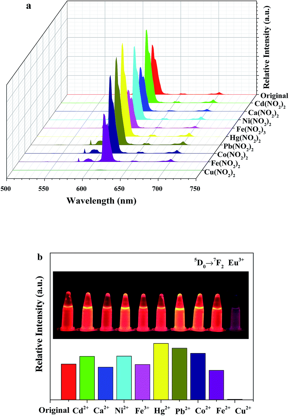 | ||
| Fig. 5 (a) Suspension-state photoluminescence spectra and (b) the relative intensities of 5D0–7F2 at 613 nm for SiO2@EuTTA@ZIF-8 dispersed in various metal ion aqueous solutions upon excitation at 396 nm. Inset are the corresponding photographs under UV-light irradiation. | ||
In Fig. 6, SiO2@EuTTA@ZIF-8 is immersed in different concentrations of Cu2+ for further luminescence studies. The luminescence intensity of SiO2@EuTTA@ZIF-8 incorporated with Cu2+ shows a strong dependence on the concentration. The luminescence intensity decreases proportionally with the concentration of Cu2+. The emission intensity of the SiO2@EuTTA@ZIF-8 solution decreases sharply as the Cu2+ concentration increases from 0 to 500 μM. Quantitatively, this decrease can be rationalized using the Stern–Volmer equation:
| I0/I = 1 + Ksv[M] |
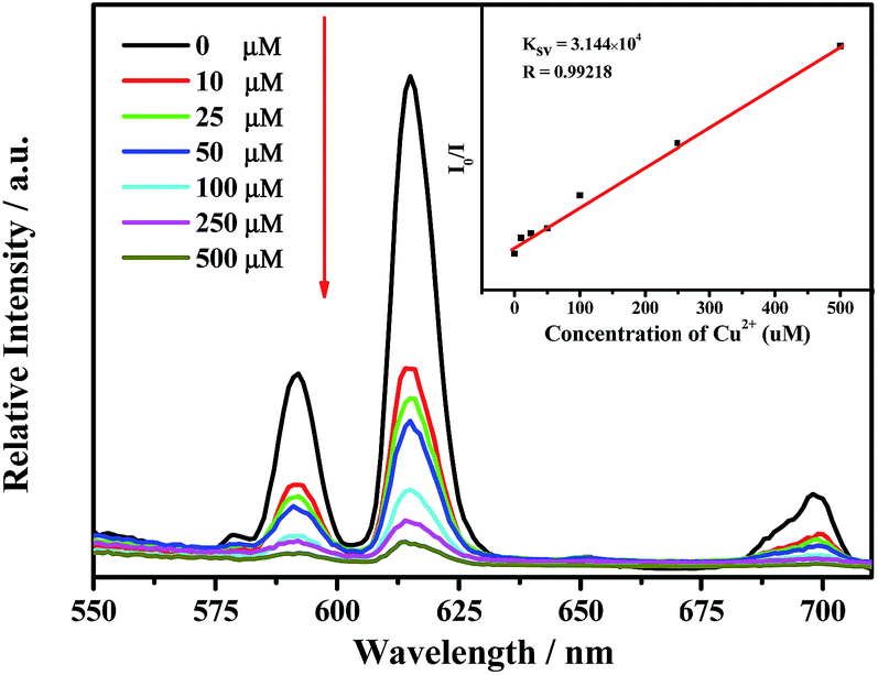 | ||
| Fig. 6 Emission spectra of SiO2@EuTTA@ZIF-8 in aqueous solutions in the presence of various concentrations of Cu2+ under excitation at 396 nm. Inset is the Ksv curve (linear relationship of the emission intensity) of SiO2@EuTTA@ZIF-8 decreased by Cu2+ ions. | ||
The values of I0 and I are the luminescence intensities of SiO2@EuTTA@ZIF-8 and SiO2@EuTTA@ZIF-8/Cu2+, respectively. Ksv is the decrease constant, and [M] is the concentration of Cu2+. The experimental data of the Ksv curve of SiO2@EuTTA@ZIF-8 with Cu2+ is illustrated in the inset of Fig. 6, and the linear correlation coefficient (R) is 0.99218, which suggest that the decrease effect of Cu2+ on the luminescence of SiO2@EuTTA@ZIF-8 agrees well with the Stern–Volmer mode. The Ksv value is calculated to be 3.144 × 10⁴ M−1, which reveals a strong decrease effect on the SiO2@EuTTA@ZIF-8 luminescence.
To examine the possible mechanism for the luminescence quenching effect by the metal ions, O 1s X-ray photoelectron spectroscopy studies are carried out on SiO2@EuTTA@ZIF-8 and SiO2@EuTTA@ZIF-8/Cd2+ (Fig. 7). The O 1s peak is shifted to 530.4 eV upon the addition of Cu2+ compared with SiO2@EuTTA@ZIF-8 at 530.0 eV. There may be a weak coordination binding of uncoordinated carbonyl to Cu2+ in SiO2@EuTTA@ZIF-8/Cd2+. Such coordination of Cu2+ ions can enhance various non-radiative activations and increase energy loss. This reduces the intraligand ligand-to-Eu energy transfer efficiency, and quenches Eu red light emission. Time-resolved fluorescence emission measurements are also used to further demonstrate the luminescence quenching effect of the Cu2+ion, and the response of the luminescence lifetime is shown in Table S1.† It is found that Cu2+-incorporated SiO2@EuTTA@ZIF-8 has a shorter emission lifetime than SiO2@EuTTA@ZIF-8, which is in agreement with the decrease in the photoluminescence intensity.
 | ||
| Fig. 7 O 1s XPS spectra of SiO2@EuTTA@ZIF-8 (black) and Cu2+-incorporated SiO2@EuTTA@ZIF-8/Cu2+ (red) dispersed in 10−2 M H2O solution of Cu(NO3)2. | ||
Finally, we examine the recyclability of the SiO2@EuTTA@ZIF-8 microspheres for sensing processes. SiO2@EuTTA@ZIF-8 microspheres can be reused several times by simply washing the chemosensors with fresh MeCN after the sensing process (Fig. 8).
 | ||
| Fig. 8 (a) On–off cycles of SiO2@EuTTA@ZIF-8 by alternating treatment of Cu2+ and MeCN at room temperature; (b) photograph of the SiO2@EuTTA@ZIF-8 microspheres recycled 1 time for Cu2+ sensing. | ||
Conclusions
In summary, heterostructured sensors are constructed using a luminescent lanthanide complex (Eu–TTA) and metal organic framework (ZIF-8) to cover the silica core. The thin heterostructured composite silica@EuTTA@ZIF-8 microsphere particles are used as a highly selective and sensitive sensor to detect Cu2+ in aqueous solution, showing a good fluorescence stability, low detection limit and broad linear range in an aqueous environment. This makes it a potential chemosensor for environmental sensing of Cu2+ due to its excellent chemical stability, low contamination and excellent recyclability ,which are regarded as necessary criteria for a new generation of sensors.Acknowledgements
This work is supported by the National Natural Science Foundation of China (21571142) and Developing Science Funds of Tongji University.Notes and references
- (a) N. Sabbatini, M. Guardigli and J. M. Lehn, Coord. Chem. Rev., 1993, 123, 201 CrossRef CAS; (b) T. Justel, H. Nikol and C. Ronda, Angew. Chem., Int. Ed., 1998, 37, 3085 CrossRef; (c) D. Parker, R. S. Dickins, H. Puschmann, C. Crossland and J. A. K. Howard, Chem. Rev., 2002, 102, 1977 CrossRef CAS PubMed; (d) A. J. Kenyon, Prog. Quantum Electron., 2002, 26, 225 CrossRef CAS.
- (a) J. C. G. Bunzli, Acc. Chem. Res., 2006, 39, 53 CrossRef PubMed; (b) S. V. Eliseeva and J. C. G. Bunzli, Chem. Soc. Rev., 2010, 39, 189 RSC.
- (a) P. A. Tanner, B. Yan and H. J. Zhang, J. Mater. Sci., 2000, 35, 4325 CrossRef CAS; (b) B. Yan, Mater. Lett., 2003, 57, 2535 CrossRef CAS; (c) B. Yan and Q. M. Wang, Opt. Mater., 2004, 27, 533 CrossRef CAS.
- (a) Q. P. Li and B. Yan, J. Colloid Interface Sci., 2012, 380, 67 CrossRef CAS PubMed; (b) Y. Y. Li and B. Yan, Dalton Trans., 2013, 42, 1678 RSC.
- (a) L. D. Carlos, R. A. S. Ferreira, V. D. Bermudez and S. J. L. Ribeiro, Adv. Mater., 2009, 21, 509 CrossRef CAS PubMed; (b) K. Binnemans, Chem. Rev., 2009, 109, 4283 CrossRef CAS PubMed.
- (a) B. Yan, RSC Adv., 2012, 2, 9304 RSC; (b) J. Feng and H. J. Zhang, Chem. Soc. Rev., 2013, 42, 387 RSC.
- L. D. Carlos, R. A. S. Ferreira, V. D. Bermudez, B. Julian-Lopez and P. Escribano, Chem. Soc. Rev., 2011, 40, 536 RSC.
- (a) A. C. Franville, D. Zambon and R. Mahiou, Chem. Mater., 2000, 12, 428 CrossRef CAS; (b) H. R. Li, J. Lin, H. J. Zhang, H. C. Li, L. S. Fu and Q. G. Meng, Chem. Commun., 2001, 1212 RSC; (c) Q. M. Wang and B. Yan, J. Mater. Chem., 2004, 14, 2450 RSC; (d) B. Yan and Q. M. Wang, Cryst. Growth Des., 2008, 8, 1484 CrossRef CAS; (e) B. Yan and H. F. Lu, Inorg. Chem., 2008, 47, 5601 CrossRef CAS PubMed.
- (a) Y. Wang, H. R. Li, Y. Feng, H. J. Zhang, G. Calzaferri and T. Z. Ren, Angew. Chem., Int. Ed., 2010, 49, 1434 CrossRef CAS PubMed; (b) P. P. Cao, Y. G. Wang, H. R. Li and X. Y. Yu, J. Mater. Chem., 2011, 21, 2709 RSC; (c) J. N. Hao and B. Yan, Dalton Trans., 2014, 43, 2810 RSC; (d) L. Chen and B. Yan, Dalton Trans., 2014, 43, 14123 RSC.
- (a) L. N. Sun, H. J. Zhang, L. S. Fu, F. Y. Liu, Q. G. Meng, C. Y. Peng and J. B. Liu, Adv. Funct. Mater., 2005, 15, 1041 CrossRef CAS; (b) X. M. Guo, H. D. Guo, L. S. Fu, R. P. Deng, W. Chen, J. Feng, S. Dang and H. J. Zhang, J. Phys. Chem. C, 2009, 113, 2603 CrossRef CAS; (c) Y. Li, B. Yan and H. Yang, J. Phys. Chem. C, 2008, 112, 3959 CrossRef CAS; (d) Y. J. Li, L. Wang and B. Yan, J. Mater. Chem., 2011, 21, 1130 RSC.
- (a) B. Yan and X. F. Qiao, J. Phys. Chem. B, 2007, 111, 12362 CrossRef CAS PubMed; (b) X. F. Qiao and B. Yan, J. Phys. Chem. B, 2008, 112, 14742 CrossRef CAS PubMed; (c) X. F. Qiao and B. Yan, J. Phys. Chem. B, 2009, 113, 11865 CrossRef CAS PubMed; (d) D. M. Wang, J. H. Zhang, Q. Lin, L. S. Fu, H. J. Zhang and B. Yang, J. Mater. Chem., 2003, 13, 2279 RSC.
- (a) B. H. Kwon, H. S. Jang, H. S. Yoo, S. W. Kim, D. S. Kang, S. Maeng, D. S. Jang, H. Kimand and D. Y. Jeon, J. Mater. Chem., 2011, 21, 12812 RSC; (b) Y. Zhao and B. Yan, Dalton Trans., 2012, 41, 5334 RSC; (c) Y. Zhao and B. Yan, J. Colloid Interface Sci., 2013, 395, 145 CrossRef CAS PubMed; (d) Q. P. Li and B. Yan, RSC Adv., 2012, 2, 10840 RSC.
- (a) J. Cuan and B. Yan, Dalton Trans., 2013, 43, 14230 RSC; (b) J. Cuan and B. Yan, RSC Adv., 2013, 3, 20077 RSC; (c) J. Cuan and B. Yan, RSC Adv., 2014, 4, 1735 RSC; (d) J. Cuan and B. Yan, Dalton Trans., 2013, 43, 14230 RSC.
- (a) Y. Mei and B. Yan, New J. Chem., 2013, 37, 2619 RSC; (b) T. W. Duan and B. Yan, CrystEngComm, 2014, 16, 3395 RSC.
- (a) M. Kurmoo, Chem. Soc. Rev., 2009, 38, 1353 RSC; (b) S. Natarajan and P. Mahata, Chem. Soc. Rev., 2009, 38, 2304 RSC; (c) O. Shekhah, J. Liu, R. A. Fischer and C. Woll, Chem. Soc. Rev., 2011, 40, 1081 RSC; (d) A. Betard and R. A. Fischer, Chem. Rev., 2012, 112, 1055 CrossRef CAS PubMed.
- (a) J. J. Perry IV, J. A. Perman and M. J. Zaworotko, Chem. Soc. Rev., 2009, 38, 1400 RSC; (b) D. Zhao, D. J. Timmons, D. Q. Yuan and H. C. Zhou, Acc. Chem. Res., 2011, 44, 123 CrossRef CAS PubMed; (c) M. O’Keeffe and O. M. Yaghi, Chem. Rev., 2012, 112, 675 CrossRef PubMed; (d) F. A. Almeida Paz, J. Klinowski, S. M. F. Vilela, J. P. C. Tome, J. A. S. Cavaleiro and J. Rocha, Chem. Soc. Rev., 2012, 41, 1088 RSC.
- (a) O. M. Yaghi, M. O’Keeffe, N. W. Ockwig, H. K. Chae, M. Eddaoudi and J. Kim, Nature, 2003, 423, 705 CrossRef CAS PubMed; (b) H. Hayashi, A. P. Cote, H. Furukawa, M. O’Keeffe and O. M. Yaghi, Nat. Mater., 2007, 6, 501 CrossRef CAS PubMed; (c) X. C. Huang, Y. Y. Lin, J. P. Zhang and X. M. Chen, Angew. Chem., Int. Ed., 2006, 45, 1557 CrossRef CAS PubMed.
- (a) J. Cravillon, S. Münzer, S. J. Lohmeier, A. Feldhoff, K. Huber and M. Wiebcke, Chem. Mater., 2009, 21, 1410 CrossRef CAS; (b) H. Bux, F. Y. Liang, Y. S. Li, J. Cravillon, M. Wiebcke and J. Caro, J. Am. Chem. Soc., 2009, 131, 16000 CrossRef CAS PubMed; (c) S. R. Venna and M. A. Carreon, J. Am. Chem. Soc., 2010, 132, 76 CrossRef CAS PubMed.
- (a) R. Ostermann, J. Cravillon, C. Weidmann, M. Wiebcke and B. M. Smarsly, Chem. Commun., 2011, 47, 442 RSC; (b) J. F. Yao, D. H. Dong, D. Li, L. He, G. S. Xu and H. T. Wang, Chem. Commun., 2011, 47, 2559 RSC; (c) G. Lu, S. Z. Li, Z. Guo, O. K. Farha, B. G. Hauser, X. Y. Qi, Y. Wang, X. Wang, S. Y. Han, X. G. Liu, J. S. DuChene, H. Zhang, Q. C. Zhang, X. D. Chen, J. Ma, S. C. J. Loo, W. D. Wei, Y. H. Yang, J. T. Hupp and F. W. Huo, Nat. Chem., 2012, 4, 310 CrossRef CAS PubMed; (d) W. W. Zhan, Q. Kuang, J. Z. Zhou, X. J. Kong, Z. X. Xie and L. S. Zheng, J. Am. Chem. Soc., 2013, 135, 1926 CrossRef CAS PubMed.
- X. Lian and B. Yan, New J. Chem., 2015, 39, 5898 RSC.
- (a) A. S. Huang, H. Bux, F. Steinbach and J. Caro, Angew. Chem., Int. Ed., 2010, 49, 4958 CrossRef CAS PubMed; (b) A. S. Huang and J. Caro, Angew. Chem., Int. Ed., 2011, 50, 4979 CrossRef CAS PubMed; (c) A. S. Huang, N. Y. Wang, C. L. Kong and J. Caro, Angew. Chem., Int. Ed., 2012, 51, 10551 CrossRef PubMed.
- (a) C. Liu and B. Yan, Eur. J. Inorg. Chem., 2015, 279 CrossRef; (b) C. Liu and B. Yan, RSC Adv., 2015, 5, 11101 RSC; (c) C. Liu and B. Yan, New J. Chem., 2015, 39, 112 Search PubMed.
- C. Jo, H. J. Lee and M. Oh, Adv. Mater., 2011, 23, 1716 CrossRef CAS PubMed.
- D. J. Macquarrie, Chem. Commun., 1996, 1961 RSC.
- Q. P. Liu, L. X. Gao, Z. W. Gao and L. Yang, Mater. Lett., 2007, 61, 4456 CrossRef CAS.
- Q. P. Li, Z. L. Yuan, J. J. Qian and S. W. Du, RSC Adv., 2015, 5, 49110 RSC.
Footnote |
| † Electronic supplementary information (ESI) available. See DOI: 10.1039/c5ra19973e |
| This journal is © The Royal Society of Chemistry 2015 |