DOI:
10.1039/C5RA19959J
(Paper)
RSC Adv., 2015,
5, 102818-102827
Solvent assisted and solvent free orientation of growth of nanoscaled lanthanide sulfides: tuning of morphology and manifestation of photocatalytic behavior†
Received
27th September 2015
, Accepted 24th November 2015
First published on 26th November 2015
Abstract
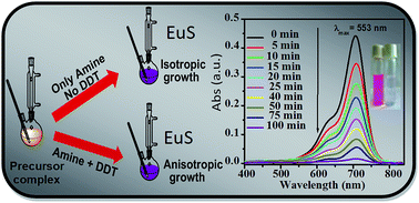
A new class of precursor complexes Ln(acda)3(phen), (where Ln = Nd, Sm, Eu, Tb and Yb, acda− is the anion of 2-aminocyclopentene-1-dithiocarboxylic acid and phen stands for 1,10-phenanthroline) have been used to obtain phase pure lanthanide sulfide nanoparticles by solution-based as well as solution-free thermal treatments at inert conditions. During solution-phase thermolysis, long chain alkyl-amine solvents have been used to promote the reaction at much lower temperature (280 °C) than the solid-phase reaction (650 °C). A contrasting growth feature is observed for nano sulfides and consequently the shape of the material is varied from isotropic cube-like morphology to anisotropic short nanofibers according to the variation of surfactants. The study confirmed that the introduction of 1-dodecanethiol as a structure-modifying capping agent along with reacting amine significantly facilitates the anisotropic growth in a preferred direction. These have been characterized by XRD, TEM, FESEM, UV-vis spectroscopy and BET surface area measurements. The optical absorption data indicated a narrow band gap energy ranging from 1.71–1.97 eV for different EuS. The material emerged as a highly active visible light-driven photocatalyst among the lanthanide sulfides towards the degradation of organic dyes. A comparative catalytic study with morphologically different EuS revealed that the degradation rate changes with varying morphology and for all the dyes it strictly follows the decreasing order of sphere-like particles > cube-like particles > nanofibers.
1. Introduction
In the last few decades, the field of nano-scaled semiconducting materials has undergone a significant expansion and become one of the most dominating research areas within the nanoscience community. There is a growing need for highly efficient, low cost materials which can be used in catalysis, electronics, photonics, energy conversion and storage, data storage and drug delivery.1–7 Due to these unique properties and promising applications in various domains, many wet and non-wet synthetic procedures have been developed, including high temperature thermolysis by utilizing single source precursors for the fabrication of semiconducting materials, tuned into different morphology nanostructures, such as nanocubes, nanoplates, nanowires, nanobelts etc.8–14 Among these, the particular solvent assisted one dimensional growth of nanoparticles is an important domain of investigation as it selectively affects the materials properties in various ways.15–19
Recently, a sincere efforts were involved towards the preparation of variety of lanthanide based nanomaterials such as oxides,20,21 fluorides,22–25 sulfides,26–37 oxy-sulfides,38–42 phosphates,43 molybdates44 and vanadates45 due to their fascinating application in the opto-electronic devices, high performance magnets biological cell imaging, drug carriers and so on.46–48 Among them, lanthanide sulfides have drawn sufficient attraction to the researchers due to their several interesting features. Although, the synthesis of LnS/Ln2S3 nanoparticles are still challenging for the material scientists as Ln3+ are too hard to combine spontaneously with the potentially soft S2−. On the other hand, as lanthanides are extremely sensitive towards the oxygen and always tend to form the oxides; even the trapped water molecules in the precursor or the moisture can spoil it.
In the present study, we report an effective synthetic routes to produce lanthanide sulfides with variable shape via thermal decomposition of a new class of precursor complex of [Ln(acda)3(phen)] (phen = 1,10-phenanthroline; Hacda = 2 aminocyclopentene-1-dithiocarboxylic acid) by simply managing the proper choice of surfactants. Both the precursor complex and nanomaterials are thoroughly characterized by the common investigating tools (UV-vis spectroscopy, FT-IR study, ESI-MS analysis and HR-XRD, TEM, FESEM, EDX respectively). Studies have shown that, all other lanthanides (Nd, Sm, Tb, Yb) except europium form pure trivalent sulfides whereas europium forms pure bivalent sulfide which have been indicated by the high resolution X-ray diffraction peaks. A major finding obtained during the course of studies of the nanomaterials is the observation of enhanced photocatalytic activity of EuS compared to the other lanthanide sulfides under visible light irradiation towards the degradation of rhodamine B, congo-red and methylene blue dyes. In order to address the morphological effects on the catalytic activity of EuS, the same catalytic experiment has been performed with EuS of three different shapes and finally a correlation is obtained.
2. Experimental
2.1. Materials
All chemicals were of reagent grade and used without further purification. Ln(NO3)3·xH2O (x = 6 for Ln = Nd, Sm, Tb and x = 5 for Ln = Eu, Yb), oleylamine (OAm), 1-dodecanethiol (DDT), hexadecylamine (HDA), rhodamine-B (RhB), congo-red (CR) and methylene blue (MB) were purchased from Sigma-Aldrich. Cyclopentanone, carbon disulfide, 1,10-phenanthroline monohydrate (phen), triethylamine, p-benzoquinone (BQ), tert-butyl alcohol (TBA) and ammonium oxalate (AO) were purchased from Spectrochem Pvt Ltd (India). Nano-sized titanium dioxide (Degussa-P25) was purchased from Degussa Company. Solvents were used as received.
2.2. Synthesis
The ligand 2-aminocyclopentene-1-dithiocarboxylic acid (Hacda) was prepared according to the previously published procedure.49
2.2.1. Synthesis of the precursor complex [Eu(acda)3(phen)]. A solution of Hacda (0.48 g, 3 mmol) and 1,10-phenanthroline monohydrate (0.2 g, 1 mmol) in dry methanol (20 mL) were added to a methanolic solution (10 mL) of europium(III) nitrate pentahydrate (0.43 g, 1 mmol). To this was slowly added with stirring a methanol solution of triethylamine (0.31 g, 3 mmol). The bright orange solid was separated immediately and the stirring was continued for another 20 min. Then, the product was filtered, washed with cold methanol and dried in vacuum; yield 0.548 g (68%). Anal. calcd for C30H32N5EuS6: C, 44.66; H, 3.97; N, 8.68. Found: C, 44.12; H, 4.16; N, 8.62. IR data (KBr pellet, cm−1): 3354 (m, br), 2941 (s, br), 1609 (s), 1465 (s), 1421 (s), 1385 (s), 1311 (s), 1219 (m), 1146 (m), 1102 (m), 1034 (m), 912 (m), 853 (m), 812 (m), 727 (m). ESI-MS (positive) in MeOH: m/z 807.97 [Eu(acda)3(phen)H]+ (35%). UV-vis [in N,N-dimethylformamide, λmax, nm (ε/M−1 cm−1)] 386 (38
200), 332 (22
400).Mixed ligand complexes of Nd3+, Sm3+, Tb3+ and Yb3+ have been prepared in the same way as stated above. Detailed characterization data for the remaining complexes were given in the ESI.†
2.2.2. Syntheses of EuS nanoparticles.
2.2.2.1. Solid-state thermolysis (1). The mixed ligand complex Eu(acda)3(phen) (0.5 g, 0.62 mmol) was taken on a graphite boat and placed inside a quartz tube furnace horizontally. The system was degassed under vacuum for 30 min. Then the sample was heated to 120 °C under vacuum for 30 min prior to strong heating. Then the whole system was filled with argon gas and the temperature was raised to 650 °C (with a heating rate 20 °C min−1) under a gentle argon flow through the tube. The heating was continued for 1 h after reaching the desired temperature and then slowly cooled down to room temperature under inert atmosphere. Highly crystalline black solid was obtained.
2.2.2.2. Solution phase thermolysis (2).
Synthesis of cube-like nanocrystals (2a).
The precursor [Eu(acda)3(phen)] (0.4 g, 0.5 mmol) was added to OAm (10 mL, 30 mmol) in a 100 mL two-necked flask at room temperature. The slurry was heated to 100 °C under vacuum to remove moisture and oxygen with vigorous magnetic stirring for several minutes in a heating mantle and finally a clear solution is obtained. Then, the mixture was heated to 280 °C quickly under argon atmosphere and kept for 1 h. As soon as it reached the temperature, the resulting solution became dark purple. After cooling to room temperature, the nanoparticles were precipitated by adding excess amount of dry ethanol. The product was collected by centrifugation (9000 rpm, 8 min) and dispersed in toluene and again reprecipitated with ethanol. The process was repeated several times to make the product free from reactants. It was then dried in vacuum.
Synthesis of nanocrystals (2b).
The synthesis was carried out under argon atmosphere. In a typical procedure, HDA (7.2 g, 30 mmol), taken in clean two-necked flask was heated to 50 °C. To this 0.4 g of precursor complex [Eu(acda)3(phen)] was added under vigorous stirring. The suspension was heated to 120 °C under vacuum for 15 min to get a clear solution. The temperature of the stirred solution was then rapidly raised to 280 °C under argon protection and maintained for 1 h. After cooling to room temperature, the product was isolated in the same way as described for cube-like particles.
Synthesis of nanofiber (2c).
To a stirred mixture of OAm (10 mL, 30 mmol) and DDT (2.4 mL, 10 mmol), 0.4 g of precursor complex was added. The temperature of the mixture was brought to 120 °C under vacuum and kept so until a clear solution was obtained. The solution was then heated to 280 °C for 1 h during which the color of the solution appeared as dark purple. After cooling to room temperature excess ethanol was added to quench the reaction followed by centrifugation (9000 rpm, 8 min). The washing procedure was identical as described in above cases.
To study the effect of the [OAm]/[DDT] ratio, the above experiment was repeated by using 15 mmol (3.6 mL) (2d) and 30 mmol (7.2 mL) (2e) DDT respectively keeping all other factors fixed. The role of amine was also studied by replacing OAm with HDA (7.2 g, 30 mmol) along with DDT (2.4 mL, 10 mmol) (2f) and by performing the experiment in DDT (7.2 mL, 30 mmol) alone (2g).
The nanoparticle synthesis were carried out with the other Ln(acda)3(phen) complexes (Ln = Nd, Sm, Tb. Yb) following the same procedure stated above.
2.3. Physical measurements
Powder XRD patterns were obtained by using Philips PW 1140 parallel beam X-ray diffractometer with monochromatic CuKα radiation (λ = 1.540598 Å). Surface morphologies were studied using a JEOL JEM-2100 transmission electron microscope (TEM) working at 200 kV. FE-SEM images were acquired with a Gemini Zeiss Supra 40VP field emission scanning electron microscope (FE-SEM) with a 20 kV accelerating voltage. Energy dispersed X-ray characterizations (EDX) were performed in JEOL JSM-7100F. FTIR spectra were obtained in the range of 4000–400 cm−1 as pressed pellets in KBr on JASCO FT-IR-460 Plus. Positive-ion electron spray ionization mass spectra (ESI-MS) were recorded on a Micromass Qtof YA 264 mass spectrometer. Thermogravimetric analysis (TGA) was carried out on Perkin-Elmer Diamond TG/DTA analyzer. Absorption spectral measurement and photocatalytic experiment were performed on a JASCO V-530 UV-vis spectrophotometer. C, H and N elemental analyses were performed on a Perkin-Elmer model 2400 analyzer. The BET (Brunaur–Emmett–Teller) surface area of the samples were analysed on Quantachrome Autosorb-1 instrument.
2.4. Photocatalytic activity measurements
Photocatalytic performance of the prepared EuS was tested by the degradation of some common hazardous organic dyes like RhB, CR, MB etc. The experiments were carried out in a double necked round bottom flask kept in a thermostated bath at 27 °C and the light source used in the measurements was a 200 W tungsten halogen lamp (≥400 nm) placed vertically on the reaction vessel at a distance of ∼10 cm. 1 M NaNO2 solution was used as UV cut-off filter.6 The catalytic experiments were carried out with 40 mL, 1 Th 10−5 M aqueous solution of RhB, CR and MB separately using 10 mg of the catalyst. Before light was turned on, the well dispersed suspensions were magnetically stirred in the dark for 30 min to confirm the establishment of the adsorption–desorption equilibrium. During irradiation, 3 mL aliquot was withdrawn from the system at a certain time intervals and centrifuged. The clear solutions of the respective dyes were analyzed by recording the variation of absorption band maximum on a UV-vis spectrophotometer. Commercial photocatalyst TiO2 (Degussa-P25) was also used as the reference to compare the photo-catalytic efficiency under the same experimental conditions. The photoactivity was also monitored in presence of different radical scavengers (ammonium oxalate as photogenerated hole scavenger, tert-butyl alcohol as a hydroxyl radical scavenger and p-benzoquinone as a superoxide radical scavenger) following the above similar procedure to investigate the effect of reactive species in reaction mechanism.
The stability of the photocatalyst during photocatalytic process was also monitored by repeated experiments of dye degradation. The catalysts were washed thoroughly by anhydrous ethanol and then air dried to use it again for the second runs of catalytic efficiency testing. The recyclability testing up to fifth run was performed to establish the catalytic stability of the material.
3. Results and discussion
3.1. Synthesis and characterization
In the last few years, several dithiocarbamate complexes of lanthanides of the type [Ln(S2CNR2)3L] (R = alkyl group; L = 2,2′-bipyridine/1,10-phenanthroline), Q+[Ln(S2CNR2)4]− (Q = quaternary ammonium/trialkyphosphonium salt) are extensively used as model precursor complexes for the preparation of different binary and ternary chalcogenide materials.26,27,32–34 In present work, we have designed a new class of molecular precursors Ln(acda)3(phen)] (Ln = Nd, Sm, Eu, Tb and Yb) (Scheme 1) which have been successfully employed to synthesize LnS/Ln2S3 by simple thermal treatment.
 |
| Scheme 1 | |
The precursor Ln(acda)3(phen) complexes have been obtained by reacting one equivalent of corresponding lanthanide salts with three equivalents of Hacda, one equivalent of phen and three equivalents of triethylamine. Because of its poor solubility in common organic solvents (except DMF and DMSO), attempts to grow suitable single crystals for X-ray structure analysis was unsuccessful even in DMF also. Nevertheless, it is evident from the chemical analysis that the compound prepared was analytically pure and characterized by IR, UV-vis spectra and ESI-MS (Fig. S1, S2 and S3 respectively in the ESI†). FT-IR spectrum reveals the presence of symmetric C–S band in the region of 1034 cm−1 and the characteristic C⋯CH bend vibration of phen at 727 cm−1. UV-vis absorption spectrum exhibits two intra-ligand charge transfer bands at 386 and 332 nm. Similarly ESI-MS spectrum of Eu(acda)3(phen) shown in Fig. S3† indicates the m/z value at 807.97 which is in excellent agreement with the mono-positive charged species [Eu(acda)3(phen)H]+, confirming the proposed composition of the complex.
3.1.1. Synthesis and structural characterization of nanoparticles. Nano-structured EuS has been successfully synthesized by simple one-pot approach employing solid state thermal and solvothermal decomposition of as prepared mixed ligand complex (Scheme 2). In case of solution based reaction, the influence of synthetic conditions on the growth and morphology of the product has been investigated by changing solvents and surfactants at elevated temperature.50,51 It was observed that the particular reagents used in this synthesis play a decisive role in the growth of nanoparticles in a specific direction.52,53
 |
| Scheme 2 | |
The crystalline phases of pure EuS were determined by powder X-ray diffraction (XRD) analysis. Fig. 1 shows the powder X-ray diffractogram of europium sulfide where all diffraction peaks were indexed to cubic phase of the material. As shown in Fig. 1, the diffraction pattern of europium sulfide synthesized via solid state thermolysis exhibits distinct sharp peaks which are in good agreement with the (111), (200), (220), (311), (222), (400), (331), (420), (422) lattice planes (JCPDS no. 26-1419, a = 5.967, space group: Fm3m) respectively. No additional signals from other phases or from impurities are identified. If we closely observe the peak pattern and their relative intensities in all cases we will find that clear peak broadening occurs in case of solvothermally decomposed products which might be due to the loss of crystallinity.
 |
| Fig. 1 XRD patterns of EuS. (a) nanoparticles synthesized by solid sate thermolysis EuS (1); (b) cube-like EuS (2a) and (c) nanotubes EuS (2f) prepared by solvothermal decomposition. | |
It is also noted that the intensities of few diffraction peaks are not similar in some cases. If we consider the peaks obtained for EuS nanofiber (Fig. S4 in ESI†), we found that the peaks corresponding to (222), (331) and (422) lattice planes are not clearly distinguishable. Apart from EuS, the other lanthanides form air stable trivalent sulfides of the type Ln2S3 (Ln = Nd, Sm, Tb, Yb), as evidenced from the powder XRD patterns (Fig. S5 in ESI†). Distinct sharp peaks were obtained in case of Nd2S3 and Sm2S3 synthesized by solid state thermal decomposition and they fit well with standard diffraction data. On the other hand, Tb2S3, Yb2S3 appears to be amorphous by XRD analysis.
The morphology and the dimension of the product were examined by TEM analysis. Typical TEM image of the product (1) are shown in Fig. 2A, indicating the formation of sphere-like particles which are of 25–35 nm in diameter. The corresponding HRTEM images in Fig. 2E reveals that the particles are composed of several layers where the interlayer distance is calculated to be 0.211 nm (220) and 0.180 nm (311). The average crystal size estimated from Scherrer equation i.e., D = 0.94λ/β
cos
θ, where β is the peak width at half maxima, is found to be ∼38 nm. In case of other lanthanides also same sphere-like morphology was obtained (Fig. S6 in ESI†) on solid state thermal treatment.
 |
| Fig. 2 TEM (A, B and D) and HRTEM (C, E, G and H) images of as-synthesized EuS nanoparticles; (A) and (E) EuS (1); (B), (C) and (G) EuS (2a); (D) and (H) EuS (2b); (F) represents SAED pattern of EuS (2a). | |
In solution based synthesis of EuS, we observed that the surface stabilizers and capping agents play a serious role in determining the shape and the morphology of the product when other factors remain fixed.53–55 It has been well observed that the surfactant molecules generally serve as structure modifying agents and easily modulate the growth of the nanoparticles in a preferred direction by interacting with various crystal facets at different extent. The TEM and HRTEM images of EuS prepared by using OAm are shown in Fig. 2B and C respectively indicating a cube-like morphology. Fig. 2G shows the clear lattice fringes with an average spacing of d = 0.291 ± 0.005 nm corresponding to (200) lattice plane of cubical phase. Selected area electron diffraction (SAED) patterns of these cube-like nanocrystals (Fig. 2F) exhibit concentric rings that can be indexed to (111), (200), (220), (311), (222), (400), (420) diffraction planes for face centred cubical phase of EuS. The purity of the cube-like EuS was further verified by EDX analysis (Fig. S7 in ESI†) and it has been confirmed that other than Eu and S no other elements are present. When HDA was used instead of OAm, the irregularly shaped nanocrystals (2b) was the outcome which has been displayed in Fig. 2D. HRTEM pattern of EuS (2b) (Fig. 2H) corresponds to the lattice spacing of 0.210 ± 0.002 nm (220).
Significant differences in the growth kinetics and the overall nanocrystal shape development can be observed when DDT was introduced along with reacting amine (OAm) in the reaction medium. The growth of nanocrystal was anisotropic resulting the formation of short nanofiber of length ∼12 nm (Fig. 3A). In the recent past, Stoll and her group have synthesized europium chalcogenide one dimensional nanowire in a drastic condition by allowing vapour phase reaction.35 Otherwise, no such anisotropic morphology has been reported for this material. But, in the present case, a simple and selective solution based technique has been adopted for the synthesis of one dimensional lanthanide sulfide nanomaterials. Controlled experiments and extensive TEM analysis were carried out to understand the solvent effect in the growth of the nanostructures. It has been documented that DDT not only serves as an effective sulfur donor as well as good reducing agents but also plays the unique role of surface active capping agents during solvothermal synthesis of sulfide based nanomaterials.55–57 As described before, we have confirmed that DDT was neither served as sulfur source nor as reducing agent here because starting from Eu(III) precursor complex EuS was obtained successfully in absence of DDT also. Then, what roles did DDT really play here? To address this question we performed several experiments by changing the reaction condition. When the mole ratio of DDT was increased (15 mmol) with respect to OAm keeping the amount of OAm fixed (30 mmol), the growth was again anisotropic with the formation of slightly thicker nanofiber (Fig. S8A in ESI†). When OAm/DDT ratio was made further high (1
:
1), network like arrangement of nanofiber was obtained (Fig. S8B in ESI†). In a close view (HRTEM image shown in Fig. S8D†), the arrangement of nets was clearly observed. If we change the combination of OAm and DDT by replacing OAm with an equal amount of HDA along with DDT, the long nanotube (Fig. 3B) was the outcome confirming the fact that the one dimensional growth was certainly governed by DDT. On the other hand, when the choice of the solvent was constrained to DDT solely, the resulting product was highly agglomerated and amorphous in nature (Fig. S8C†). In this case, no nanofibers or nanorods were obtained. These results reveal that the use of either reacting amine or DDT does not induce the anisotropic growth. It is a synergistic effect originating from the combination of these two where DDT guides the nanocrystallites to grow in a one dimensional manner in presence of reacting amine which are basically the activating agents. The difference in shape between the two cases (presence of DDT and the absence of DDT) shows that the binding affinity of DDT on different crystal facets is not uniform leading to unequal growth rates in different directions which are responsible for the anisotropic morphologies. It acts not only to regulate the nucleation but also behaves as passivating ligands, inducing the preferential growth along certain facets. Alternately, when no DDT was present in the medium, the reacting amine (OAm or HDA) show a strong ability to bind selectively to different growth directions to instigate isotropic shape control. The results obtained for EuS holds good for other lanthanide sulfides also where DDT successfully induces the anisotropic growth (Fig. S9 in ESI†).
 |
| Fig. 3 (A) and (C) TEM and HRTEM images of EuS nanofibre (2c); (B) and (D) TEM and HRTEM images of EuS nanotube (2f). (C) Inset: SAED pattern of EuS nanofiber (2c). | |
The effect of temperature on the nanoparticle growth has not been investigated here because the precursor decomposition and consequently nanoparticle nucleation which was evident from the sharp color change of the reaction mixture (Fig. S10 in ESI†) would not be initiated at the temperature below 280 °C.
FESEM images of EuS (2a) and EuS (2b) has been shown in Fig. S11A and B in ESI.† The cube-like morphology of EuS has been confirmed from FESEM investigation also (Fig. S11A inset in ESI†). Spherical shape of Yb2S3 synthesized in solution free method was also evident from the FESEM study (Fig. S11C in ESI†).
3.2. Optical properties
Optical properties of EuS nanoparticles have great importance as it provides information for the application of the material in fabrication of photo-active devices. The UV-vis absorption spectra of as synthesized europium(II) sulfides (shown in Fig. 4 and S12 in the ESI†) were recorded using the sample solution in toluene. The absorption spectra of EuS exhibit a broad maximum in the range of 520–550 nm and a sharp rise in the low wavelength region (shown in Fig. 4 and S12 in the ESI†). The former one can be attributed to the 4f7 (8S7/2) → 4f6 (7FJ) 5d (t2g)1 transition whereas the latter one is assigned to characteristic charge transfer from S2− to Eu2+. From the recorded optical spectra, the band gap energies (Eg) of EuS have been estimated using the Tauc's relation,58where parameters a, Eg, hν and A are the absorption coefficient, band gap, photonic energy and a constant respectively. Band gap value has been calculated by extrapolating the linear region of a plot of (ahν)1/2 versus hν. In the previous study, the band gap of bulk EuS was found to be 1.65 ev, but there was an ambiguity in determining the actual band gap for EuS nanocrystallites. The ambiguity arises due to the inconsistency in peak assignments during band gap determination.59 Guntherodt et al. showed that at 300 K there were two major peaks, one was at ∼517 nm (2.4 eV) and second one at ∼270 nm (4.6 eV).60 However, most of the reports agreed with the fact that the band in the visible region is attributed to the characteristic 4f7 → 4f65d1 (t2g) transition and actually responsible for band gap of EuS. In our study, we also observed that for all cases the band gap values vary within the range of 1.71–1.97 eV (considering visible region absorption edges) depending on the size and the morphology. The justification of this band gap values lie on the fact of dependency of Eg on particle size and consequent confinement effect. When the dimensions of the particles become comparable to the bulk exciton Bohr radius (aB), quantum confinement effect predominates resulting the increase of the energy gap.61,62 Bulk exciton Bohr radius was calculated using the relation, |
 | (2) |
where a0 is the hydrogen atom Bohr radius (∼0.529 Å), ε is the optical dielectric constant of the material, m0 is the mass of a free electron and the μ is the exciton reduced mass. The exciton Bohr radius for EuS was obtained as 1.2 nm (diameter 2.4 nm). In our case, the dimensions of the nanoparticles are much higher than that of aB. Hence, the confinement effect is practically insignificant and it is consistent with the low energy gap values (<2.0 eV) obtained for EuS. Although, a slight variation in the energy gap has been noticed with the change of size and morphology, may be attributed to the extremely weak effect of quantum confinement (Fig. 4 and S12 in ESI†). The UV-vis absorption spectra and the corresponding energy gap of other lanthanides sulfides (Fig. S13 and S14 in ESI†) were also measured by using their toluene solution (Fig. S15†) and the values obtained shows good agreement with the previous literature values.63,64
 |
| Fig. 4 Room temperature UV-vis spectra and corresponding band gap of EuS: (a) (1) and (b) (2a) and (c) (2c) in toluene medium. | |
3.3. Photocatalytic activity
As we have seen that the band gap of the material lies in the range of 1.71–1.97 eV, hence it is a quite appropriate material for the application in the visible light induced photocatalytic reactions. Therefore, the photocatalytic performance of EuS nanomaterials was explored by the photo-decomposition of some well known organic contaminants and dyes such as RhB, CR and MB in aqueous solution under visible light irradiation at room temperature and ambient pressure. In this article, we have studied the role of EuS in the photocatalytic mechanism in comparison with the other lanthanides analogues. The comparative study was also extended with morphologically different EuS synthesized by solution free as well as solution based technique. The temporal changes in the concentration of the dyes were monitored by examining the variations in the intensity of the absorption maxima in the UV-vis spectra of the respective dyes. The absorption spectrum of tetraethylated RhB shows a absorption maxima at 553 nm, which decreases rapidly and almost disappears after 100 min irradiation in presence of sphere-like EuS (1) (Fig. 5C). Similarly, visible light irradiation of the aqueous CR/EuS (1) and MB/EuS (1) dispersion causes an apparent decrease of the absorption maxima appeared at 335 and 498 nm (in case of CR) and 288 and 660 nm (in case of MB) respectively (Fig. 5A and E). The time dependent relative concentration changes of the dyes in presence of catalyst, TiO2–P25 (used as the reference) and in absence of catalyst are studied to establish the catalytic importance of the material (Fig. 5B, D and F). The study ensures that, in the absence of catalyst, the self-degradation of all the dyes is almost negligible. The decomposition kinetics have been modeled as a pseudo-first order reaction expressed by the equation ln(C0/Ct) = kt, where C0 represents the initial concentration, Ct denotes the concentration at a given reaction time ‘t’, and k is the reaction rate constant. The apparent pseudo-first-order linear relationship is obtained by the plots of ln(C0/Ct) vs. time (t) following the Langmuir–Hinshelwood model (Fig. 5B, D and F insets). The reaction rate constants and the half-life values have been calculated respectively and are given in Table S1 and S2 in ESI.† It is eventually striking to note that the catalytic activities of EuS nanoparticles are significantly better than TiO2 (Table S2 in ESI†). Moreover, it has also been observed that EuS (1) synthesized by solid state thermolysis emerged as a better catalyst than the cube-shaped variety (2a) as well as the short nanofiber (2c). The difference in the rate has been expected due to the change in BET surface area (given in Table S1 in ESI†) of the nanoparticles that fluctuate the dye adsorption on the nanocatalyst surface and hence affect the carrier transformation for the degradation.
 |
| Fig. 5 Photocatalytic degradation profile for (A) CR, (C) RhB, (E) MB; pseudo-first order kinetics plot for (B) CR degradation, (D) RhB degradation, (F) MB degradation: (a) without catalyst in dark, (b) without catalyst in light, (c) TiO2 (Digussa P25) in light, (d) EuS (1) in light. Inset: the corresponding kinetic plots. | |
Another interesting fact we could notice that the degradation rate becomes remarkably slow when EuS is replaced by other lanthanides analogues (Nd2S3, Sm2S3, Tb2S3, Yb2S3) (Table S2 in ESI†). The enhanced photocatalytic activity of EuS is attributed to its stable bivalent state and low standard reduction potential value [E0(Eu3+/Eu2+) = −0.36 V]. So, in the case of europium, photogenerated electrons are easily available to react with O2 to form reactive oxygen species O2˙− and consequently Eu2+ is oxidized to Eu3+. During this redox process, more hydroxyl radical is generated leading to enhanced catalytic efficiency and in due course Eu3+ is further reduced back to Eu2+. But in the case of other lanthanides (where they are in trivalent state), Ln4+/Ln3+ reduction potential value is extremely high which does not allow this redox transformation and subsequently disfavors the formation of reactive oxygen species (O2˙−). Hence, the degradation rate becomes insignificant.65,66 Controlled experiments using different radical scavengers are performed with RhB to probe the influence of reactive species in the reaction mechanism of dye degradation. As evidenced from Fig. S16 in ESI,† the addition of p-benzoquinone (BQ) which has the ability to trap O2˙− significantly suppresses the photocatalytic reaction rate.67 On the other hand, the addition of ammonium oxalate (AO) as hole scavenger68 does not affect the decomposition rate at all. Although, the addition of tert-butyl alcohol (TBA) as OH˙ radical scavenger68,69 provokes partial inhibition of photodegradation. To interpret the above observations, it can be concluded that the oxidation of organic pollutants is triggered by the participation of O2˙− mainly and to some extent by the contribution of OH˙ radical. The stability of the photocatalyst was also evaluated. As shown in Fig. 6, there is no significant loss in degradation efficiency for all three dyes even after five successive cycles. It reflects that the photocatalyst is of good stability and reusability.
 |
| Fig. 6 Recyclability testing of photocatalytic activity of EuS (1) towards the degradation of CR, RhB, MB under ambient conditions. | |
4. Conclusion
In summary, we have developed a facile synthetic route for the preparation LnS/Ln2S3 of nano dimension with controllable shape by solid state and solution-based thermal decomposition of a potential single source precursor. On changing the reacting amines and the surfactants the shape and the morphology of EuS has been changed from cube-like particles to short nanofiber to nanotube selectively. The study revealed that the anisotropic growth of europium sulfide was attained due to the combined effect of reacting amine and DDT where the later one was believed to play the pivotal role in governing the nanoparticle growth. Moreover, this study successfully opens up an avenue for the synthesis of one dimensional lanthanide sulfide nanomaterial in a simple and selective way. In addition, EuS has emerged as highly active visible light driven photocatalyst among all other lanthanide sulfides. Among the different EuS, the catalytic efficiency of nanoparticles follows the order of sphere-like particles > cube-like particles > short-nanofibers, which is in good agreement with the decreasing surface area of the nanoparticles. To the best of our knowledge, it is the first report on photocatalytic activity of europium sulfide which will encourage the material scientists to explore the other photo-generated properties of the material as well as to apply it as a promising candidate in the design of photo-active devices.
Acknowledgements
Authors are thankful to Prof. K. Nag, Department of Inorganic Chemistry, IACS, Kolkata, India, for helpful discussion. The authors also acknowledge CSIR (India) (scheme no. 01(2749)/13/EMR-II) for research funding. A. Sarkar is also thankful to UGC (BSR), India, for research fellowship [F. no. F.7-223/2009 (BSR)]. N. S is indebted to DST-INSPIRE (IF 130593), India. Authors also thank Dr Goutam Kumar Patra, Professor, Dept. of Chemistry, Guru Ghasidas University, Bilaspur for the FESEM analysis. The authors are also grateful to the MHRD (India) and the Department of Chemistry, IIEST, Shibpur for providing instrumental facilities.
References
- Y. Xia, P. Yang, Y. Sun, Y. Wu, B. Mayers, B. Gates, Y. Yin, F. Kim and H. Yan, Adv. Mater., 2003, 15, 353–389 CrossRef CAS.
- Y. He, D. Li, G. Xiao, W. Chen, Y. Chen, M. Sun, H. Huang and X. Fu, J. Phys. Chem. C, 2009, 113, 5254–5262 CAS.
- H. Zhong, Z. Bai and B. Zou, J. Phys. Chem. Lett., 2012, 3, 3167–3175 CrossRef CAS PubMed.
- S. K. Bhunia and N. R. Jana, ACS Appl. Mater. Interfaces, 2014, 6, 20085–20092 CAS.
- D. J. Sirbuly, M. Law, H. Q. Yan and P. D Yang, J. Phys. Chem. B, 2005, 109, 15190–15213 CrossRef CAS PubMed.
- S. Jana and A. Mondal, ACS Appl. Mater. Interfaces, 2014, 6, 15832–15840 CAS.
- H. Cheng, B. Huang, X. Cin, X. Zhang and Y. Dai, Chem. Commun., 2012, 48, 97–99 RSC.
- L. Chen, Y. B. Chen and L. M. Yu, J. Am. Chem. Soc., 2004, 126, 16334–16335 CrossRef CAS PubMed.
- T. H. Larsen, M. Sigman, A. Ghezelbash, R. C. Doty and B. A. Korgel, J. Am. Chem. Soc., 2003, 125, 5638–5639 CrossRef CAS PubMed.
- J. Kim, E. J. Lee, J. Lee, H. J. Yu, C. B. Kim, K. An, Y. Hwang, H. C Shin, G. J Park, J. Kim and T. Hyeon, J. Am. Chem. Soc., 2006, 128, 7130–7131 CrossRef PubMed.
- J. Yuan, W. N. Li, S. Gomez and S. L. Suib, Chem. Mater., 2005, 127, 14184–14185 CAS.
- S. Shuling, Y. Zhang, L. Peng, B. Xu, Y. Du, M. Deng, H. Xu and Q. Wang, CrystEngComm, 2011, 13, 4572–4579 RSC.
- T. Lu, Y. O. Ti, P. K. Loh and J. J. Vittal, J. Mater. Chem., 2006, 16, 272–277 RSC.
- Z. Gu, T. Zhai, B. Gao, X. Sheng, Y. Wang, H. Fu, Y. Ma and J. Yao, J. Phys. Chem. B, 2006, 110, 23829–23836 CrossRef CAS PubMed.
- L. Carbone, C. Nobile, M. D. Giorgi, F. D. Sala, G. Morello, P. Pompa, M. Hytch, E. Snoeck, A. Fiore, I. R. Franchini, M. Nadasan, A. F. Silvestre, L. Chiodo, S. Kudera, R. Cingolani, R. Krahne and L. Manna, Nano Lett., 2007, 7, 2942–2950 CrossRef CAS PubMed.
- C. Wang, D. Zhang, L. Xu, Y. Jiang, F. Dong, B. Yang, K. Yu and Q. Lin, Angew. Chem., Int. Ed., 2011, 50, 7587–7591 CrossRef CAS PubMed.
- X. Gou, F. Cheng, Y. Shi, L. Zhang, S. Peng, J. Chen and P. Shen, J. Am. Chem. Soc., 2006, 128, 7222–7229 CrossRef CAS PubMed.
- W. Wang, H. Cheng, B. Huang, X. Lin, X. Qin, X. Zhang and Y. Dai, J. Colloid Interface Sci., 2013, 402, 34–39 CrossRef CAS PubMed.
- C. Zou, L. Zhang, L. Zhai, D. Lin, J. Gao, Q. Li, Y. Yang, X. Chen and S. Huang, Chem. Commun., 2011, 47, 5256–5258 RSC.
- C. Y. Cao, J. Am. Chem. Soc., 2004, 126, 7456–7457 CrossRef PubMed.
- T. Yu, J. Joo, I. Y. Park and T. Hyeon, Angew. Chem., Int. Ed., 2005, 44, 7411–7414 CrossRef CAS PubMed.
- H. J. Burns, Inorg. Chem., 1965, 4, 881–886 CrossRef.
- G. S. Yi and M. G. Chow, Chem. Mater., 2007, 19, 341–343 CrossRef CAS.
- W. Y. Zhang, X. Sun, R. Si, P. L. You and H. C. Yan, J. Am. Chem. Soc., 2005, 127, 3260–3261 CrossRef PubMed.
- C. J. Boyer, F. Vetrone, A. L. Cuccia and A. J. Capobianco, J. Am. Chem. Soc., 2006, 128, 7444–7445 CrossRef PubMed.
- T. Mirkovic, M. A. Hines, P. S. Nair and G. D. Scholes, Chem. Mater., 2005, 17, 3451–3456 CrossRef CAS.
- M. D. Regulacio, N. Tomson and S. L. Stoll, Chem. Mater., 2005, 17, 3114–3121 CrossRef CAS.
- S. Thongchan, Y. Hasegawa, Y. Wada and S. Yanagida, J. Phys. Chem. B, 2003, 107, 2193–2196 CrossRef.
- F. Zhao, H. L. Sun, S. Gao and G. Su, J. Mater. Chem., 2005, 15, 4209–4214 RSC.
- A. Tanaka, H. Kamikubo, M. Kataoka, Y. Hasegawa and T. Kawai, Langmuir, 2011, 27, 104–108 CrossRef CAS PubMed.
- Y. Hasegawa, M. Kumagai, A. Kawashima, T. Nakanishi, K. Fujita, K. Tanaka and K. Fushimi, J. Phys. Chem. C, 2012, 116, 19590–19596 CAS.
- F. Zhao, H. L. Sun, G. Su and S. Gao, Small, 2006, 2, 244–248 CrossRef CAS PubMed.
- M. D. Regulacio, S. Kar, E. Zuniga, G. Wang, N. R Dollahon, G. T. Yee and S. L. Stoll, Chem. Mater., 2008, 20, 3368–3376 CrossRef CAS.
- Y. Hasegawa, Y. Okada, T. Kataoka, T. Sakata, H. Mori and Y. Wada, J. Phys. Chem. B, 2006, 110, 9008–9011 CrossRef CAS PubMed.
- W. L. Boncher, N. Rosa, S. Kar and S. L. Stoll, Chem. Mater., 2014, 26, 3144–3150 CrossRef CAS.
- W. Boncher, H. Dalafu, N. Rosa and S. Stoll, Coord. Chem. Rev., 2015, 289, 279–288 CrossRef.
- Y. Hasegawa, M. Maeda, T. Nakanishi, Y. Doi, Y. Hinatsu, K. Fujita, K. Tanaka, H. Koizumi and K. Fushimi, J. Am. Chem. Soc., 2013, 135, 2659–2666 CrossRef CAS PubMed.
- F. Zhao, M. Yuan, W. Zhang and S. Gao, J. Am. Chem. Soc., 2006, 128, 11758–11759 CrossRef CAS PubMed.
- F. Zhao and S. Gao, J. Mater. Chem., 2008, 18, 949–953 RSC.
- W. He, M. E. Osmulski, J. Lin, D. S. Koktysh, J. R. Mcbride, J. H. Park and J. H. Dickerson, J. Mater. Chem., 2012, 22, 16728–16731 RSC.
- H. Peng, S. Huang, F. You, J. Chang, S. Lu and L. Cao, J. Phys. Chem. B, 2005, 109, 5774–5778 CrossRef CAS PubMed.
- M. Pokhrel, A. G. Kumar and D. K. Sardar, J. Mater. Chem. A, 2013, 1, 11595–11606 CAS.
- S. Heer, O. Lehman, M. Hasse and U. H. Gudel, Angew. Chem., Int. Ed., 2003, 42, 3179–3182 CrossRef CAS PubMed.
- N. Zhang, W. Bu, Y. Xu, D. Jiang and J. Shi, J. Phys. Chem. C, 2007, 111, 5014–5019 CAS.
- A. Huignard, V. Buissette, G. Laurent, T. Gacoin and P. J. Boilot, Chem. Mater., 2002, 14, 2264–2269 CrossRef CAS.
- F. Wang, D. Banerjee, Y. Liu, X. Chen and X. Liu, Analyst, 2010, 135, 1839–1854 RSC.
- G. Wang, Q. Peng and Y. Li, Acc. Chem. Res., 2011, 44, 322–332 CrossRef CAS PubMed.
- D. Yang, P. Ma, Z. Hou, Z. Cheng, C. Li and J. Lin, Chem. Soc. Rev., 2015, 44, 1416–1448 RSC.
- K. Nag and D. S. Joardar, Inorg. Chim. Acta, 1975, 14, 133–141 CrossRef CAS.
- S. Mourdikoudis and L. M. L. Marzan, Chem. Mater., 2013, 25, 1465–1476 CrossRef CAS.
- B. L. Cushing, V. L. Kolesnichenko and C. J. O'Connor, Chem. Rev., 2004, 104, 3893–3946 CrossRef CAS PubMed.
- M. F. Sarac, W. C. Wu and J. B. Tracy, Chem. Mater., 2014, 26, 3057–3064 CrossRef CAS.
- F. Gao, Q. Lu, S. Xie and D. Zhao, Adv. Mater., 2002, 14, 1537–1540 CrossRef CAS.
- M. T. Ng, C. Boothroyd and J. J. Vittal, Chem. Commun., 2012, 48, 7289–7291 RSC.
- X. Peng, L. Manna, W. Yang, A. Wickham, J. Biswas, E. Scher, A. Kadavanich and A. P. Alivasatos, Nature, 2000, 404, 59–60 CrossRef CAS PubMed.
- M. T. Ng, C. Boothroyd and J. J. Vittal, J. Am. Chem. Soc., 2006, 128, 7118–7119 CrossRef CAS PubMed.
- Y. Zhu, T. Mei, Y. Wang and Y. Qian, J. Mater. Chem., 2011, 21, 11457–11463 RSC.
- J. Tauc, A. Menth and D. L. Wood, Phys. Rev. Lett., 1970, 25, 749 CrossRef CAS.
- M. D. Regulacio, K. Bussmann, B. Lewis and S. L. Stoll, J. Am. Chem. Soc., 2006, 128, 11173–11179 CrossRef CAS PubMed.
- G. Guntherodt, J. Schoenes and P. Wachter, J. Appl. Phys., 1970, 41, 1083–1084 CrossRef.
- B. C. Murray, J. D. Norris and G. M. Bawendi, J. Am. Chem. Soc., 1993, 115, 8706–8715 CrossRef.
- L. S. Cumberland, M. K. Hanif, A. Javier, A. G. Khitrov, F. G. Strouse, M. S. Woessner and S. C. Yun, Chem. Mater., 2002, 14, 1576–1584 CrossRef.
- C. M. Forster and W. B. White, Mater. Res. Bull., 2006, 41, 448–454 CrossRef CAS.
- C. M. Marine, L. Wang, J. R. Brewer, W. N. Mei and C. L. Cheung, J. Alloys Compd., 2013, 563, 293–299 CrossRef.
- H. Jia, W. He, W. G. Warmer, X. Han, B. Zhang, S. Zhang, Z. Zheng, Y. Xiang and J.-J. Yin, J. Phys. Chem. C, 2014, 118, 21447–21456 CAS.
- M. A. Fox and M. T. Dulay, Chem. Rev., 1993, 93, 341–357 CrossRef CAS.
- S. P. Rao and E. Hayon, Biochem. Biophys. Res. Commun., 1973, 51, 468–473 CrossRef PubMed.
- L. S. Zhang, K. H. Wong, H. Y. Yip, C. Hu, J. C. Yu, C. Y. Chan and P. K. Wong, Environ. Sci. Technol., 2010, 44, 1392 CrossRef CAS PubMed.
- A. A. Khodja, A. Boulkamh and C. Richard, Appl. Catal., B, 2005, 59, 147 CrossRef.
Footnote |
† Electronic supplementary information (ESI) available: Characterization data for Ln(acda)3(phen) (Ln = Nd, Sm, Tb, Yb), Fig. S1–S16 and Tables S1 and S2. See DOI: 10.1039/c5ra19959j |
|
This journal is © The Royal Society of Chemistry 2015 |
Click here to see how this site uses Cookies. View our privacy policy here.