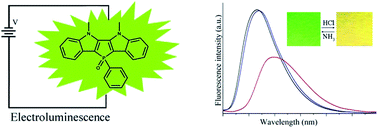
Electroluminescence and fluorescence response towards acid vapors depending on the structures of indole-fused phospholes

Peng Gonga, Kaiqi Ye*a, Jingbo Suna, Peng Chenb, Pengchong Xuea, Hao Yanga and Ran Lu*a
aState Key Laboratory of Supramolecular Structure and Materials, College of Chemistry, Jilin University, Changchun 130012, P. R. China. E-mail: yekq@jlu.edu.cn; luran@mail.jlu.edu.cn
bKey Laboratory of Functional Inorganic Material Chemistry (MOE), School of Chemistry and Materials Science, Heilongjiang University, No. 74, Xuefu Road, Nangang District, Harbin, P. R. China

Received 25th September 2015 , Accepted 29th October 2015

First published on 30th October 2015


Abstract

New isomers of phosphole heteroacenes 2-DIPO and 3-DIPO, in which indoles were fused with phospholes in different manners, have been synthesized. It is found that 2-DIPO containing the 5,6-dihydrophospholo[3,2-b:4,5-b′]diindole skeleton is highly emissive in solution and in solid state, but the emission of 3-DIPO bearing 5,7-dihydrophospholo[2,3-b:5,4-b′]diindole is weak because the emission of 2-DIPO and 3-DIPO is originated from π*–π and π*–n transitions, respectively. The OLED using 2-DIPO as emitter exhibits better performance than that based on 3-DIPO on account of the poor emission of 3-DIPO. It is interesting that the filter paper strip containing 2-DIPO could detect gaseous strong acid by naked eye. For example, the emitting color of the test paper changes from green to yellow upon exposure to HCl vapor, and can recover upon exposure to gaseous NH3. This is the first example of a fluorescence sensor probe based on phosphole to detect acid vapors. However, acid cannot induce the change of the emission for 3-DIPO due to the steric effect of neighboring groups of N–CH3 and benzene around P[double bond, length as m-dash]O to prevent the formation of H-bonding between acid and P[double bond, length as m-dash]O in 3-DIPO.


Introduction

Organic π-conjugated systems have gained increasing interest from both fundamental and technological viewpoints due to their promising applications in electronic and optoelectronic devices.1,2 Many fused heterocyclic substructures have been used in organic functional materials for tailoring their functionalities.3,4 In particular, N-fused heterocyclic compounds usually exhibit excellent carrier mobility, strong fluorescence emission and good self-assembling properties, so that they have been used as hole transport materials, light harvesting antenna, fluorescence sensors and building blocks in supramolecular assemblies.5–7 For example, indole derivatives have been designed to show good performance in electrochromic devices (ECDs), organic light-emitting diode (OLED) and anion probing.8–12 Liu et al. synthesized ladder-type diindolo[3,2-b:4,5-b′]thiophene, which exhibited low-lying energy level of highest occupied molecular orbital (HOMO) and large band gap, had potential application in organic field effect transistor (OFET).13 We have synthesized indolocarbazole derivatives, from which emitting nanofibers were generated via organogelation to detect TNT.6a,b It is worth noting that the aromatic rings-fused phospholes have received much attention on account of their unique optoelectronic properties, good thermal stability, stability against oxidant and easy structural modification.14,15 However, to the best of our knowledge, there is no example of bisindole-fused phospholes, although benzothiophene-fused, benzofuran-fused and indole-fused phospholes have been prepared.16–18 In order to enrich the members of the family of phospholes, which are still reported limitedly, and to reveal their structure–property relationship, we designed two new isomers of indole-fused phospholes 2-DIPO and 3-DIPO, in which indoles were fused with phospholes in different manners. It was found that 2-DIPO containing 5,6-dihydrophospholo[3,2-b:4,5-b′]diindole skeleton gave strong emission in solution and in solid state. The OLED using 2-DIPO as emitter exhibited moderate performance. Notably, the filter paper strip containing 2-DIPO could sense gaseous strong acid by naked eyes reversibly. This is the first example for the fluorescence sensory material based on phosphole towards acid vapors. Although the emission of 3-DIPO bearing 5,7-dihydrophospholo[2,3-b:5,4-b′]diindol was weak in solution, it was also emissive in solid states. The performance of OLED based on 3-DIPO was poor due to its low fluorescence quantum yield. Meanwhile, acid vapor could not induce the change of the emission of 3-DIPO because of the steric effect of the neighboring groups of N–CH3 and benzene ring around O[double bond, length as m-dash]P to forbid the formation of H-bonding between acid and O[double bond, length as m-dash]P in 3-DIPO. Therefore, the fused manner of indoles to phospholes played a key role on the properties of phosphole derivatives. It provided a strategy for designing novel phospholes with desired functionalities.

Results and discussion

Synthesis

The synthetic routes for indole-fused phosphole derivatives 2-DIPO and 3-DIPO were shown in Scheme 1. Firstly, 3,3′-dibromo-1,1′-dimethyl-1H,1′H-2,2′-biindole 1 and 1,1′-dimethyl-1H,1′H-3,3′-biindole 2 were synthesized according to the reported literatures.12,14,19 The ring-forming reaction of compound 1 with DCPP (dichlorophenyl phosphine) catalyzed by n-BuLi, followed by the oxidation with H2O2 afforded 2-DIPO in a yield of 60%. The bromination reaction of compound 2 with NBS yielded compound 3, which could be transformed into 3-DIPO via similar ring-forming reaction. 2-DIPO and 3-DIPO were characterized by 1H NMR, 13C NMR, 31P NMR, FT-IR and MALDI/TOF MS (Fig. S13–S21). In 31P NMR spectra, the chemical shifts of phosphorus in 2-DIPO and 3-DIPO appeared at 9.00 ppm and 6.79 ppm, respectively, indicating higher density of electron cloud around phosphorus in 3-DIPO than in 2-DIPO since two nitrogen atoms were located at α-positions of phosphole ring in 3-DIPO.
image file: c5ra19867d-s1.tif
Scheme 1 Synthetic routes for 2-DIPO and 3-DIPO.

UV-vis absorption and fluorescence emission spectra

The UV-vis absorption and fluorescence emission spectra of 2-DIPO and 3-DIPO in THF were shown in Fig. 1. It was clear that 2-DIPO gave a strong absorption band at ca. 261 nm with shoulders at ca. 279 nm, 316 m and 331 nm, as well as a broad absorption band at 409 nm. The density functional theory (DFT) calculation at the B3LYP/6-31G level revealed that the absorption at 409 nm for 2-DIPO was attributed to a HOMO–LUMO transition (π–π* transition, Fig. S1). Table S1 showed that the molar absorption coefficient (ε) at 409 nm was 1.2 × 10⁴ L mol−1 cm−1. Compound 3-DIPO exhibited the strong absorption at ca. 265 nm and weak absorption at ca. 317 nm and 430 nm. The absorption at 430 nm was also due to HOMO–LUMO transition (n–π* transition) confirmed by DFT calculations (Fig. S1), but its ε value was as low as 11 L mol−1 cm−1 (Table S1). Additionally, we found that 2-DIPO and 3-DIPO gave emission bands at 494 nm and 548 nm, respectively, in THF when excited at 408 nm. The fluorescence quantum yields of 2-DIPO and 3-DIPO were 70% and 1.8% in THF, respectively, using 9,10-diphenylanthracene in benzene as reference. Since the emission of 2-DIPO and 3-DIPO was originated from π*–π and π*–n transitions, respectively, it was understandable that the emission intensity of 2-DIPO was much higher than 3-DIPO and a red-shift of the emission of 3-DIPO compared with 2-DIPO was observed. Although the emission of 3-DIPO in THF was weak, its fluorescence quantum yield in solid state reached 0.13, meaning that it was emissive in solid state. It should be noted that the emission band of 3-DIPO red-shifted to 574 nm in solid state from 548 nm in THF (Fig. S2) due to π–π interaction. Similarly, the emission of 2-DIPO also red-shifted to 530 nm in solid state compared with that in THF, and the solid fluorescence quantum yield was as high as 53%, which made it possible to be used as emitter in OLED.
image file: c5ra19867d-f1.tif
Fig. 1 UV-vis absorption and normalized fluorescence emission (λex = 408 nm) spectra of 2-DIPO (5.0 × 10⁻⁶ M) and 3-DIPO (5.0 × 10⁻⁵ M) in THF.

Single crystal structures of 2-DIPO and 3-DIPO

Furthermore, the single crystals of 2-DIPO and 3-DIPO were obtained by slow evaporation of the solutions in ethanol/CH2Cl2 and methanol/CH2Cl2, respectively. As shown in Fig. 2, we could find that the indolo-fused phosphole unit in both molecules exhibited planar structure and the benzene ring connected with phosphorus atom was almost perpendicular to the indolo-fused phosphole ring. The bond angle of C–P–C in five-membered ring was 91.3°, and the bond lengths of P–C were all 1.8 Å in 2-DIPO (Fig. 2a). In the single crystal of 2-DIPO the solvent molecules of ethanol were involved. The indolo-fused phosphole units in the adjacent molecules were in a parallel arrangement with a distance of 3.4 Å, and the H-bonding of P[double bond, length as m-dash]O⋯H–OC2H5 was formed with a distance of 1.9 Å. As shown in Fig. 2b, the molecular packing of 3-DIPO in single crystal was different from that of 2-DIPO. Benzene rings linked to P atom in the adjacent molecules were arranged in a face to face mode, and the distance between the two benzene rings was 3.1 Å. We also find the H-bonding of P[double bond, length as m-dash]O⋯H–OCH3 with a distance of 1.7 Å in single crystal of 3-DIPO. Therefore, we deduced that the π–π interaction in the crystal of 2-DIPO might be stronger than that in the crystal of 3-DIPO. Meanwhile, we found the formation of C–H⋯π and the face to face stacking of two benzene rings in the single crystal 3-DIPO, which led to the higher fluorescence quantum yield of 3-DIPO in solid state than in solution.20
image file: c5ra19867d-f2.tif
Fig. 2 Single crystal structures of 2-DIPO (a) and 3-DIPO (b).

Electrochemical properties and DFT calculations

The electrochemical properties of 2-DIPO and 3-DIPO were investigated using cyclic voltammetry with SCE as the reference electrode in CH2Cl2. 2-DIPO showed one reversible oxidation peak and another irreversible oxidation peak with onset potential at around 1.19 V and 1.63 V, respectively. 3-DIPO showed the similar CV curves with onset potential at ca. 0.90 V and 1.45 V, respectively (Fig. S3). We deduced that the appearance of two oxidation peaks for each compound might be originated from the successive oxidation processes of the two indolo rings, and 2-DIPO exhibited higher oxidation stability. The HOMO energy levels of 2-DIPO and 3-DIPO calculated from the oxidation onset potential using the ferrocene calibrated formula of EHOMO = −(Eoxonset + 4.15) eV were −5.33 eV, −5.04 eV, respectively (Table 1). Since the reduction peaks were out of our scan range, the LUMO energy levels of 2-DIPO and 3-DIPO were calculated from HOMO energy levels and the absorption band gaps, and were −2.68 eV and −2.64 eV, respectively. Moreover, the molecular orbitals and the optimized configurations of the two compounds were calculated by the B3LYP/6-31G level using the Gaussian 09w program. As shown in Fig. 3, the HOMO and LUMO for 2-DIPO and 3-DIPO were mainly delocalized through the entire molecules. Based on the calculation results, it was clear that HOMO and LUMO energy levels of 2-DIPO were lower than those of 3-DIPO on account of the better conjugated degree of 2-DIPO than 3-DIPO.
Table 1 Electrochemical data and HOMO/LUMO energy levels of 2-DIPO and 3-DIPO
Compound Eoxonseta (V) HOMOb (eV) LUMOb (eV) Egc (eV) HOMOd (eV) LUMOd (eV) Egd (eV)
a Eoxonset (V) = onset oxidation potential.b EHOMO = −(Eoxonset + 4.15) eV; ELUMO = EHOMO + Eg.c Determined from the onset of the absorption at the lower energy band edge (Eg = 1240/λonset).d Calculated data.
2-DIPO 1.19 −5.33 −2.68 2.65 −5.14 −1.54 3.60
3-DIPO 0.90 −5.04 −2.64 2.40 −4.82 −1.28 3.54


image file: c5ra19867d-f3.tif
Fig. 3 The molecular orbital plots of HOMO and LUMO for 2-DIPO and 3-DIPO calculated by the B3LYP/6-31G method using the Gaussian 09w program.

Electroluminescence of 2-DIPO and 3-DIPO

Since 2-DIPO and 3-DIPO could emit fluorescence in solid states, we employed them as emitters in OLEDs, and fabricated the OLEDs with the configuration of ITO/NPB (40 nm)/emitter (30 nm)/TPBi (30 nm)/LiF (1.0 nm)/Al (100 nm), in which NPB was N,N′-dinaphtho-1-yl-N,N′-diphenylbenzidine, emitter was 2-DIPO (named as device A) or 3-DIPO (named as device B) and TPBi was 1,3,5-tris(N-phenylbenzimidazol-2-yl)benzene. From the electroluminescence spectra (Fig. 4), we could find that devices A and B gave the emission located at 540 nm and 578 nm, respectively, which showed a small red-shift compared with their solid-state photoluminescence. Device A could be turned on at about 1.5 V (1 cd m−2) and the current density of device A increased with the driving voltage. The brightness of device A increased with the driving voltage in the range of 0–7.5 V, and the brightness reached maximum of 2636 cd m−2 at the driving voltage of 7.5 V. The maximum current efficiency and maximum power efficiency of device A were 1.59 cd A−1 and 1.35 cd W−1, respectively (Table 2, Fig. S4 and S5). The performance of device B was poorer than device A. In the case of device B, the turned on voltage, maximum brightness, maximum current efficiency and maximum power efficiency were 2.5 V (1 cd m−2), 2228 cd m−2 (at 8.0 V), 0.22 cd A−1 and 0.3 cd W−1, respectively (Table 2, Fig. S6 and S7). In order to evaluate the thermal stability of the emitters, DSC and TGA curves for 2-DIPO and 3-DIPO were shown in Fig. S8. As to 2-DIPO, the melting point was as high as 334 °C, and the decomposition temperature was above 405 °C. The melting point of 3-DIPO was at 307 °C and the decomposition temperature was at 348 °C. It indicated that 2-DIPO and 3-DIPO exhibited good thermal stabilities, which was a critical issue for device stability. Therefore, the indole-fused phosphole derivatives could be used as emitting materials applied in OLED, and the performance of OLED device based on 2-DIPO was better than 3-DIPO on account of its strong emission in solid state. Meanwhile, we found that device A showed better device performance than the non-doped benzothiophene-fused phospholes-based OLED,16 illustrating that bisindole-fused phospholes were promising emitting materials.
image file: c5ra19867d-f4.tif
Fig. 4 Normalized electroluminescence spectra of devices A and B.
Table 2 EL performance of devices A and B
Devicea λELb (nm) Vonc (V) Lmaxd (cd m−2) ηce (cd A−1) ηpf (cd W−1)
a Device A and B: ITO/NPB(40 nm)/emitter(30 nm)/TPBI(30 nm)/LiF(1 nm)/Al.b Electroluminescence emission peak.c Turn-on voltage.d Maximum brightness.e Maximum current efficiency.f Maximum power efficiency.
A 540 1.5 2636 1.59 1.35
B 578 2.5 2228 0.22 0.30


Fluorescence response towards acid vapors

On the other hand, we prepared the filter paper strips containing 2-DIPO and 3-DIPO. It was interesting that the emission band of 2-DIPO located at 517 nm red-shifted to 550 nm, and the emitting color changed from green to yellow upon exposed to HCl vapor (Fig. 5). Moreover, the emission could recover to green when the above filter paper strip was exposed to NH3 vapor. Thus, the filter paper strip based on 2-DIPO could be used as test paper to sense HCl vapor by naked eyes. Meanwhile, we found that the response reversibility of the test paper was good (Fig. S9). However, HCl vapor could not induce any change of the emission of 3-DIPO in filter paper strip, meaning 3-DIPO could not sense acid. In order to reveal the different fluorescence sensory behaviors of 2-DIPO and 3-DIPO towards gaseous acid, the sensory mechanism should be proposed. In UV-vis absorption spectra titration experiment, trifluoromethanesulfonic acid (TfOH) was selected as analyst due to its good solubility in toluene. As shown in Fig. S10, we only found slight red-shift and slight decrease of the intensity of the absorption for 2-DIPO when 10 equiv. of TfOH was added, illustrating that a weak interaction between 2-DIPO and TfOH happened and no new species was formed. In 1H NMR titration experiment, slight downfield shifts of Ha and Hb in 2-DIPO were detected when 1.0 equiv. of TfOH was added (Fig. S11), further suggesting the occurrence of weak interaction between 2-DIPO and TfOH. In this case, we deduced that the formation of H-bonding of CF3SO2–O–H⋯O[double bond, length as m-dash]P in 2-DIPO led to the significant changes of the emission of 2-DIPO. For example, the emission of 2-DIPO decreased and red-shifted gradually with the increasing of the concentration of TfOH, and it shifted to 550 nm from 497 nm when 20 equiv. of TfOH was added. Additionally, we found that the test paper also exhibited reversible fluorescence response to gaseous strong acids of HBr, HNO3, trifluoroacetic acid (TFA) and TfOH (Fig. S12). However, the weak acid vapors of formic acid and acetic acid could not be detected by the test paper based on 2-DIPO. To the best of our knowledge, this is the first example of the fluorescence probe based on phosphole derivative to detect acid vapors. Furthermore, similar to the fact that 3-DIPO could not response towards vapor acid in filter paper strip, the emission of 3-DIPO could not change upon the addition of TfOH in toluene. We suggested that the P[double bond, length as m-dash]O in 3-DIPO was shield by the neighboring groups of N–CH3 and benzene ring, so that the formation of H-bonding of CF3SO2–O–H⋯O[double bond, length as m-dash]P in 3-DIPO would be forbidden.
image file: c5ra19867d-f5.tif
Fig. 5 Fluorescence emission spectra of 2-DIPO in filter paper strip before (black) and after upon exposed to vapors of HCl (red) and NH3 (blue) in sequence (λex = 365 nm). Inset: photos of filter paper strips based on 2-DIPO upon exposed with HCl and NH3 vapors repeatingly under 365 light.

Conclusions

In summary, we have synthesized two new isomers of phospholes 2-DIPO and 3-DIPO, in which indoles were fused with phospholes in different manners. It is found that the OLED using 2-DIPO as the emitter exhibited moderate performance, but the performance of OLED based on 3-DIPO was poor due to its weak emission in solid state. Notably, the filter paper strip containing 2-DIPO could sense gaseous strong acid by naked eyes, and the emitting color of the test paper changed from green to yellow upon exposed to HCl vapor. This is the first report on the fluorescence probe based on phosphole for detecting acid vapors. 3-DIPO could not probe acid on account of the steric effect of neighboring groups of O[double bond, length as m-dash]P to prevent the formation of H-bonding. Therefore, the optoelectronic properties of phosphole derivatives could be tuned by the fused manner of heterocyclic rings to phospholes. It provided a strategy for design novel phospholes with desired functionalities.

Experimental section

General information

The synthetic procedures were performed under N2 atmosphere. THF was refluxed with Na until the color of solvent turned into atropurpureus using benzophenone as indicator. CH2Cl2 and CCl4 were refluxed with CaH2 overnight. 1H NMR, quantitative 13C NMR and 31P NMR spectra were recorded with a Bruker Avance III 400 MHz, 100 MHz and 162 MHz, respectively, using CDCl3 and DMSO-d6 as the solvents. The chemical shifts in 1H NMR and 13C NMR spectra were reported in parts per million (ppm) relative to Si(CH3)4 as external standard. The 31P NMR signals were expressed with a positive sign, and the chemical shifts were given in ppm relative to external H3PO4 (85%). Mass spectra were performed on Agilent 1100 MS series and AXIMA CFR MALDI/TOF (matrix assisted laser desorption ionization/time-of-flight) MS (COMPACT). FT-IR spectrum was measured with a Nicolet-360 FT-IR spectrometer by incorporation of sample in KBr disk. DSC measurements were taken by NETZSCH DSC 204 F1 phoenix. TGA measurements were run on a TA Q500 system under N2 purge with a heating rate of 10 °C min−1. UV-vis absorption spectra were determined on a Shimadzu UV-1601PC Spectrophotometer. Fluorescence emission spectra were carried out on a Shimadzu RF-5301 Luminescence Spectrometer. The solid fluorescence quantum yields were measured using Edinburgh Instrument FLS920. Single crystal data were collected with a Rigaku R-AXIS RAPID. Cyclic voltammetry (CV) was performed using a CHI 604C voltammetric analyzer and the measurements were carried out containing 0.1 M Bu4NBF4 as a supporting electrolyte. A platinum button was used as a working electrode and a platinum plate as a counter electrode. All potentials were recorded versus SEC as a reference electrode (using ferrocene as an internal standard). The scan rate was maintained at 50 mV s−1. The optimized configurations and the orbital plots of HOMO and LUMO were calculated by the DFT (B3LYP/6-31G) method on the Gaussian 09W software.

The devices were grown on a glass, which was pre-coated with indium tin oxide (ITO) having a sheet resistance equal to 20 Ω cm−2. The ITO glass was routinely cleaned by ultrasonic treatment in detergent solutions, followed by rinsing with acetone, boiling in isopropanol, rinsing in methanol, and then in deionized water. The glass was dried in a vacuum oven between each cleaning step above. To reduce the possibility of electrical shorts on the ITO anode and increase the value of its work function, the ITO substrate was treated using a Plasma Cleaner (PDC-32G-2, 100 W) with the oxygen ambient. Prior to the deposition, all of the organic materials were further purified by the sublimation method. The organic layer was sequentially deposited onto the substrate without breaking vacuum at a pressure of about 10−4 Pa. A very thin layer of LiF could enhance electron injection from the aluminum cathode. A shadow mask with 2 × 3 mm2 openings was used to define the cathodes. The EL spectra, brightness and the current–brightness–voltage characteristics of the devices were measured with a rapid scan system using a spectrophotometer (PR-650, Photo Research) and a computer-controlled, programmable, direct current (DC) source (Keithley 2400). Luminance–voltage and current–voltage characteristics were measured at room temperature under an ambient atmosphere.

The filter paper strips containing 2-DIPO and 2-DIPO were prepared by dipping the filter paper strips in the CH2Cl2 solutions, followed by evaporating the solvent naturally.

Synthesis

3,3′-Dibromo-1,1′-dimethyl-1H,1′H-2,2′-biindole 1 and 1,1′-dimethyl-1H,1′H-3,3′-biindole 2 were synthesized according to the reported literatures.12,14,19
5,6-Dimethyl-11-phenyl-5,6-dihydrophospholo[3,2-b:4,5-b′]diindole 11-oxide 2-DIPO. Under N2 atmosphere, n-BuLi (4.21 mL, 10.52 mmol, 2.5 mol L−1 in n-hexane) was added dropwise to dry THF solution of 3,3′-dibromo-1,1′-dimethyl-1H,1′H-2,2′-biindole (2.0 g, 4.78 mmol) at −78 °C. After stirring for 30 min, PhPCl2 (0.72 mL, 5.26 mmol) was added and the resulting suspension was allowed to warm quickly to room temperature. After stirring for 2 h at room temperature, diluted H2O2 (10 mL of 30% H2O2 in 40 mL H2O) was added to the mixture. After stirring for another 2 h, the mixture was slowly poured into saturated solution of Na2S2O3 in water. The precipitation was filtered to afford crude product, which was purified by column chromatography using acetone/petroleum (v/v = 1/3) as eluent to afford 2-DIPO (1.10 g, 60%) as a bright yellow green solid. Mp 334.0 °C (obtained from DSC result). 1H NMR (400 MHz, DMSO-d6) δ (ppm): 7.76 (dd, J = 13.2, 7.1 Hz, 2H, ArH), 7.67 (d, J = 8.2 Hz, 2H, ArH), 7.53–7.40 (m, 5H, ArH), 7.23 (t, J = 7.3 Hz, 2H, ArH), 7.15 (t, J = 7.4 Hz, 2H, ArH), 4.26 (s, 6H, NCH3) (see Fig. S13). Quantitative 13C NMR (100 MHz, DMSO-d6) δ (ppm): 143.46, 143.21, 141.97, 141.86, 134.69, 133.57, 132.05, 132.03, 130.87, 130.76, 129.34, 129.22, 126.12, 126.03, 122.94, 122.68, 119.26, 113.73, 112.48, 112.39, 34.54 (see Fig. S14). 31P NMR (162 MHz, DMSO-d6) δ (ppm): 9.00 (see Fig. S15). MS, m/z: cal.: 382.1, found: 383.0 [M + H]+ (see Fig. S16). IR (KBr, cm−1): 3053, 3011, 2945, 1609, 1570, 1525, 1481, 1460, 1438, 1400, 1364, 1338, 1263, 1204, 1174, 1121, 1106, 1069, 1016, 983, 761, 751, 741, 734, 710, 696, 658, 651, 589, 575, 551, 538, 525, 499, 464, 431.
2,2′-Dibromo-1,1′-dimethyl-1H,1′H-3,3′-biindole 3. 1,1′-Dimethyl-1H,1′H-3,3′-biindole (2 g, 7.69 mmol) and NBS (1.64 g, 9.22 mmol) were dissolved in dry CCl4. After refluxed for 5 h, the mixture was cooled to room temperature, and the precipitation was removed by vacuum filtration. The filtrate was evaporated under reduced pressure and the residual solids were recrystallized in ethyl acetate to afford 3 as a light brown solid. Mp. 110.0–112.0 °C. 1H NMR (400 MHz, CDCl3) (ppm): 7.36 (m, 4H, ArH), 7.25 (m, 2H, ArH), 7.08 (m, 2H, ArH), 3.88 (s, 6H, NCH3) (see Fig. S17). IR (KBr, cm−1): 3051, 2937, 2872, 1495, 1463, 1417, 1379, 1352, 1329, 1307, 1228, 1156, 1128, 1097, 1081, 1020, 1011, 913, 802, 742, 583, 550, 428.
5,7-Dimethyl-6-phenyl-5,7-dihydrophospholo[2,3-b:5,4-b′]diindole 6-oxide 3-DIPO. By following the synthetic procedure for 2-DIPO except using 3 (2.0 g, 4.78 mmol), n-BuLi (2.5 mol L−1 in n-hexane, 4.21 mL, 10.52 mmol), PhPCl2 (0.72 mL, 5.26 mmol) and diluted H2O2 (10 mL of 30% H2O2 in 40 mL H2O) as reagents. The crude product was purified by column chromatography using acetone/petroleum (v/v = 1/3) as eluent to afford 3-DIPO (0.86 g, 47%) as a deep yellow solid. Mp. 308.0 °C (obtained from DSC). 1H NMR (400 MHz, DMSO-d6) δ (ppm): 8.07 (d, J = 7.9 Hz, 2H, ArH), 7.75 (dd, J = 13.5 Hz, J = 7.5 Hz, 2H, ArH), 7.66 (t, J = 7.1 Hz, 1H, ArH), 7.53 (d, J = 8.2 Hz, 4H, ArH), 7.28 (dt, J = 14.8, J = 7.2 Hz, 4H, ArH), 3.72 (s, 6H, NCH3) (see Fig. S18). Quantitative 13C NMR (100 MHz, DMSO-d6) δ (ppm): 141.99, 141.91, 133.86, 133.54, 133.52, 132.58, 131.17, 131.05, 130.15, 130.07, 130.03, 128.98, 124.41, 123.07, 122.88, 121.78, 121.56, 121.31, 121.21, 112.20, 32.69 (see Fig. S19). 31P NMR (162 MHz, DMSO-d6) δ (ppm): 6.79 (see Fig. S20). MS, m/z: cal.: 382.1, found: 383.1 [M + H]+ (see Fig. S21). IR (KBr, cm−1): 3055, 3012, 2934, 2795, 2689, 1611, 1585, 1519, 1497, 1473, 1435, 1405, 1377, 1345, 1309, 1289, 1235, 1202, 1156, 1135, 1098, 1067, 1033, 1011, 933, 914, 826, 811, 780, 736, 714, 697, 646, 637, 606, 584, 568, 549, 496, 449, 439, 425, 406.

Acknowledgements

This work was financially supported by the National Natural Science Foundation of China (21374041) and the Open Project of State Key Laboratory of Supramolecular Structure and Materials (SKLSSM2015014).

Notes and references

  1. (a) F. J. M. Hoeben, P. Jonkheijm, E. W. Meijer and A. P. H. J. Schenning, Chem. Rev., 2005, 105, 1491–1546 CrossRef CAS PubMed; (b) C. L. Wang, H. L. Dong, W. P. Hu, Y. Q. Liu and D. B. Zhu, Chem. Rev., 2012, 112, 2208–2267 CrossRef CAS PubMed; (c) J. Roncali, Chem. Soc. Rev., 2005, 34, 483–495 RSC; (d) L. Zang, Y. K. Che and J. S. Moore, Acc. Chem. Res., 2008, 41, 1596–1608 CrossRef CAS PubMed; (e) S. L. Xu, N. Ai, J. Zheng, N. Zhao, Z. G. Lan and L. R. Wen, RSC Adv., 2015, 5, 8340–8344 RSC; (f) Y. S. Guan, Y. K. Qin, Y. H. Sun, C. Wang, W. Xu and D. B. Zhu, Chem. Commun., 2015, 51, 12182–12184 RSC.
  2. (a) F. S. Kim, G. Q. Ren and S. A. Jenekhe, Chem. Mater., 2011, 23, 682–732 CrossRef CAS; (b) P. A. Korevaar, T. F. A. D. Greef and E. W. Meijer, Chem. Mater., 2014, 26, 576–586 CrossRef CAS; (c) V. Coropceanu, J. Cornil, D. A. da Silva Filho, Y. Olivier, R. Silbey and J. L. Brédas, Chem. Rev., 2007, 107, 926–952 CrossRef CAS PubMed; (d) L. Ding, S. C. Dong, Z. Q. Jiang, H. Chen and L. S. Liao, Adv. Funct. Mater., 2015, 25, 645–650 CrossRef CAS; (e) S. T. Zhang, L. Yao, Q. M. Peng, W. J. Li, Y. Y. Pan, R. Xiao, Y. Gao, C. Gu, Z. M. Wang, P. Lu, F. Li, S. J. Su, B. Yang and Y. G. Ma, Adv. Funct. Mater., 2015, 25, 1755–1762 CrossRef CAS.
  3. (a) T. Baumgartner and R. Réau, Chem. Rev., 2006, 106, 4681–4727 CrossRef CAS PubMed; (b) D. Joly, D. Tondelier, V. Deborde, B. Geffroy, M. Hissler and R. Réau, New J. Chem., 2010, 34, 1603–1611 RSC; (c) H. C. Su, O. Fadhel, C. J. Yang, T. Y. Cho, C. Fave, M. Hissler, C. C. Wu and R. Réau, J. Am. Chem. Soc., 2006, 128, 983–995 CrossRef CAS PubMed.
  4. C. Fave, T. Y. Cho, M. Hissler, C. W. Chen, T. Y. Luh, C. C. Wu and R. Réau, J. Am. Chem. Soc., 2003, 125, 9254–9255 CrossRef CAS PubMed.
  5. (a) S. Y. Cai, G. J. Tian, X. Li, J. H. Su and H. Tian, J. Mater. Chem. A, 2013, 1, 11295–11305 RSC; (b) J. Yu, J. Luo, Q. Chen, K. He, F. Meng, X. Deng, Y. Wang, H. Tan, H. Jiang and W. Zhu, Tetrahedron, 2014, 70, 1246–1251 CrossRef CAS.
  6. (a) P. Gong, P. C. Xue, C. Qian, Z. Q. Zhang and R. Lu, Org. Biomol. Chem., 2014, 12, 6134–6144 RSC; (b) P. Gong, J. B. Sun, P. C. Xue, C. Qian, Z. Q. Zhang, J. B. Sun and R. Lu, Dyes Pigm., 2015, 118, 27–36 CrossRef CAS; (c) G. H. Hong, J. B. Sun, C. Qian, P. C. Xue, P. Gong, Z. Q. Zhang and R. Lu, J. Mater. Chem. C, 2015, 3, 2371–2379 RSC.
  7. J. Peng, J. B. Sun, P. Gong, P. C. Xue, Z. Q. Zhang, G. H. Zhang and R. Lu, Chem.–Asian J., 2015, 10, 1717–1724 CrossRef CAS PubMed.
  8. (a) G. M. Nie, L. J. Zhou, Q. F. Guo and S. S. Zhang, Electrochem. Commun., 2010, 12, 160–163 CrossRef CAS; (b) Y. J. Na, W. Song, J. Y. Lee and S. H. Hwang, Org. Electron., 2015, 22, 92–97 CrossRef CAS.
  9. (a) C. W. Lee and J. Y. Lee, Adv. Mater., 2013, 25, 5450–5454 CrossRef CAS PubMed; (b) M. S. Park, D. H. Choi, B. S. Lee and J. Y. Lee, J. Mater. Chem., 2012, 22, 3099–3104 RSC.
  10. (a) L. Zou, B. Yan, D. Pan, Z. Tan and X. Bao, Spectrochim. Acta, Part A, 2015, 148, 78–84 CrossRef CAS PubMed; (b) F. M. Pfeffer, K. F. Lim and J. Sedgwick, Org. Biomol. Chem., 2007, 5, 1795–1799 RSC.
  11. (a) K. J. Chang, D. Moon, M. S. Lah and K. S. Jeong, Angew. Chem., Int. Ed., 2005, 117, 8140–8143 CrossRef; (b) J. L. Sessler, D. G. Cho and V. Lynch, J. Am. Chem. Soc., 2006, 128, 16518–16519 CrossRef CAS PubMed.
  12. M. Barboiu, Eur. J. Inorg. Chem., 2015, 1112–1125 CrossRef CAS.
  13. T. Qi, W. Qiu, Y. Liu, H. Zhang, X. Gao, Y. Liu, K. Lu, C. Y. Du, G. Yu and D. B. Zhu, J. Org. Chem., 2008, 73, 4638–4643 CrossRef CAS PubMed.
  14. M. Stolar, J. Borau-Garcia, M. Toonen and T. Baumgartner, J. Am. Chem. Soc., 2015, 137, 3366–3371 CrossRef CAS PubMed.
  15. S. Furukawa, S. Haga, J. Kobayashi and T. Kawashima, Org. Lett., 2014, 16, 3228–3231 CrossRef CAS PubMed.
  16. H. Chen, W. Delaunay, J. Li, Z. Y. Wang, P. A. Bouit, D. Tondelier, B. Geffroy, F. Mathey, Z. Duan, R. Réau and M. Hissler, Org. Lett., 2013, 15, 330–333 CrossRef CAS PubMed.
  17. Y. Dienes, M. Eggenstein, T. Kárpáti, T. C. Sutherland, L. Nyulászi and T. Baumgartner, Chem.–Eur. J., 2008, 14, 9878–9889 CrossRef CAS PubMed.
  18. Y. Xu, Z. Wang, Z. Gan, Q. Xi, Z. Duan and F. Mathey, Org. Lett., 2015, 17, 1732–1734 CrossRef CAS PubMed.
  19. P. W. Zhang, R. Y. Liu and J. M. Cook, Tetrahedron Lett., 1995, 36, 3103–3106 CrossRef CAS.
  20. (a) Y. Hong, J. W. Y. Lam and B. Z. Tang, Chem. Commun., 2009, 4332–4353 RSC; (b) P. C. Xue, B. Q. Yao, J. B. Sun, Z. Q. Zhang and R. Lu, Chem. Commun., 2014, 50, 10284–10286 RSC; (c) P. C. Xue, R. Lu, P. Zhang, J. H. Jia, Q. X. Xu, T. R. Zhang, M. Takafuji and H. Ihara, Langmuir, 2013, 29, 417–425 CrossRef CAS PubMed; (d) X. L. Liu, D. F. Xu, R. Lu, B. Li, C. Qian, P. C. Xue, X. F. Zhang and H. P. Zhou, Chem.–Eur. J., 2011, 17, 1660–1669 CrossRef CAS PubMed; (e) P. Chen, R. Lu, P. C. Xue, T. H. Xu, G. J. Chen and Y. Y. Zhao, Langmuir, 2009, 25, 8395–8399 CrossRef CAS PubMed; (f) C. Bao, R. Lu, M. Jin, P. C. Xue, C. H. Tan, G. F. Liu and Y. Y. Zhao, Chem.–Eur. J., 2006, 12, 3287–3294 CrossRef CAS PubMed.

Footnote

Electronic supplementary information (ESI) available: 1H NMR, 13C NMR, 31P NMR and MALDI/TOF MS spectra; CV, DSC and TGA curves; deposition numbers of the single crystals of 2-DIPO and 3-DIPO are CCDC 1052683 and 1042193. For ESI and crystallographic data in CIF or other electronic format see DOI: 10.1039/c5ra19867d

This journal is © The Royal Society of Chemistry 2015
Click here to see how this site uses Cookies. View our privacy policy here.