Graphene oxide-based Fe3O4 nanoparticles as a novel scaffold for the immobilization of porcine pancreatic lipase

Yongbo Shao, Tao Jing*, Jingzhi Tian and Yongjie Zheng
College of Chemistry and Chemical Engineering, Qiqihar University, Qiqihar 161006, China. E-mail: jtkr@163.com

Received 18th September 2015 , Accepted 18th November 2015

First published on 19th November 2015


Abstract

Nano- and hybrid materials have recently emerged as a new approach for improving enzyme activity and stability, and are suitable for commercial applications. In this paper, graphene oxide-based magnetic hybrids were prepared successfully. A chloropropyl-functionalized graphene oxide decorated with Fe3O4 nanoparticles was made, denoted as CPS/GO-Fe3O4@MCM-41. This was characterized by X-ray powder diffraction, scanning electron microscopy, transmission electron microscopy, Fourier transform infrared spectroscopy, vibrating sample magnetometry, thermogravimetry and N2 adsorption/desorption. Then, porcine pancreas lipase (PPL) was immobilized onto the graphene oxide-based magnetic nanoparticles via covalent bonding. The results show that the novel supporting material CPS/GO-Fe3O4@MCM-41 was the best for PPL immobilization compared to two other nanomaterials (GO-Fe3O4@MCM-41 and GO-Fe3O4). The supporting material CPS/GO-Fe3O4@MCM-41 exhibited enhanced immobilization efficiency (up to 98%), maximum relative activity (up to 97.9%), high stability and reusability (85% after 56 d and 87% after 10 cycles respectively, both at 30 °C). Additionally, it offered some other advantages, such as easy recycling and reuse, complying with the requirements of green chemistry. Therefore, it is proposed that CPS/GO-Fe3O4@MCM-41 provides a new approach for commercial applications.

1 Introduction

Graphene, a two-dimensional (2D) honeycomb lattice, is regarded as the ‘thinnest material in the universe’. It offers outstanding thermal conductivity, mechanical strength, chemical stability, biocompatibility and non-toxicity, therefore, it has a great future in industrial applications.1–5 Graphene oxide, a precursor of graphene, has a large surface area and plenty of functional groups, and exhibits good biocompatibility. Therefore, it is a good candidate for grafting other nanoparticles and immobilizing a large amount of enzymes. However, it is difficult to separate graphene oxide from an aqueous solution due to its strong hydrophilicity. Another difficulty is that nanosheets of graphene oxide are inclined to agglomerate because of the strong π–π interactions between the nanosheets, which leads to a loss of surface area, so that many functional groups could be covered.6

Fe3O4 nanoparticles can offer convenience of recovery due to their response to a magnetic force; they have been adopted in catalytic, biological and drug delivery fields.7–12 If graphene oxide is integrated with Fe3O4 nanoparticles, the combined supporting material can be separated rapidly with the addition of an external magnetic field. Meanwhile, Fe3O4 nanoparticles can prevent the agglomeration of graphene oxide, so that the large surface area and the functional groups can be retained.

On the other hand, enzyme-catalyzed reactions have been applied extensively in industry due to their high efficiency, mild reaction conditions, non-toxicity, fewer by-products and environmentally friendly properties.13

Among all commercialized enzymes, porcine pancreatic lipase (PPL) is one of the more common, cheaper and easily used, as it offers many advantages including high thermostability and water solubility. PPL is a small globular protein, which consists of a single chain of 449 amino acids, and has a three-dimensional structure (molecular volume) of 4.6 nm × 2.6 nm × 1.1 nm. Its unique crystal structure looks like a lid that covers the catalytic site, which contains a catalytic triad: Ser, Asp and His at positions 153, 177 and 264, respectively.14

Water-soluble enzymes show some drawbacks, such as instability, easy deactivation and the need for additional efforts to separate the enzymes from a product mixture. Therefore, immobilized enzyme technology has been adopted and developed.15–17

To date, PPL has been immobilized on various supports, and applied to various purposes. Unfortunately, some supports have low surface areas and barely any functional groups or binding sites, which is inappropriate for industrial applications. Therefore, it is still necessary to design a more efficient support for immobilizing enzymes.

In this regard, we designed a new supporting material, which integrates graphene oxide with Fe3O4 nanoparticles, for better enzyme immobilization. Meanwhile, we explored the relative activity, reusability and storage stability after the enzyme was immobilized on the new supporting material.

2 Experimental

2.1 Materials and methods

2.1.1 Materials. FeCl3·6H2O, cetyltrimethylammonium bromide (CTAB), acetone, ethylene glycol, sodium acetate, absolute ethanol, absolute toluene, potassium permanganate, sodium nitrate, hydrogen peroxide, concentrated sulfuric acid and porcine pancreas lipase (PPL) powder (EC 3.1.1.3, 3000 U mL−1, BR), were purchased from Sinopharm Chemical Reagent (Shanghai, China). Tetraethylorthosilicate (TEOS), ammonia (25 wt%), 3-chloropropyltriethoxysilane (CPS) and olive oil were purchased from Aladdin (Shanghai, China). Natural flake graphite was purchased from Qingdao Golden days of graphite company (Qingdao, China). All chemicals were of analytical grade and used without further purification.
2.1.2 Methods.
2.1.2.1 Preparation of Fe3O4 nanoparticles. The preparation of Fe3O4 and Fe3O4@MCM-41 was carried out according to work described previously.18
2.1.2.2 Preparation of GO. Graphene oxide (GO) was prepared using the Hummers method.19 Natural flake graphite (0.5 g) and sodium nitrate (2.5 g) were added into a round-bottom flask, which was placed in an ice-water bath, then concentrated sulfuric acid (115 mL, 36 N) was added and stirred slowly. Afterwards, potassium permanganate (15 g) was added and mixed. The mixture was kept in the ice-water bath for 2 h, then the flask was transferred into a thermostat water bath, and stirred for 30 min at 35 °C. After that, the flask was transferred into the ice-water bath again, and deionized water (230 mL) was added gradually. The temperature would be increased. The flask was transferred into the thermostat water bath again, and stirred for 20 min at 98 °C. The reaction mixture was then cooled down naturally to room temperature. Subsequently, hydrogen peroxide (250 mL) was added. By now, a bright yellow solution was obtained, a precipitate was collected by centrifugation and washed with diluted hydrochloric acid; the supernatant was collected and detected with barium chloride solution. Then, the precipitate was washed with distilled water until the pH of the supernatant reached neutral.
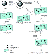
2.1.2.3 Preparation of CPS/GO-Fe3O4@MCM-41. Fe3O4@MCM-41 (0.3 g) was dispersed in absolute ethanol (60 mL) and sonicated for 30 min. Meanwhile, GO was also dispersed in distilled water (0.5 mg mL−1). The two solutions (3[thin space (1/6-em)]:[thin space (1/6-em)]4 v/v) prepared above were mixed and stirred vigorously for 1 h. Then, 3-chloropropyltriethoxysilane (CPS) (1 mL) was dropped into the solution generated above, which was then stirred vigorously at 120 °C, refluxed for 10 h, washed with absolute ethanol several times and freeze-dried. The product was noted as CPS/GO-Fe3O4@MCM-41. For comparison, GO-Fe3O4 and GO-Fe3O4@MCM-41 were also prepared by the same process except without the addition of 3-chloropropyltriethoxysilane (Fig. 1).
image file: c5ra19276e-f1.tif
Fig. 1 Schematic representation of the formation of (a) GO-Fe3O4–PPL, (b) GO-Fe3O4@MCM-41–PPL and (c) CPS/GO-Fe3O4@MCM-41–PPL.

2.1.2.4 Immobilization of PPL on CPS/GO-Fe3O4@MCM-41, GO-Fe3O4@MCM-41 and GO-Fe3O4. CPS/GO-Fe3O4@MCM-41 (0.36 g) was equilibrated in PBS buffer (10.0 mL; pH 7.5, 0.05 M) at 25 °C for 1 h, which was named the supporting solution. PPL (0.036 g) was dissolved in PBS buffer (10.0 mL; pH 7.5, 0.05 M) to obtain a solution, which was then added into the supporting solution mentioned above. The resulting mixture was shaken for 12 h at 150 rpm (25 °C). Afterwards, the immobilized PPL material was collected, washed with enough PBS buffer and stored at 4 °C until use. The other two immobilized PPL materials were obtained according to the same process mentioned above.
2.1.2.5 Immobilization efficiency of PPL. Immobilization efficiency of PPL on nanoparticles was determined by the Bradford method as described previously.20 Briefly, the absorbance of a solution was measured at 595 nm using UV-visible spectroscopy first, and then the percentage of the immobilized PPL was calculated by subtracting the PPL content in the supernatant from the total as follows.
Immobilization efficiency (%) = (total PPL content (mg mL−1) − PPL content in the supernatant (mg mL−1))/total PPL content × 100%.

The optimal concentrations of the enzyme were 0.35 mg mL−1 for CPS/GO-Fe3O4@MCM-41–PPL, 0.30 mg mL−1 for GO-Fe3O4@MCM-41–PPL, and 0.25 mg mL−1 for GO-Fe3O4–PPL.


2.1.2.6 Activity assay. The activities of PPL and immobilized-PPL were determined by use of olive oil as the substrate.18 One unit (U) of PPL activity was defined as the amount of lipase releasing 1 μmol of fatty acid per minute. The initial activity (3000 U mL−1) of PPL was defined as the maximum activity (100%).
2.1.2.7 The effect of temperature on PPL relative activity. The effect of temperature on relative activity was investigated as follows: immobilized PPL and non-immobilized PPL (3.6 mg mL−1 for both) were added into PBS buffer (pH 7.5, 0.05 M) and kept for 1 hour at temperatures of 25 °C, 30 °C, 35 °C, 40 °C, 45 °C, 50 °C, 55 °C and 60 °C.
2.1.2.8 The effect of pH on PPL relative activity. The effect of pH on PPL relative activity was investigated under the conditions of 30 °C for 1 h, and the PBS buffer pH values were adjusted to 5.5, 6.0, 6.5, 7.0, 7.5, 8.0, 8.5 and 9.0.
2.1.2.9 Reusability and storage stability of immobilized PPL. Reusability was studied as follows. The relative activities of the immobilized PPLs (3.6 mg mL−1) were measured after each run and up to ten runs under the conditions of 30 °C, pH 7.5 and 1 hour reaction time. After each run, the immobilized PPL was recovered with the aid of a magnet and washed with PBS buffer (pH 7.5, 0.05 M) three times. The storage stabilities of the immobilized PPL and non-immobilized PPL were determined based on the days that the activity lasted for. The activity was tested every 7 days under the conditions of 30 °C at pH 7.5.
2.1.3 Characterizations. X-ray diffraction (XRD) spectra of the samples were recorded using a Bruker D8-FOCUS diffractometer equipped with Cu/Kα radiation at a scanning rate of 4–8° min−1. Fourier transform infrared spectroscopy (FTIR) (Spectrum One B, PE) was used for detecting the functional groups of materials in the range of 400–4000 cm−1. Magnetic susceptibility was measured using a vibrating sample magnetometer (VSM) (VersaLab VL-072, Quantum Design) in the magnetic field sweeps of −30[thin space (1/6-em)]000 Oe to 30[thin space (1/6-em)]000 Oe at room temperature. Morphological characteristics of the samples were measured using a Hitachi S-4300 scanning electron microscope (SEM) and a Hitachi S-7650 transmission electron microscope (TEM). For investigating the surface area and pore size, the samples were first degassed in a vacuum at 77 K for 3 h, and then tested with a Surface Area & Pore Size Analyzer (AUTOSORB-1, Quantachrome). Thermogravimetric analysis (TGA) was conducted using a NETZSCH STA 449 F3 Jupiter thermo-analyzer in a temperature range of 0–800 °C at a heating rate of 10 °C min−1. The surface composition inspection and the valence states of the samples were measured by X-ray photoelectron spectroscopy (XPS) (ESCALAB250Xi, Thermo Company).

3 Results and discussion

3.1 Materials characterizations

3.1.1 Morphological characteristics observed by SEM and TEM images. The SEM and TEM images for Fe3O4, Fe3O4@MCM-41, GO, GO-Fe3O4, GO-Fe3O4@MCM-41 and CPS/GO-Fe3O4@MCM-41 are shown in Fig. 2. Before coating, the shape of the Fe3O4 nanoparticles was spherical, their size was about 300 nm, and they displayed good dispersancy (a and c). After coating with mesoporous silicon (MCM-41), the shape of Fe3O4@MCM-41 was also spherical, but the size was increased to 500 nm, and it showed an orderly and close arrangement (b and d). Compared to Fe3O4, the surface of Fe3O4@MCM-41 became smoother (b). Morphologically, the Fe3O4 nanoparticles were coated by mesoporous silicon evenly.21 The morphology of GO is shown in Fig. 2(e), in which a four layered structure is observed. It is thin, transparent, and is accompanied with several micrometer-long wrinkles. As shown in Fig. 2(f–h), Fe3O4 and Fe3O4@MCM-41 nanoparticles located on the surfaces of GO layers were tightly wrapped by wrinkled and ultrathin GO sheets.22 These results show that the materials used in the experiments were prepared successfully.
image file: c5ra19276e-f2.tif
Fig. 2 SEM images of (a) Fe3O4 and (b) Fe3O4@MCM-41, and TEM images of (c) Fe3O4; (d) Fe3O4@MCM-41; (e) GO; (f) GO-Fe3O4; (g) GO-Fe3O4@MCM-41 and (h) CPS/GO-Fe3O4@MCM-41.
3.1.2 Determination of crystal structure by XRD analysis. The crystal structures of Fe3O4, Fe3O4@MCM-41, GO and CPS/GO-Fe3O4@MCM-41 were determined by powder X-ray diffraction (XRD) (Fig. 3). The structures of Fe3O4@MCM-41 and CPS/GO-Fe3O4@MCM-41 were determined by small angle X-ray analysis in the range of 1–6°. The result from the small angle X-ray analysis for CPS/GO-Fe3O4@MCM-41 was exactly the same as the result for Fe3O4@MCM-41 (Fig. 3(c)), the only characteristic peaks were observed at a 2θ of 2.0°, which were attributed to the short-range ordering features of mesopore alignment on the spherical surface.18 The wide angle analysis of XRD revealed six characteristic peaks for Fe3O4, which were located at 2θ of 30.2°, 35.4°, 42.9°, 53.5°, 57.0° and 62.4°. These peaks were assigned to the crystal planes (220), (311), (400), (422), (511) and (440), respectively. For Fe3O4@MCM-41, the peak at a 2θ of 18–24° represented the amorphous phase of SiO2. The XRD pattern of GO is shown in Fig. 3(d). A unique characteristic peak was observed at a 2θ of 10.5°, which was assigned to the crystal plane (001). The XRD pattern of CPS/GO-Fe3O4@MCM-41 is shown in Fig. 3(e), where the characteristic diffraction peak of GO is not observed, however, the six characteristic peaks of Fe3O4 are observed.23 These results indicate that Fe3O4, Fe3O4@MCM-41, GO and CPS/GO-Fe3O4@MCM-41 were prepared successfully.
image file: c5ra19276e-f3.tif
Fig. 3 XRD patterns of (a) Fe3O4 and (b) Fe3O4@MCM-41, (c) small angle X-ray analyses of Fe3O4@MCM-41 and CPS/GO-Fe3O4@MCM-41, (d) GO and (e) CPS/GO-Fe3O4@MCM-41.
3.1.3 X-ray photoelectron spectroscopy (XPS) analysis. The surface compositions and the valence states of the samples were investigated by XPS spectroscopy, as shown in Fig. 4. The wide scan XPS spectra of the samples are shown in Fig. 4(a), and display sharp peaks at the binding energies of 285, 530, 711, 101 and 199 eV, which were assigned to C 1s, O 1s, Fe 2p, Si 2p and Cl 2p, respectively. The results indicate the existence of C, O, Fe, Si and Cl in the corresponding samples. The high-resolution XPS spectra of the Fe 2p scan are shown in Fig. 4(b). The peaks at 711.2 eV and 725.6 eV could be assigned to Fe 2p3/2 and Fe 2p1/2, respectively. The peak at around 720 eV of γ-Fe2O3 was not observed, suggesting that only the form of Fe3O4 existed in GO-Fe3O4, GO-Fe3O4@MCM-41 and CPS/GO-Fe3O4@MCM-41.24 The Si 2p spectra of GO-Fe3O4@MCM-41 are shown in Fig. 4(c). The two peaks at 101.3 eV and 102.3 eV were assigned to Si–O–C and Si–O–Si, respectively, which suggested that there were two chemical states of Si. This result was consistent with the conclusion from FTIR analysis (Fig. 5). The C1s spectra of GO, GO-Fe3O4, GO-Fe3O4@MCM-41 and CPS/GO-Fe3O4@MCM-41 are shown in Fig. 4(d–g). The deconvoluted peaks of GO are displayed at four positions corresponding to C[double bond, length as m-dash]C sp2 (284.4 eV), C–C sp3 (285.1 eV), C–OH and/or C–O–C (286.9 eV), and C[double bond, length as m-dash]O (288.9 eV).24 The peak of C–C sp3 in GO-Fe3O4, GO-Fe3O4@MCM-41 and CPS/GO-Fe3O4@MCM-41 shifted to a higher binding energy by 0.1 eV, 0.1 eV and 0.6 eV, respectively. In addition, the intensities of the C[double bond, length as m-dash]C sp2 peak in GO-Fe3O4, GO-Fe3O4@MCM-41 and CPS/GO-Fe3O4@MCM-41 were increased compared to that in GO. Together, it was believed that the magnetic mesoporous silicon was grafted onto the surface of the GO sheet.24 The peaks of C–OH and/or C–O–C in GO-Fe3O4 and GO-Fe3O4@MCM-41 greatly decreased compared to those in GO. They were even decreased more when CPS was grafted onto the surface of the GO sheet. These results suggested that GO was partly reduced in the modified nanomaterials.23,24 The N 1s spectra of PPL and CPS/GO-Fe3O4@MCM-41–PPL are shown in Fig. 4(h and i). The sharp peak at a binding energy of 399.8 eV was assigned to N 1s for PPL. The peak in CPS/GO-Fe3O4@MCM-41–PPL shifted to a higher binding energy by 0.5 eV. The result indicated that PPL was bound to CPS/GO-Fe3O4@MCM-41 by covalent attachment.25,26 All the results from the X-ray photoelectron spectroscopy (XPS) analysis demonstrate that the supporting materials were prepared successfully.
image file: c5ra19276e-f4.tif
Fig. 4 XPS spectra scans; (a) wide scan XPS spectra of the supporting materials, (b) high-resolution XPS spectra of Fe 2p scan, (c) Si 2p spectra of GO-Fe3O4@MCM-41, C 1s spectra of (d) GO, (e) GO-Fe3O4, (f) GO-Fe3O4@MCM-41, (g) CPS/GO-Fe3O4@MCM-41 and N 1s spectra of (h) PPL and (i) CPS/GO-Fe3O4@MCM-41–PPL.

image file: c5ra19276e-f5.tif
Fig. 5 FTIR spectra for the supporting materials and the immobilized PPL.
3.1.4 Functional groups by FT-IR spectra analysis. FT-IR spectra of GO-Fe3O4@MCM-41 (a), GO-Fe3O4 (b), GO (c), CPS/GO-Fe3O4@MCM-41 (d) and Fe3O4 (e) are shown in Fig. 5. For the results of Fe3O4 (e), the peaks at 582 cm−1, 1629 cm−1 and 3423 cm−1 are assigned to vibration of the Fe–O bond, the bending vibration of the O–H bond and the stretching vibration of the O–H group, respectively. In the curve of GO (c), the peaks at 1733 cm−1, 1621 cm−1, 1226 cm−1 and 1061 cm−1 were assigned to C[double bond, length as m-dash]O stretching vibrations of the carbonyl and carboxyl groups located at the edges of the GO networks, C[double bond, length as m-dash]C vibration of the skeleton, C–OH vibration and C–O stretching vibration of an epoxide group, respectively.27 For the GO-Fe3O4 spectrum (b), the peak of C[double bond, length as m-dash]O was observed with a lower intensity compared to the GO spectrum (c), suggesting that the GO sheet was partly reduced, which is consistent with the XPS results (Fig. 4). In addition, the wavelength of the Fe–O bond was shifted to a lower wavelength of 576 cm−1, compared to 582 cm−1 in the Fe3O4 nanoparticles. It was suggested that the Fe3O4 nanoparticles were grafted onto the GO sheet.28 In the spectra of GO-Fe3O4@MCM-41 (a) and CPS/GO-Fe3O4@MCM-41 (d), the peaks at 470 cm−1 and 801 cm−1 were attributed to the characteristic vibrations of the mesoporous framework (Si–O–Si). The vibration peaks at 1095 cm−1 and 1041 cm−1 were attributed to the Si–O–Si and Si–O–C groups, suggesting that magnetic mesoporous silicon was grafted on the surface of GO by covalent attachment.29 After modification with mesoporous silicon, the intensities of the peaks at 2925 cm−1 and 2854 cm−1 were increased, which were attributed to the symmetric and asymmetric stretching of –CH3 and –CH2 groups, respectively. The peak of the O–H bond at 3423 cm−1 was decreased and this was assigned to the fact that the GO sheet was decorated with magnetic mesoporous silicon.30 In the curve of CPS/GO-Fe3O4@MCM-41, the vibration peak at 691 cm−1 was attributed to the C–Cl bond. The new bonds at 1645 cm−1 and 1573 cm−1 were attributed to the fact that 3-chloropropyltriethoxysilane (CPS) was bound to –COO on the GO sheet.29,30 For the immobilized PPL spectrum, the peak at 2939 cm−1, specific to the N–H bond, was observed in all the curves (GO-Fe3O4@MCM-41–PPL (f), GO-Fe3O4–PPL (g), PPL (h) and CPS/GO-Fe3O4@MCM-41–PPL (i)), which confirmed that PPL remained in the supporting materials. The O–H peak at 3390 cm−1 was decreased when the enzyme was adsorbed onto GO-Fe3O4@MCM-41 and GO-Fe3O4. These results suggest that the N–H groups of PPL interact with the O–H groups of the supporting materials via hydrogen bonding.31 In addition, the GO sheet was partly reduced in the process (Fig. 4), so we propose that PPL was immobilized onto the surface (GO-Fe3O4@MCM-41 and GO-Fe3O4) mainly in the form of hydrogen bonding with hydroxyl groups. For the CPS/GO-Fe3O4@MCM-41–PPL spectrum (i), the peak for a C–N bond was not observed because it was overlapped by strong Si–O–Si and Si–O–C bonds at 1200–900 cm−1.32 However, the C–Cl bond in CPS/GO-Fe3O4@MCM-41–PPL disappeared, suggesting that the enzyme was immobilized on the supporting materials. All the results from the FT-IR spectra indicate that the supporting materials were prepared well, and that PPL was immobilized on them successfully.
3.1.5 Magnetic properties by VSM analysis. The magnetic properties of the differently synthesized nanoparticles were measured using a vibrating sample magnetometer at room temperature (Fig. 6). The saturation magnetizations (Ms) for GO-Fe3O4, GO-Fe3O4@MCM-41 and CPS/GO-Fe3O4@MCM-41 were 81 emu g−1, 66 emu g−1, and 45 emu g−1, respectively. The Ms of the samples were gradually weakened, suggesting that the Fe3O4 nanoparticles were coated by mesoporous silica, and further modified with CPS. These phenomena were consistent with the results from FTIR, SEM, TEM and XRD. Fig. 6(b) shows the magnified magnetic field of the samples ranging from −3000 Oe to 3000 Oe, the remanent magnetization and coercivity were very small, suggesting that all the samples exhibit ferromagnetic behavior. The contract figure of CPS/GO-Fe3O4@MCM-41 with the magnet is displayed in Fig. 6(c). It shows that the introduction of the magnetic field made the separation of the supporting materials from the solution phase efficient.
image file: c5ra19276e-f6.tif
Fig. 6 VSM curves for GO-Fe3O4, GO-Fe3O4@MCM-41 and CPS/GO-Fe3O4@MCM-41 (a) in a magnetic field from −30[thin space (1/6-em)]000 Oe to 30[thin space (1/6-em)]000 Oe and (b) in a magnetic field from −3000 Oe to 3000 Oe, and (c) the contract figures of CPS/GO-Fe3O4@MCM-41 with the magnetic field.
3.1.6 Thermal behavior by TGA analysis. The thermal behavior of GO and CPS/GO-Fe3O4@MCM-41 was characterized using TGA in a nitrogen environment, as shown in Fig. 7. Based on the curve of GO, the initial weight loss occurred at lower temperatures (below 100 °C) because residual water was lost at the beginning. The second weight loss occurred at temperatures from 150 °C to 300 °C, which was attributed to the decomposition of the oxygen-containing groups of GO.32 The third weight loss (from 300 °C to 650 °C) was assigned to the breakdown of the –COOH group in GO.33 For the CPS/GO-Fe3O4@MCM-41 curve, the weight loss below 150 °C was assigned to the loss of residual water and absorbed solvent. The second weight loss occurred at temperatures ranging from 150 °C to 417 °C, and was attributed to the burning of the residual oxygen functional groups of CPS/GO-Fe3O4@MCM-41.23 The third weight loss at 417–700 °C was ascribed to the decomposition of the chloropropyl groups and carbon frame of the graphene.34 From the results of TGA, the loading amount of CPS occupied 10 wt% among CPS/GO-Fe3O4@MCM-41.
image file: c5ra19276e-f7.tif
Fig. 7 TGA curves of GO and CPS/GO-Fe3O4@MCM-41.
3.1.7 Pore structure and distribution by nitrogen adsorption/desorption isotherm analysis. Nitrogen adsorption/desorption isotherm analysis was performed on the materials to investigate their porous structure and surface area, as shown in Fig. 8. For the pure Fe3O4 (a), the isotherm exhibited type II behavior according to IUPAC classification without a hysteresis loop.35 The isotherms for the other four nanomaterials were all close to the type IV isotherm with a hysteresis loop in the 0.4–1.0 range of relative pressure, which is a typical characteristic of a mesoporous structure. The surface area was 484.10 m2 g−1 for GO (b), 234.32 m2 g−1 for GO-Fe3O4 (c), 200.09 m2 g−1 for GO-Fe3O4@MCM-41 (d) and 191.58 m2 g−1 for CPS/GO-Fe3O4@MCM-41 (e). The results were attributed to the fact that the Fe3O4 nanocomposites easily aggregated together.35 In addition, the surface area of the graphene oxide-based Fe3O4 nanoparticles increased compared to that of the pure Fe3O4 nanoparticles (89.51 m2 g−1). The pore volume and pore size of GO, GO-Fe3O4, GO-Fe3O4@MCM-41 and CPS/GO-Fe3O4@MCM-41 gradually decreased when the Fe3O4 nanocomposites and 3-chloropropyltriethoxysilane (CPS) were anchored on the GO sheets (Table 1), which was consistent with another study.35
image file: c5ra19276e-f8.tif
Fig. 8 Nitrogen adsorption/desorption isotherms and pore size distributions of (a) Fe3O4, (b) GO, (c) GO-Fe3O4, (d) GO-Fe3O4@MCM-41 and (e) CPS/GO-Fe3O4@MCM-41.
Table 1 Surface area, pore volume and pore size
Samples Surface area (m2 g−1) Pore volume (cm3 g−1) Pore size (nm)
Fe3O4 89.51 0.11 1.85
GO 484.10 0.52 3.70
GO-Fe3O4 234.32 0.25 3.00
GO-Fe3O4@MCM-41 200.09 0.20 2.93
CPS/GO-Fe3O4@MCM-41 191.58 0.13 2.65


3.2 PPL immobilization

The amount of PPL immobilized on CPS/GO-Fe3O4@MCM-41, GO-Fe3O4@MCM-41 and GO-Fe3O4 was investigated using an ultraviolet-visible spectrophotometer, as shown in Fig. 9. The results indicate that the amount of PPL immobilized on different supporting materials varied. Under the optimal conditions, 98% of PPL was immobilized onto CPS/GO-Fe3O4@MCM-41, 88% of PPL was immobilized onto GO-Fe3O4@MCM-41, and 76% of PPL was immobilized onto GO-Fe3O4 (based on the equation mentioned in the Methods section). It was clear that CPS/GO-Fe3O4@MCM-41–PPL was better than the other two supporting materials. It was speculated that 3-chloropropyltriethoxysilane (CPS), as a spacer, could reduce steric hindrance, so that more enzymes could bind to the surface of the supporting materials.36 Secondly, the covalent attachment also played an important role in the performance. Thirdly, magnetic mesoporous silicon was deposited on the surface of graphene oxide, which provided more surface area and more chemical functional groups that could absorb PPL.37
image file: c5ra19276e-f9.tif
Fig. 9 Effect of the concentration of PPL on immobilization efficiency.

3.3 Relative activity of the immobilized PPL

3.3.1 The effect of the immobilized PPL concentration on the relative activity. The relative activity varied with the concentration of non-immobilized PPL and immobilized PPL (Fig. 10). The highest relative activity was obtained when the concentrations of CPS/GO-Fe3O4@MCM-41–PPL, GO-Fe3O4@MCM-41–PPL, GO-Fe3O4–PPL and non-immobilized PPL were 0.35 mg mL−1, 0.30 mg mL−1, 0.30 mg mL−1 and 0.25 mg mL−1, respectively. As shown in Fig. 10, it was clear that the relative activity of CPS/GO-Fe3O4@MCM-41–PPL (97.9% of initial activity) was the highest when the enzyme concentration was kept at an optimum. Again, this could be mainly attributed to CPS/GO-Fe3O4@MCM-41–PPL providing more space for interactions between the enzyme molecules and substrates, because PPL is quite big (the molecular weight ∼50[thin space (1/6-em)]000 Da). It needs room to be bound on the surface and also needs room to bind the substrates.38 The relative activity of GO-Fe3O4@MCM-41–PPL was lower than that of CPS/GO-Fe3O4@MCM-41–PPL, but was better than that of GO-Fe3O4–PPL and PPL. This was because GO-Fe3O4@MCM-41 had more hydroxyl groups on the surface of magnetic mesoporous silicon for adsorption of PPL. The relative activity of GO-Fe3O4–PPL ranked third, which was attributed to the partial reduction of GO so that less functional groups were present to adsorb PPL. The relative activity of the non-immobilized enzyme was the lowest because the non-immobilized PPL easily suffered aggregation and denaturation.
image file: c5ra19276e-f10.tif
Fig. 10 Effect of concentration on the activity of PPL and immobilized PPL.
3.3.2 The effect of pH on the relative activity of PPL. The relative activities of non-immobilized PPL and immobilized PPL (3.6 mg mL−1) were influenced by different pHs of the PBS buffer solution, under the conditions of 30 °C for 1 h. As shown in Fig. 11, the best pH was 8.0 for CPS/GO-Fe3O4@MCM-41–PPL with a maximum activity of 97% of initial activity. The optimum pH was 7.5 for GO-Fe3O4@MCM-41–PPL, GO-Fe3O4–PPL and non-immobilized PPL. The immobilization method used for CPS/GO-Fe3O4@MCM-41–PPL was covalent attachment, which may reduce the conformational change of PPL, and further increase the stability of immobilized PPL at a higher pH.39 GO-Fe3O4@MCM-41–PPL and GO-Fe3O4–PPL had better stability compared to non-immobilized PPL, it was believed that the microenvironment around PPL changed after immobilization, which was beneficial to the nucleophiles (usually Lys) in the enzyme for binding the substrates.40 Compared to non-immobilized PPL, immobilized PPL has advantages in terms of stability at different pH values, which was similar to previous reports.35​–37
image file: c5ra19276e-f11.tif
Fig. 11 Effect of pH on the activity of PPL and immobilized PPL.
3.3.3 The effect of temperature on PPL relative activity. Temperature could also affect the relative activity of immobilized PPL and non-immobilized PPL. The results show that the optimum temperatures were 40 °C, 35 °C, 35 °C and 35 °C, respectively, for CPS/GO-Fe3O4@MCM-41–PPL, GO-Fe3O4@MCM-41–PPL, GO-Fe3O4–PPL and PPL. As shown in Fig. 12, CPS/GO-Fe3O4@MCM-41–PPL shows the best relative activity in the whole temperature range from 25 °C to 60 °C (83% of the initial activity at 60 °C). Similarly, covalent attachment may play an important role in thermal stability. Secondly, the spacer arm increases the rigidity of the enzyme structure so that the most active sites are “effectively fixed”.40​–42 GO-Fe3O4@MCM-41–PPL exhibits the second best relative activity (72% of the initial activity at 60 °C), the third best one was that of GO-Fe3O4–PPL (69% of the initial activity at 60 °C), which was attributed to the physical adsorption being weak so that the form of the interactions between the enzyme and support was unstable.42 The relative activity of the non-immobilized PPL decreased rapidly (33% of the initial activity at 60 °C) with an increase in temperature, due to denaturation.
image file: c5ra19276e-f12.tif
Fig. 12 Effect of temperature on the activity of PPL and immobilized PPL.
3.3.4 Reusability of immobilized PPL. The reusability of the three immobilized PPLs was analyzed and the results are displayed in Fig. 13. The relative activity of PPL immobilized on CPS/GO-Fe3O4@MCM-41 remained at 87.4% of the initial activity after ten cycles, which was better than the results previously reported.43,44 This could be attributed to the excellent mechanical properties of graphene oxide. Also, the covalent attachment between PPL and CPS/GO-Fe3O4@MCM-41 could prevent desorption of PPL from the supporting material so that the relative activity remained high.45
image file: c5ra19276e-f13.tif
Fig. 13 Reusability of the immobilized PPL.
3.3.5 Storage stability of immobilized PPL and non-immobilized PPL. The storage stability of the non-immobilized and immobilized PPL (3.6 mg mL−1) was investigated at 30 °C and pH 7.5 (0.05 M) for 56 days, and a stability test was conducted every seven days (Fig. 14). The relative activity of CPS/GO-Fe3O4@MCM-41–PPL remained at over 84% of the initial activity after 56 days, which was better than the results previously reported.46 As speculated, non-immobilized PPL showed the poorest storage stability, and the activity was only 19% under the same conditions. Once again, the excellent mechanical properties of graphene oxide may play an important role in the stability.
image file: c5ra19276e-f14.tif
Fig. 14 Storage stability of PPL and immobilized PPL.

4 Conclusion

In this work, we carried out a specific immobilization strategy using Fe3O4@MCM-41, GO, CPS and PPL, which showed an excellent performance in terms of enzymology properties such as relative activity, reusability and storage ability. The unique properties of the supporting materials such as nanoscale particles, large surface area, excellent mechanical properties and good dispersion ability, provided a synergetic effect on enzyme stabilization and activity. In addition, steric hindrance between the enzyme and supporting materials was avoided, which had a positive effect on catalysis. Based on the results obtained from this study, we propose that CPS/GO-Fe3O4@MCM-41 is a promising carrier for enzyme immobilization.

Acknowledgements

The authors are grateful for the Project supported by the National Natural Science Foundation of China (Grant No. 51473046).

References

  1. A. K. Geim and K. S. Novoselov, The rise of graphene, Nat. Mater., 2007, 6, 183–191,  DOI:10.1038/nmat1849.
  2. V. Singh, D. Joung, L. Zhai, S. Das, S. Khondaker and S. Seal, Graphene based materials: past, present and future, Prog. Mater. Sci., 2011, 56, 1178–1271,  DOI:10.1016/j.pmatsci.2011.03.003.
  3. H. Bi, X. Xie, K. Yin, Y. Zhou, S. Wan, L. He, F. Xu, F. Banhart, L. Sun and R. S. Ruoff, Spongy graphene as a highly efficient and recyclable sorbent for oils and organic solvents, Adv. Funct. Mater., 2012, 22, 4421–4425,  DOI:10.1002/adfm.201200888.
  4. X. Huang, Z. Yin, S. Wu, X. Qi, Q. He, Q. Zhang, Q. Yan, F. Boey and H. Zhang, Graphene-based materials: synthesis, characterization, properties, and applications, Small, 2011, 7, 1876–1902,  DOI:10.1002/smll.201002009.
  5. W. Xu, X. Ling, J. Xiao, M. Dresselhaus, J. Kong, H. Xu, Z. Liu and J. Zhang, Surface enhanced Raman spectroscopy on a flat graphene surface, Proc. Natl. Acad. Sci. U. S. A., 2012, 109, 9281–9286,  DOI:10.1073/pnas.1205478109.
  6. I. T. Kim, A. Magasinski, K. Jacob, G. Yushin and R. Tannenbaum, Synthesis and electrochemical performance of reduced graphene oxide/maghemite composite anode for lithium ion batteries, Carbon, 2013, 52, 56–64,  DOI:10.1016/j.carbon.2012.09.004.
  7. W. W. Tang, G. M. Zeng, J. L. Gong, J. Liang, P. Xu, C. Zhang and B. B. Huang, Impact of humic/fulvic acid on the removal of heavy metals from aqueous solutions using nanomaterials: a review, Sci. Total Environ., 2014, 468, 1014–1027,  DOI:10.1016/j.scitotenv.2013.09.044.
  8. P. Xu, G. M. Zeng, D. L. Huang, C. L. Feng, S. Hu, M. H. Zhao, C. Lai, Z. Wei, C. Huang, G. X. Xie and Z. F. Liu, Use of iron oxide nanomaterials in wastewater treatment: a review, Sci. Total Environ., 2012, 424, 1–10,  DOI:10.1016/j.scitotenv.2012.02.023.
  9. P. Xu, G. M. Zeng, D. L. Huang, C. Lai, M. H. Zhao, Z. Wei, N. J. Li, C. Huang and G. X. Xie, Adsorption of Pb(II) by iron oxide nanoparticles immobilized Phanerochaete chrysosporium: equilibrium, kinetic, thermodynamic and mechanisms analysis, Chem. Eng. J., 2012, 203, 423–431,  DOI:10.1016/j.cej.2012.07.048.
  10. D. Zhang, C. Zhou, Z. Sun, L. Wu, C. Tung and T. Zhang, Magnetically recyclable nanocatalysts (MRNCs): a versatile integration of high catalytic activity and facile recovery, Nanoscale, 2012, 4, 6244–6255,  10.1039/c2nr31929b.
  11. M. Mazur, A. Barras, V. Kuncser, A. Galatanu, V. Zaitzev, K. V. Turcheniuk, P. Woisel, J. Lyskawa, W. Laure, A. Siriwardena, R. Boukherroub and S. Szunerits, Iron oxide magnetic nanoparticles with versatile surface functions based on dopamine anchors, Nanoscale, 2013, 5, 2692–2702,  10.1039/c3nr33506b.
  12. M. B. Gawande, P. S. Branco and R. S. Varma, Nano-magnetite (Fe3O4) as a support for recyclable catalysts in the development of sustainable methodologies, Chem. Soc. Rev., 2013, 42, 3371–3393,  10.1039/c3cs35480f.
  13. J. Garcia, Y. Zhang, H. Taylor, O. Cespedes, M. E. Webb and D. J. Zhou, Multilayer enzyme-coupled magnetic nanoparticles as efficient, reusable biocatalysts and biosensors, Nanoscale, 2011, 3, 3721–3730,  10.1039/c1nr10411j.
  14. A. A. Mendes, P. C. Oliveira and H. F. de Castro, Properties and biotechnological applications of porcine pancreatic lipase, J. Mol. Catal. B: Enzym., 2012, 78, 119–134,  DOI:10.1016/j.molcatb.2012.03.004.
  15. A. Moradzadegan, S. O. Ranaei-Siadat, A. Ebrahim-Habibi, M. Barshan-Tashnizi, R. Jalili, S. F. Torabi and K. Khajeh, Immobilization of acetylcholinesterase in nanofibrous PVA/BSA membranes by electrospinning, Eng. Life Sci., 2010, 10, 57–64,  DOI:10.1002/elsc.200900001.
  16. J. L. Gómez, J. Bastida, M. F. Máximo, M. C. Montiel, M. D. Murcia and S. Ortega, Solvent-free polyglycerol polyricinoleate synthesis mediated by lipase from Rhizopus arrhizus, Biochem. Eng. J., 2011, 54, 111–116,  DOI:10.1016/j.bej.2011.02.007.
  17. M. Fernández-Fernández, M. Á. Sanromán and D. Moldes, Recent developments and applications of immobilized laccase, Biotechnol. Adv., 2013, 31, 1808–1825,  DOI:10.1016/j.biotechadv.2012.02.013.
  18. Y. B. Shao, T. Jing, J. Z. Tian, Y. J. Zheng and M. H. Shang, Characterization and optimization of mesoporous magnetic nanoparticles for immobilization and enhanced performance of porcine pancreatic lipase, Chem. Pap., 2015, 69, 1298–1311,  DOI:10.1515/chempap-2015-0142.
  19. W. S. Jr Hummers and R. E. Offeman, Preparation of graphitic oxide, J. Am. Chem. Soc., 1958, 80, 1339,  DOI:10.1021/ja01539a017.
  20. M. M. Bradford, A rapid and sensitive method for the quantitation of microgram quantities of protein utilizing the principle of protein-dye binding, Anal. Biochem., 1976, 72, 248–254,  DOI:10.1016/0003-2697(76)90527-3.
  21. J. Zhu, M. Xu, X. Meng, K. Shang, H. Fan and S. Ai, Electro-enzymatic degradation of carbofuran with the graphene oxide–Fe3O4–hemoglobin composite in an electrochemical reactor, Process Biochem., 2012, 47, 2480–2486,  DOI:10.1016/j.procbio.2012.10.006.
  22. Q. Liu, J. Shi, M. Cheng, G. Li, D. Cao and G. Jiang, Preparation of graphene-encapsulated magnetic microspheres for protein/peptide enrichment and MALDI-TOF MS analysis, Chem. Commun., 2012, 48, 1874–1876,  10.1039/c2cc16891j.
  23. X. Li, H. Zhu, J. Feng, J. Zhang, X. Deng, B. Zhou, H. Zhang, D. Xue, F. Li, N. J. Mellors, Y. Li and Y. Peng, One-pot polylol synthesis of graphene decorated with size-and density-tunable Fe3O4 nanoparticles for porcine pancreatic lipase immobilization, Carbon, 2013, 60, 488–497,  DOI:10.1016/j.carbon.2013.04.068.
  24. N. A. Zubir, C. Yacou, J. Motuzas, X. W. Zhang and J. C. D. A. Costa, Structural and functional investigation of graphene oxide–Fe3O4 nanocomposites for the heterogeneous Fenton-like reaction, Sci. Rep., 2014, 4, 4594,  DOI:10.1038/srep04594.
  25. A. P. Dementjev, A. de Graaf, M. C. M. van de Sanden, K. I. Maslakov, A. V. Naumkin and A. A. Serov, X-ray photoelectron spectroscopy reference data for identification of the C3N4 phase in carbon–nitrogen films, Diamond Relat. Mater., 2000, 9, 1904–1907,  DOI:10.1016/s0925-9635(00)00345-9.
  26. M. Matsuoka, S. Isotani, R. D. Mansano, W. Sucasaire, R. A. C. Pinto, J. C. R. Mittani, K. Ogata and N. Kuratani, X-Ray Photoelectron Spectroscopy and Raman Spectroscopy Studies on Thin Carbon Nitride Films Deposited by Reactive RF Magnetron Sputtering, World J. Nano Sci. Eng., 2012, 2, 92,  DOI:10.4236/wjnse.2012.22012.
  27. A. K. Patra, A. Dutta and A. Bhaumik, Highly Ordered Mesoporous TiO2–Fe2O3 Mixed Oxide Synthesized by Sol–Gel Pathway: An Efficient and Reusable Heterogeneous Catalyst for Dehalogenation Reaction, ACS Appl. Mater. Interfaces, 2012, 4, 5022–5028,  DOI:10.1021/am301394u.
  28. L. Z. Bai, D. L. Zhao, Y. Xu, J. M. Zhang, Y. L. Gao, L. Y. Zhao and J. T. Tang, Inductive heating property of graphene oxide–Fe3O4 nanoparticles hybrid in an AC magnetic field for localized hyperthermia, Mater. Lett., 2012, 68, 399–401,  DOI:10.1016/j.matlet.2011.11.013.
  29. X. Peng, F. Xu, W. Zhang, J. Wang, C. Zeng, M. Niu and E. Chmielewska, Magnetic Fe3O4@silica–xanthan gum composites for aqueous removal and recovery of Pb2+, Colloids Surf., A, 2014, 443, 27–36,  DOI:10.1016/j.colsurfa.2013.10.062.
  30. Y. Dong, H. Zhang, Z. U. Rahman, L. Su, X. J. Chen, J. Hu and X. G. Chen, Graphene oxide–Fe3O4 magnetic nanocomposites with peroxidase-like activity for colorimetric detection of glucose, Nanoscale, 2012, 4, 3969–3976,  10.1039/c2nr12109c.
  31. H. Ma, J. He, D. G. Evans and X. Duan, Immobilization of lipase in a mesoporous reactor based on MCM-41, J. Mol. Catal. B: Enzym., 2004, 30, 209–217,  DOI:10.1016/j.molcatb.2004.04.007.
  32. J. Yang, Y. Hu, L. Jiang, B. Zou, R. Jia and H. Huang, Enhancing the catalytic properties of porcine pancreatic lipase by immobilization on SBA-15 modified by functionalized ionic liquid, Biochem. Eng. J., 2013, 70, 46–54,  DOI:10.1016/j.bej.2012.09.016.
  33. W. Zhang, X. Shi, Y. Zhang, W. Gu, B. Li and Y. Xian, Synthesis of water-soluble magnetic graphene nanocomposites for recyclable removal of heavy metal ions, J. Mater. Chem. A, 2013, 1, 1745–1753,  10.1039/c2ta00294a.
  34. Z. Li, K. Xie and R. C. T. Slade, High selective catalyst CuCl/MCM-41 for oxidative carbonylation of methanol to dimethyl carbonate, Appl. Catal., A, 2001, 205, 85–92,  DOI:10.1016/s0926-860x(00)00546-9.
  35. J. Su, M. Cao, L. Ren and C. Hu, Fe3O4–graphene nanocomposites with improved lithium storage and magnetism properties, J. Phys. Chem. C, 2011, 115, 14469–14477,  DOI:10.1021/jp201666s.
  36. D. H. Zhang, Y. Q. Li, L. J. Peng and N. Chen, Lipase immobilization on magnetic microspheres via spacer arms: Effect of steric hindrance on the activity, Biotechnol. Bioprocess Eng., 2014, 19, 838–843,  DOI:10.1007/s12257-013-0495-x.
  37. T. W. Lin, C. S. Dai and K. C. Hung, High Energy Density Asymmetric Supercapacitor Based on NiOOH/Ni3S2/3D Graphene and Fe3O4/Graphene Composite Electrodes, Sci. Rep., 2014, 4, 7274,  DOI:10.1038/srep07274.
  38. G. Ozyilmaz, The effect of spacer arm on hydrolytic and synthetic activity of Candida rugosa lipase immobilized on silica gel, J. Mol. Catal. B: Enzym., 2009, 56, 231–236,  DOI:10.1016/j.molcatb.2008.05.008.
  39. T. Tang, H. Fan, S. Ai, R. Han and Y. Qiu, Hemoglobin (Hb) immobilized on amino-modified magnetic nanoparticles for the catalytic removal of bisphenol A, Chemosphere, 2011, 83, 255–264,  DOI:10.1016/j.chemosphere.2010.12.075.
  40. C. Mateo, J. M. Palomo, G. Fernandez-Lorente, J. M. Guisan and R. Fernandez-Lafuente, Improvement of enzyme activity, stability and selectivity via immobilization techniques, Enzyme Microb. Technol., 2007, 40, 1451–1463,  DOI:10.1016/j.enzmictec.2007.01.018.
  41. K. Chattopadhyay and S. Mazumdar, Structural and conformational stability of horseradish peroxidase: effect of temperature and pH, Biochemistry, 2000, 39, 263–270,  DOI:10.1021/bi990729o.
  42. C. Garcia-Galan, Á. Berenguer-Murcia, R. Fernandez-Lafuente and R. C. Rodrigues, Potential of different enzyme immobilization strategies to improve enzyme performance, Adv. Synth. Catal., 2011, 353, 2885–2904,  DOI:10.1002/adsc.201100534.
  43. Y. Xu, G. Zhou, C. Wu, T. Li and H. Song, Improving adsorption and activation of the lipase immobilized in amino-functionalized ordered mesoporous SBA-15, Solid State Sci., 2011, 13, 867–874,  DOI:10.1016/j.solidstatesciences.2011.03.003.
  44. C. Wang, G. Zhou, Y. Xu and J. Chen, Porcine pancreatic lipase immobilized in amino-functionalized short rod-shaped mesoporous silica prepared using poly(ethylene glycol) and triblock copolymer as templates, J. Phys. Chem. C, 2011, 115, 22191–22199,  DOI:10.1021/jp206836v.
  45. R. K. Sharma, Y. Monga and A. Puri, Magnetically separable silica@Fe3O4 core–shell supported nano-structured copper(II) composites as a versatile catalyst for the reduction of nitroarenes in aqueous medium at room temperature, J. Mol. Catal. A: Chem., 2014, 393, 84–95,  DOI:10.1016/j.molcata.2014.06.009.
  46. G. H. Zhang, J. Ma, J. Wang, Y. Li, G. L. Zhang, F. B. Zhang and X. B. Fan, Lipase immobilized on graphene oxide as reusable biocatalyst, Ind. Eng. Chem. Res., 2014, 53, 19878–19883,  DOI:10.1021/ie503596j.

This journal is © The Royal Society of Chemistry 2015
Click here to see how this site uses Cookies. View our privacy policy here.